The Eye: Vision & Processing
 Call Kai
 Learn
Learn Practice Test
 Spaced Repetition
 Match
Match Flashcards
 Knowt Play
1/58

There's no tags or description
Looks like no tags are added yet.
Name | Mastery | Learn | Test | Matching | Spaced | Call with Kai | Chat |
|---|
No analytics yet
Send a link to your students to track their progress
59 Terms
The range of electromagnetic radiation that the human eye can detect, approximately 400–700 nanometres in wavelength, is referred to as ______.
Visible light.
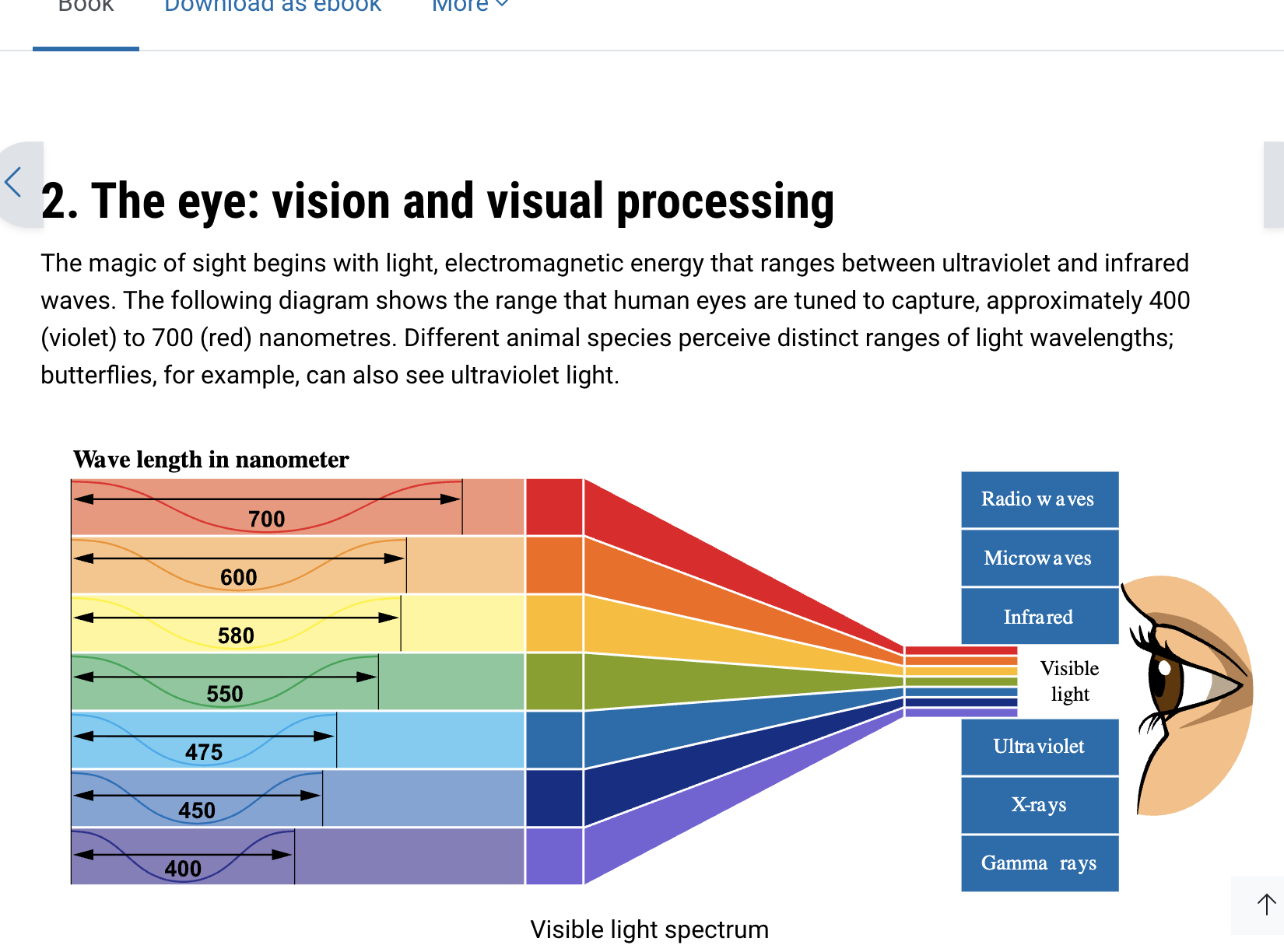
Absorption
The process by which an object takes in light energy rather than reflecting it is referred to as
Black
An object that absorbs nearly all visible light appears ______.
White
An object that reflects nearly all visible light appears ______.
Red, green and blue
The three hues that human eyes are naturally attuned to are ______, ______ and ______.
White Light
Mixing equal amounts of red, green and blue light produces ______ light.
Subtractive Color Mixing
The process of combining pigments, where more wavelengths are absorbed and darker colours result, is referred to as ______.
Fovea
The region of the retina responsible for sharp, detailed central vision is called the ______
Cones
he photoreceptors that are most densely concentrated in the fovea are ______.
Sensitivity
The high concentration of rods in the peripheral retina provides greater ______ to light.
Optic Nerve
The blind spot occurs where information exits the eye through the ______.
Cornea → pupil → lens → retina/fovea
What is the correct pathway of light through the eye?
It controls how much light enters the eye
What is the function of the pupil in vision?
Fovea
Where does incoming light converge for the sharpest vision?
Rods and Cones
Which photoreceptors detect light in the retina?
Bipolar cells
After rods and cones detect light, which cells receive and relay this information?
They relay information from rods and cones to retinal ganglion cells.
What is the main function of bipolar cells?
Which cells receive information from bipolar cells?
Retinal Ganglion Cells
They relay visual information from the retina toward the brain.
What is the role of retinal ganglion cells?
Rods/cones → bipolar cells → retinal ganglion cells → optic nerve → brain
What is the sequence of neural information after light reaches the retina?
It carries visual information from the eye to the brain.
Why is the optic nerve essential for vision?
Light → cornea → pupil → lens → retina → rods/cones → bipolar cells → retinal ganglion cells → optic nerve → brain.
What is the overall journey of visual information from light entering the eye to reaching the brain?
Photoreceptors (rods and cones) → bipolar cells → retinal ganglion cells.
Which three types of retinal cells form the main pathway for visual information?
A: Photoreceptors → Bipolar cells → Retinal ganglion cells
Hook: "P-B-G, Please Be Going" (out the optic nerve)
What is the 3-neuron chain of the retina (in order)?
A: Cones
Hook: "Rods = Rods in the dark; Cones = Cones for Color"
Rods vs Cones — which one is responsible for color vision, needs bright light, and is concentrated in the fovea?
Retinal ganglion cells (earlier cells use graded potentials only, since their axons form the optic nerve for long-distance signaling)
Which retinal cells are the ONLY ones that fire classic all-or-nothing action potentials?
The X-shaped crossing point at the base of the brain (near the pituitary) where nasal retina fibers cross to the opposite side; temporal fibers stay uncrossed
Q: What is the optic chiasm?
Bitemporal hemianopia (outer/temporal field loss, both eyes)
Damage AT the optic chiasm classically causes which visual field loss?
Half cross over to the opposite side; the rest stay on their original side
Hook: "Chiasm = choice point — cross or stay"
At the optic chiasm, what happens to the axons?
Nasal fibres (closer to the nose)
Which retinal fibres CROSS OVER at the chiasm — nasal or temporal?
Temporal fibres (closer to the temple)
Which retinal fibres stay on the SAME side (don't cross) at the chiasm?
The right hemisphere
Hook: "Opposite sides — Left field, Right brain"
Which hemisphere processes the LEFT visual field?
The thalamus (its visual region — the LGN)
Q: After the chiasm, most axons connect to which brain structure?
Visual form, location, and motion
What does the pathway to the PARIETAL lobe process?
Visual form and colour
What does the pathway to the TEMPORAL lobe process?
Multiple input signals from several cells coming together to be processed by a single output cell
Hook: "Convergence = Crowd into one"
What is "convergence" in visual processing?
126 million rods and cones → about 1 million retinal ganglion cells
Hook: "Huge army of soldiers, small group of generals"
Roughly how many rods and cones are in the retina, and how many retinal ganglion cells do they funnel into?
Because of their much higher convergence — many rods pooling signals into one ganglion cell amplifies faint light
Why are rods so much more sensitive to light than cones?
A: About 120 rods
Hook: "120 Rods Report to one boss"
Q: Roughly how many rods converge onto a single retinal ganglion cell?
About 6 cones
Hook: "Cones are Choosier — only 6 to one"
Roughly how many cones converge onto a single retinal ganglion cell (outside the fovea)?
A: One-to-one — each cone connects to its own single retinal ganglion cell
Hook: "Fovea = FOR-one-only" (no sharing)
Q: What is the convergence ratio at the fovea?
Cross
Nasal fibres cross or stay?
Stay
Temporal fibres cross or stay?
Optic nerve → optic chiasm → thalamus → V1 → V2 → parietal/temporal pathways.
What is the basic pathway of visual information through the brain?
Primary visual cortex — the first major cortical area to process visual information.
What is V1?
Secondary visual cortex — receives information from V1 for further visual processing.
What is V2?
first stop in the visual cortex
V1 =
second stop
V2 =
Visual form and colour
What does the temporal pathway process?
Macula
The central part of the retina that allows us to see detail
Where the eye changes the shape of the lens to focus near objects clearly on the retina.
What is the accommodation process of the eye?
Ciliary muscles contract → suspensory ligaments relax → lens becomes thicker/rounder → refractive power increases.
What happens when focusing on a near object?
Ciliary muscles relax → suspensory ligaments become taut (tight) → lens becomes flatter → refractive power decreases.
What happens when focusing on a distant object?
They change the shape of the lens to help the eye focus on objects at different distances.
What is the role of the ciliary muscles?
A photoreceptor cell in the retina that is highly sensitive to light and helps us see in low-light conditions.
What is a rod?
Night vision
Rod =
No. Rods mainly provide black-and-white/greyscale vision.
Do rods detect colour?
A photoreceptor cell in the retina that detects colour and provides sharp, detailed vision, especially in bright light.
What is a cone?
= Colour + Clarity 🌈👁
Cones