Nervous System (description)

0.0(0)
Studied by 8 people
call kaiCall Kai
Locked
learnLearn
examPractice Test
spaced repetitionSpaced Repetition
heart puzzleMatch
flashcardsFlashcards
GameKnowt Play
Card Sorting

1/11

encourage image

There's no tags or description

Looks like no tags are added yet.

Last updated 1:48 AM on 8/19/26
Name
Mastery
Learn
Test
Matching
Spaced
Call with Kai
Chat

No analytics yet

Send a link to your students to track their progress

12 Terms
1
New cards
term image

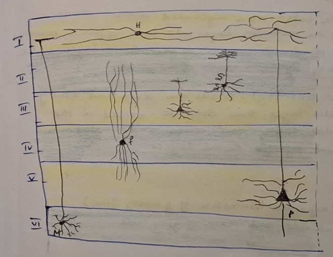
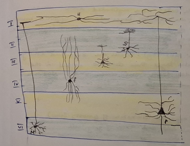
Cerebrum - HE

1. Molecular layer

  • right under pia mater; few cells

  • horizontal cells

  • dendrites

  • axons

• 2. External granular layer

  • small pyramidal and granular/stellate cells

• 3. External pyramidal layer

• medium and large pyramid cells

• Martinotti cells

4. internal granular layer

  • small granular and star-shaped cells

  • outer band of Baillarger

5. internal pyramidal layer (ganglionalis) -

  • large pyramid cells

  • inner band of Baillarger

6. Multiform layer

  • spindle cells - cells of Martinotti

  • modified pyramid cells


<p>Cerebrum - HE</p><p><strong>1. Molecular layer</strong></p><ul><li><p>right under pia mater; few cells</p></li><li><p>horizontal cells</p></li><li><p>dendrites</p></li><li><p>axons</p></li></ul><p><strong>• 2. External granular layer</strong></p><ul><li><p>small pyramidal and granular/stellate cells</p></li></ul><p><strong>• 3. External pyramidal layer</strong></p><p>• medium and large pyramid cells</p><p>• Martinotti cells</p><p><strong>4. internal granular layer</strong></p><ul><li><p>small granular and star-shaped cells</p></li><li><p>outer band of Baillarger</p></li></ul><p><strong>5. internal pyramidal layer (ganglionalis)</strong> -</p><ul><li><p>large pyramid cells</p></li><li><p>inner band of Baillarger</p></li></ul><p><strong>6. Multiform layer</strong></p><ul><li><p>spindle cells - cells of Martinotti</p></li><li><p>modified pyramid cells</p></li></ul><p></p>
2
New cards
term image

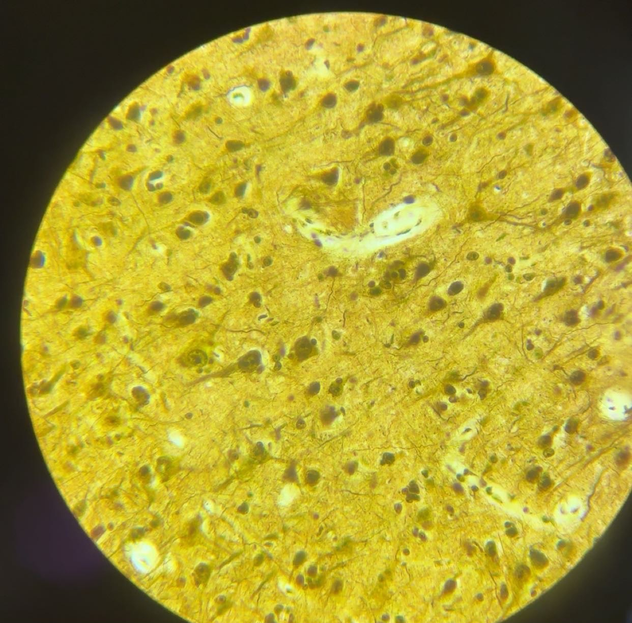
Cerebrum - AgNO3

1. Molecular layer

  • right under pia mater; few cells

  • horizontal cells

  • dendrites

  • axons

• 2. External granular layer

  • small pyramidal and granular cells

• 3. External pyramidal layer

• medium and large pyramid cells

• Martinotti cells

4. internal granular layer

  • small granular and star-shaped cells

  • outer band of Baillarger

5. internal pyramidal layer (ganglionalis) -

  • large pyramid cells

  • inner band of Baillarger

6. Multiform layer

  • spindle cells - cells of Martinotti

  • modified pyramid cells


<p>Cerebrum - AgNO3</p><p><strong>1. Molecular layer</strong></p><ul><li><p>right under pia mater; few cells</p></li><li><p>horizontal cells</p></li><li><p>dendrites</p></li><li><p>axons</p></li></ul><p><strong>• 2. External granular layer</strong></p><ul><li><p>small pyramidal and granular cells</p></li></ul><p><strong>• 3. External pyramidal layer</strong></p><p>• medium and large pyramid cells</p><p>• Martinotti cells</p><p><strong>4. internal granular layer</strong></p><ul><li><p>small granular and star-shaped cells</p></li><li><p>outer band of Baillarger</p></li></ul><p><strong>5. internal pyramidal layer (ganglionalis)</strong> -</p><ul><li><p>large pyramid cells</p></li><li><p>inner band of Baillarger</p></li></ul><p><strong>6. Multiform layer</strong></p><ul><li><p>spindle cells - cells of Martinotti</p></li><li><p>modified pyramid cells</p></li></ul><p></p>
3
New cards
<p></p>


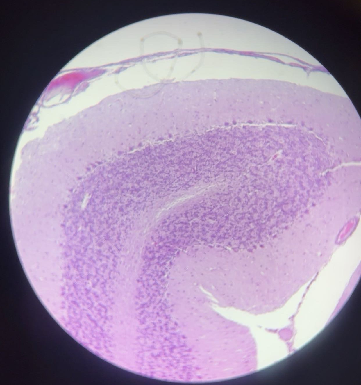
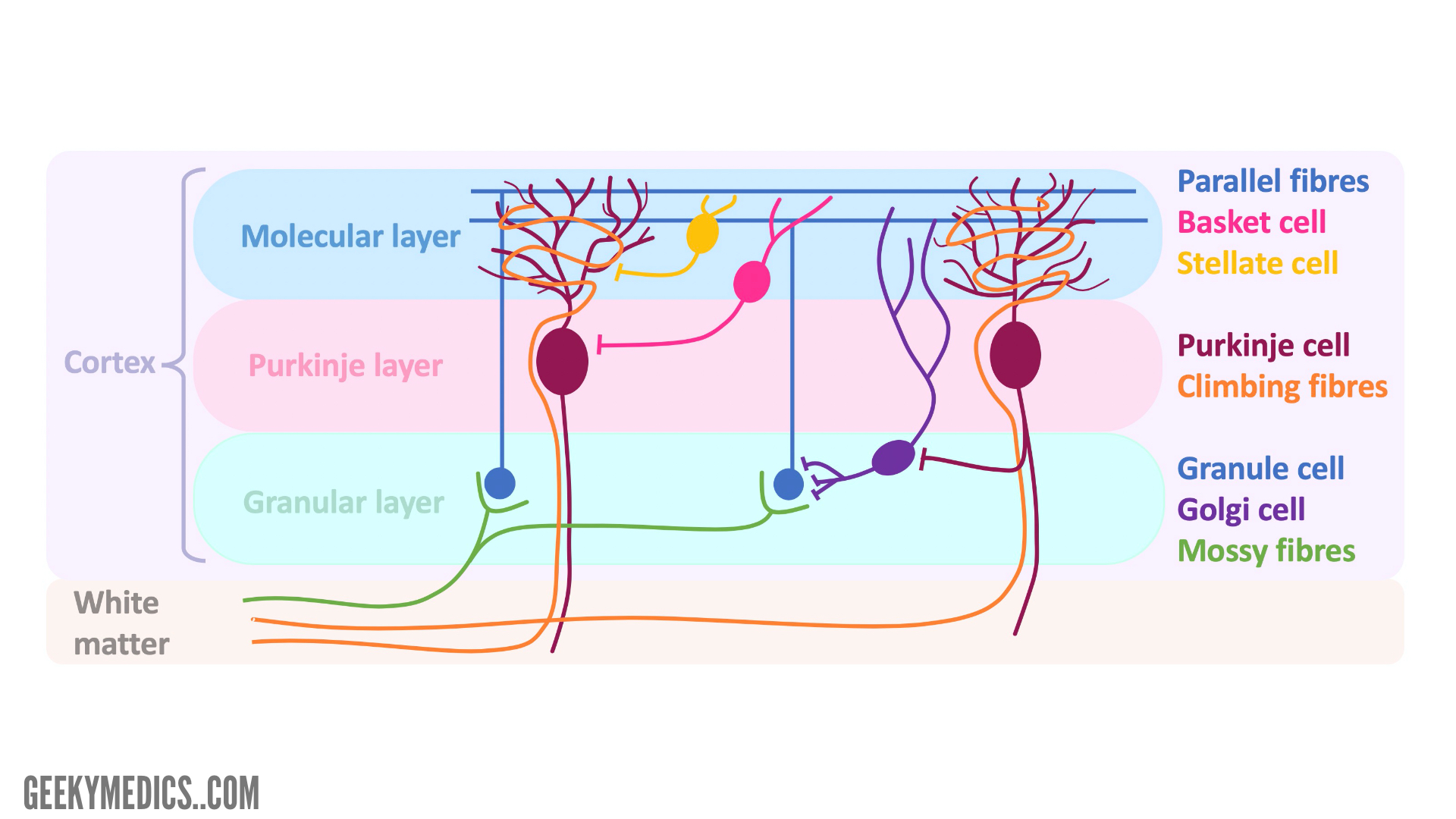
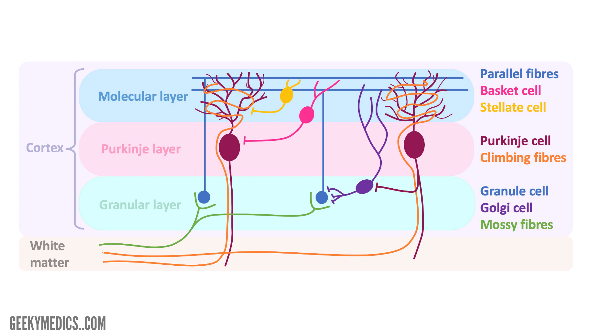
Cerebellum - HE


3 Layers:


1) Molecular Layer (unmyelinated)

  • mostly dendrites of Purkinje cells and axons of granular cells

  • some stellate and basket cells

2) Purkinje Cell Layer (myelinated)

  • single layer of Purkinje cells

  • dendrites branch to molecular layer

  • axons travel to white matter and are GABA-ergic

3) Granular Layer

  • granule cells

  • golgi cells

  • glomerulus: dendrites of granule cells and axons of golgi cells, synapse occurs here


White Matter (Corpus Medullare):

  • fibriae proprae: connect cerebellar region

  • projection fibres: connect cerebellum to other region

  • axon of purkinje cells

  • mossy fibers

  • climbing fibers


<p>Cerebellum - HE</p><p class="ds-markdown-paragraph"></p><p class="ds-markdown-paragraph">3 Layers:</p><p class="ds-markdown-paragraph"></p><p class="ds-markdown-paragraph">1) Molecular Layer (unmyelinated)</p><ul><li><p class="ds-markdown-paragraph">mostly dendrites of Purkinje cells and axons of granular cells</p></li><li><p class="ds-markdown-paragraph">some stellate and basket cells</p></li></ul><p class="ds-markdown-paragraph">2) Purkinje Cell Layer (myelinated)</p><ul><li><p class="ds-markdown-paragraph">single layer of Purkinje cells</p></li><li><p class="ds-markdown-paragraph">dendrites branch to molecular layer</p></li><li><p class="ds-markdown-paragraph">axons travel to white matter and are GABA-ergic</p></li></ul><p class="ds-markdown-paragraph">3) Granular Layer </p><ul><li><p class="ds-markdown-paragraph">granule cells</p></li><li><p class="ds-markdown-paragraph">golgi cells</p></li><li><p class="ds-markdown-paragraph">glomerulus: dendrites of granule cells and axons of golgi cells, synapse occurs here</p></li></ul><p class="ds-markdown-paragraph"></p><p class="ds-markdown-paragraph">White Matter (Corpus Medullare):</p><ul><li><p class="ds-markdown-paragraph">fibriae proprae: connect cerebellar region</p></li><li><p class="ds-markdown-paragraph">projection fibres: connect cerebellum to other region</p></li><li><p class="ds-markdown-paragraph">axon of purkinje cells</p></li><li><p class="ds-markdown-paragraph">mossy fibers</p></li><li><p class="ds-markdown-paragraph">climbing fibers</p></li></ul><p></p>
4
New cards
term image

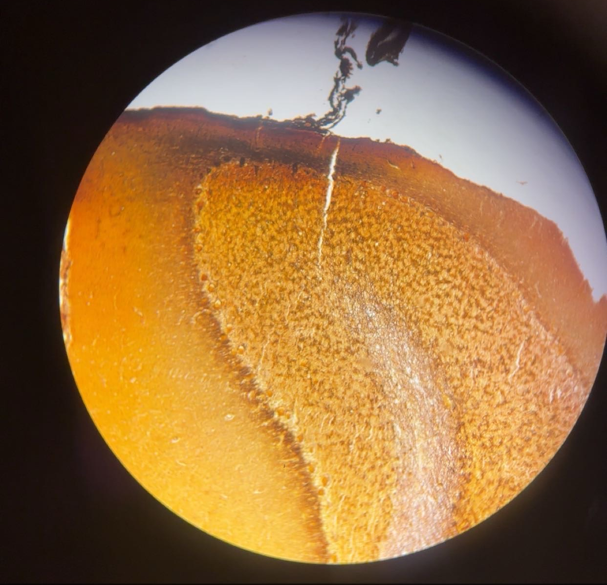
Cerebellum- AgNO3


3 Layers:


1) Molecular Layer (unmyelinated)

  • mostly dendrites of Purkinje cells and axons of granular cells

  • some stellate and basket cells

2) Purkinje Cell Layer (myelinated)

  • single layer of Purkinje cells

  • dendrites branch to molecular layer

  • axons travel to white matter and are GABA-ergic

3) Granular Layer

  • granule cells

  • golgi cells

  • glomerulus: dendrites of granule cells and axons of golgi cells, synapse occurs here


White Matter (Corpus Medullare):

  • fibriae proprae: connect cerebellar region

  • projection fibres: connect cerebellum to other region

  • axon of purkinje cells

  • mossy fibers

  • climbing fibers


<p>Cerebellum- AgNO3</p><p></p><p>3 Layers:</p><p></p><p>1) Molecular Layer (unmyelinated)</p><ul><li><p class="ds-markdown-paragraph">mostly dendrites of Purkinje cells and axons of granular cells</p></li><li><p class="ds-markdown-paragraph">some stellate and basket cells</p></li></ul><p class="ds-markdown-paragraph">2) Purkinje Cell Layer (myelinated)</p><ul><li><p class="ds-markdown-paragraph">single layer of Purkinje cells</p></li><li><p class="ds-markdown-paragraph">dendrites branch to molecular layer</p></li><li><p class="ds-markdown-paragraph">axons travel to white matter and are GABA-ergic</p></li></ul><p class="ds-markdown-paragraph">3) Granular Layer </p><ul><li><p class="ds-markdown-paragraph">granule cells</p></li><li><p class="ds-markdown-paragraph">golgi cells</p></li><li><p class="ds-markdown-paragraph">glomerulus: dendrites of granule cells and axons of golgi cells, synapse occurs here</p></li></ul><p class="ds-markdown-paragraph"></p><p class="ds-markdown-paragraph">White Matter (Corpus Medullare):</p><ul><li><p class="ds-markdown-paragraph">fibriae proprae: connect cerebellar region</p></li><li><p class="ds-markdown-paragraph">projection fibres: connect cerebellum to other region</p></li><li><p class="ds-markdown-paragraph">axon of purkinje cells</p></li><li><p class="ds-markdown-paragraph">mossy fibers</p></li><li><p class="ds-markdown-paragraph">climbing fibers</p></li></ul><p></p>
5
New cards
term image

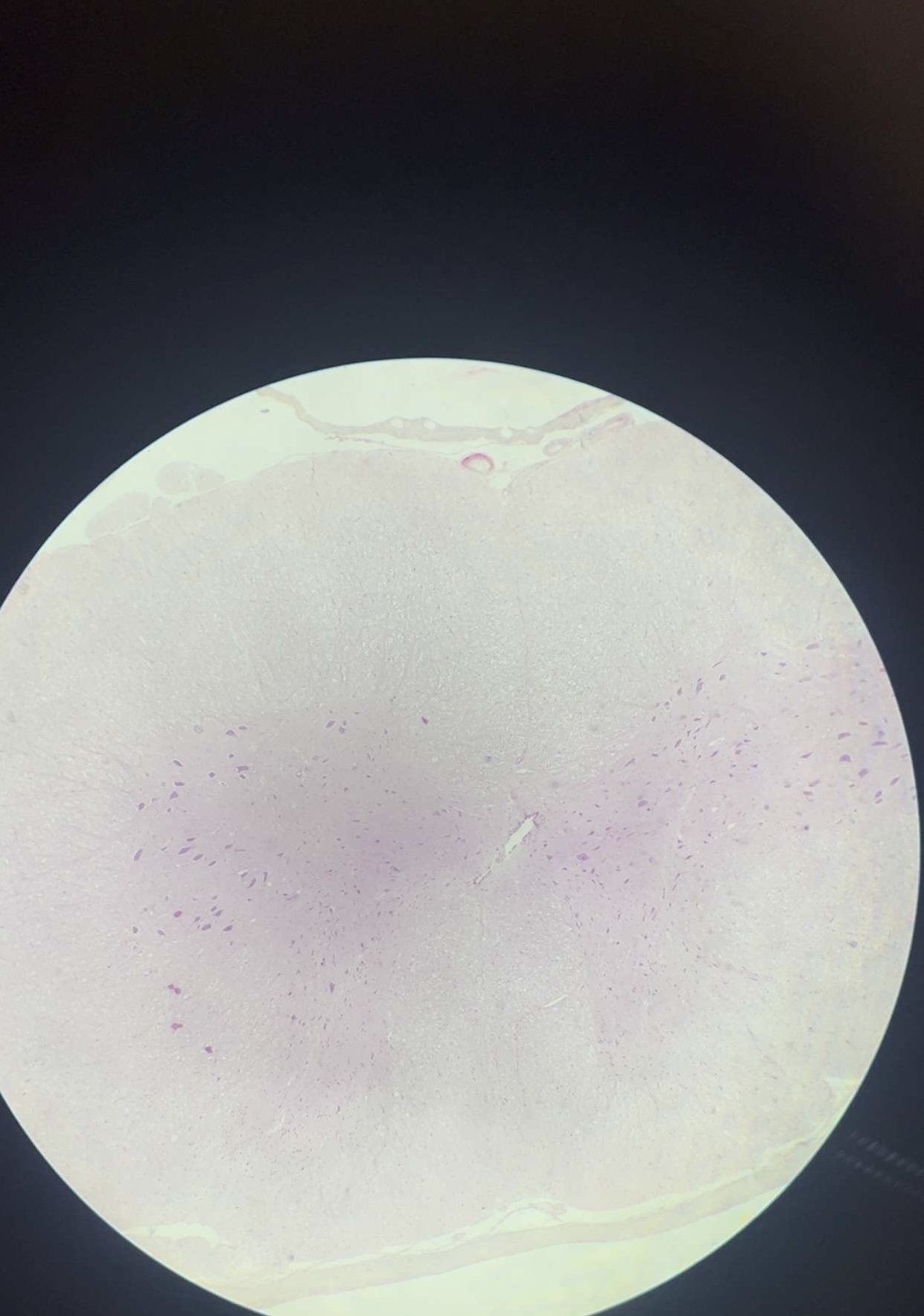
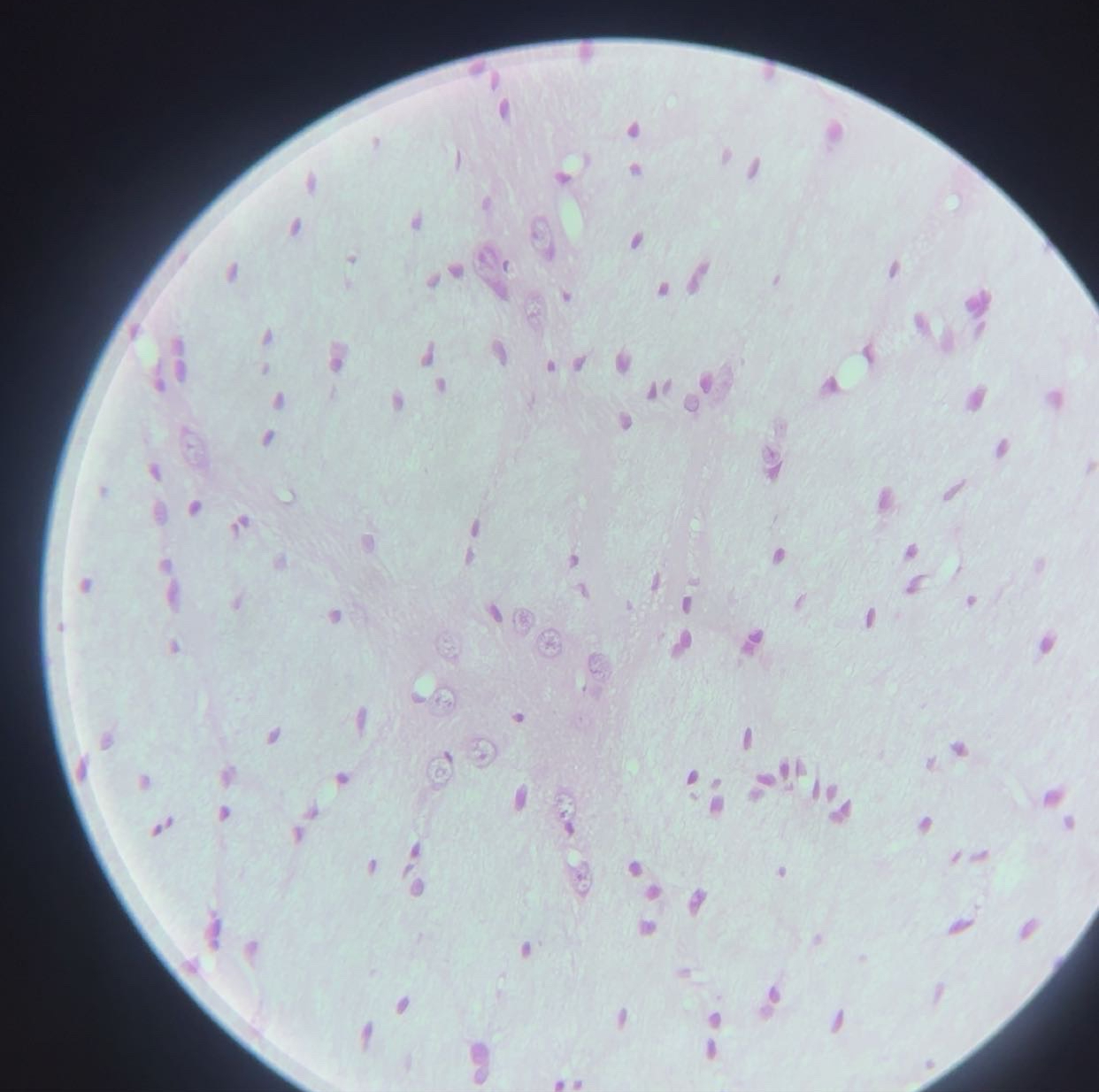
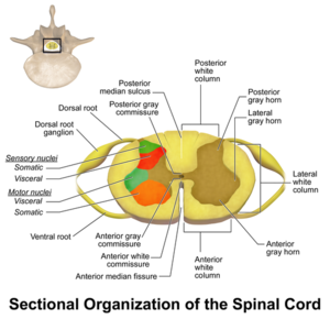
Medulla spinalis - HE


1. Gray Matter (Internal "H/Butterfly")

  • Anterior Horn: Large, rounded; contains multipolar motor neurons & Nissl bodies.

  • Posterior Horn: Slender; contains interneurons for sensory processing.

  • Lateral Horn: (T1-L2 only); contains autonomic (sympathetic) neurons.

  • Central Canal: Center of gray commissure; lined with ciliated ependymal cells.

2. White Matter (External Columns)

  • Organization: Divided into Anterior, Lateral, and Posterior Funiculi.

  • Composition: Bundles of myelinated axons (tracts) + neuroglia (oligodendrocytes/astrocytes).

  • Appearance: Myelin appears as "clear halos" around central axonal dots.

3. Landmarks

  • Anterior: Deep/wide Anterior Median Fissure.

  • Posterior: Narrow Posterior Median Septum.

  • Covering: Pia mater (innermost meninx) adheres to the surface.

Stain:

Shows large motor neuron cell bodies and blue nuclei of glia clearly.

<p>Medulla spinalis - HE</p><p></p><p><strong>1. Gray Matter (Internal "H/Butterfly")</strong></p><ul><li><p><strong>Anterior Horn:</strong> Large, rounded; contains <strong>multipolar motor neurons</strong> &amp; <strong>Nissl bodies</strong>.</p></li><li><p><strong>Posterior Horn:</strong> Slender; contains interneurons for sensory processing.</p></li><li><p><strong>Lateral Horn:</strong> (T1-L2 only); contains autonomic (sympathetic) neurons.</p></li><li><p><strong>Central Canal:</strong> Center of gray commissure; lined with <strong>ciliated ependymal cells</strong>.</p></li></ul><p><strong>2. White Matter (External Columns)</strong></p><ul><li><p><strong>Organization:</strong> Divided into <strong>Anterior, Lateral, and Posterior Funiculi</strong>.</p></li><li><p><strong>Composition:</strong> Bundles of myelinated axons (tracts) + neuroglia (<strong>oligodendrocytes/astrocytes</strong>).</p></li><li><p><strong>Appearance:</strong> Myelin appears as "clear halos" around central axonal dots.</p></li></ul><p><strong>3. Landmarks</strong></p><ul><li><p><strong>Anterior:</strong> Deep/wide <strong>Anterior Median Fissure</strong>.</p></li><li><p><strong>Posterior:</strong> Narrow <strong>Posterior Median Septum</strong>.</p></li><li><p><strong>Covering:</strong> <strong>Pia mater</strong> (innermost meninx) adheres to the surface.</p></li></ul><p>Stain: </p><p>Shows large motor neuron cell bodies and blue nuclei of glia clearly.</p><p></p>
6
New cards
term image

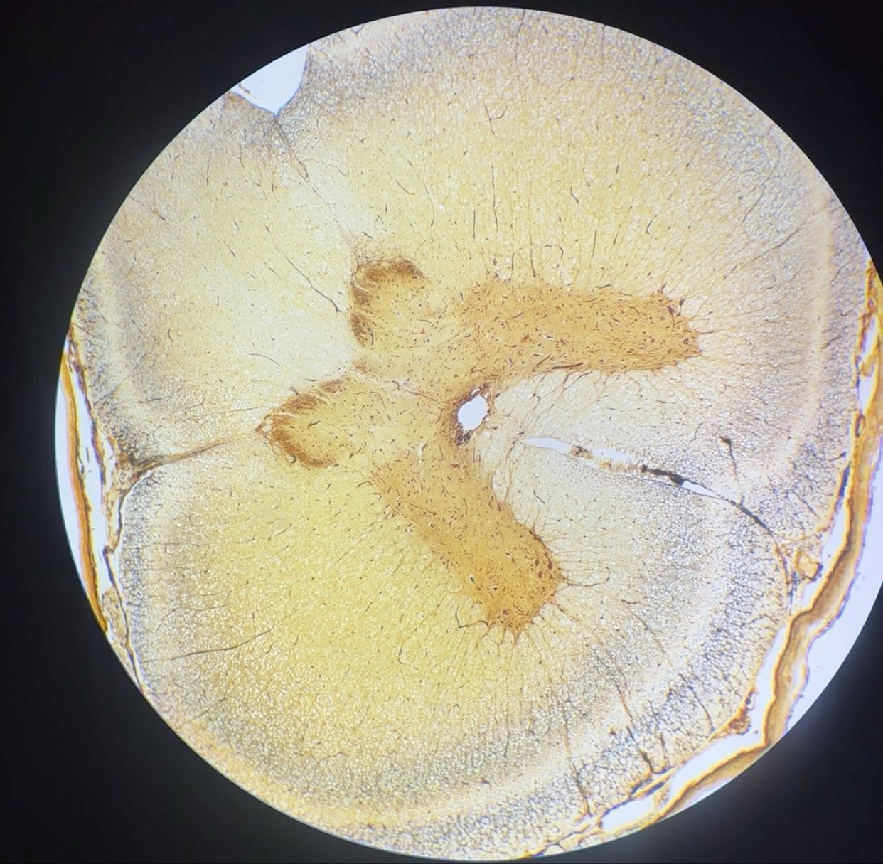
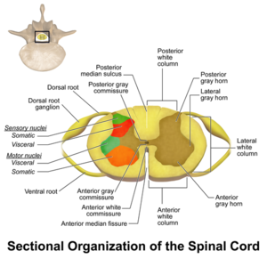
Medulla spinalis - AgNO3


1. Gray Matter (Internal "H/Butterfly")

  • Anterior Horn: Large, rounded; contains multipolar motor neurons & Nissl bodies.

  • Posterior Horn: Slender; contains interneurons for sensory processing.

  • Lateral Horn: (T1-L2 only); contains autonomic (sympathetic) neurons.

  • Central Canal: Center of gray commissure; lined with ciliated ependymal cells.

2. White Matter (External Columns)

  • Organization: Divided into Anterior, Lateral, and Posterior Funiculi.

  • Composition: Bundles of myelinated axons (tracts) + neuroglia (oligodendrocytes/astrocytes).

  • Appearance: Myelin appears as "clear halos" around central axonal dots.

3. Landmarks

  • Anterior: Deep/wide Anterior Median Fissure.

  • Posterior: Narrow Posterior Median Septum.

  • Covering: Pia mater (innermost meninx) adheres to the surface.


Stain:

Highlights the dense network of fibers (neuropil) and branching dendrites.

<p>Medulla spinalis - AgNO3</p><p></p><p class="ds-markdown-paragraph"><strong>1. Gray Matter (Internal "H/Butterfly")</strong></p><ul><li><p><strong>Anterior Horn:</strong> Large, rounded; contains <strong>multipolar motor neurons</strong> &amp; <strong>Nissl bodies</strong>.</p></li><li><p><strong>Posterior Horn:</strong> Slender; contains interneurons for sensory processing.</p></li><li><p><strong>Lateral Horn:</strong> (T1-L2 only); contains autonomic (sympathetic) neurons.</p></li><li><p><strong>Central Canal:</strong> Center of gray commissure; lined with <strong>ciliated ependymal cells</strong>.</p></li></ul><p><strong>2. White Matter (External Columns)</strong></p><ul><li><p><strong>Organization:</strong> Divided into <strong>Anterior, Lateral, and Posterior Funiculi</strong>.</p></li><li><p><strong>Composition:</strong> Bundles of myelinated axons (tracts) + neuroglia (<strong>oligodendrocytes/astrocytes</strong>).</p></li><li><p><strong>Appearance:</strong> Myelin appears as "clear halos" around central axonal dots.</p></li></ul><p><strong>3. Landmarks</strong></p><ul><li><p><strong>Anterior:</strong> Deep/wide <strong>Anterior Median Fissure</strong>.</p></li><li><p><strong>Posterior:</strong> Narrow <strong>Posterior Median Septum</strong>.</p></li><li><p><strong>Covering:</strong> <strong>Pia mater</strong> (innermost meninx) adheres to the surface.</p></li></ul><p></p><p>Stain:</p><p>Highlights the dense network of fibers (<strong>neuropil</strong>) and branching dendrites.</p><p></p>
7
New cards
term image

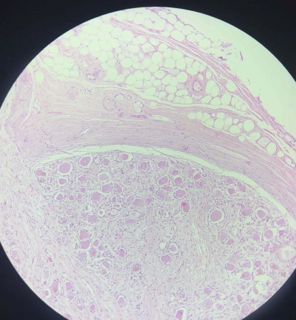
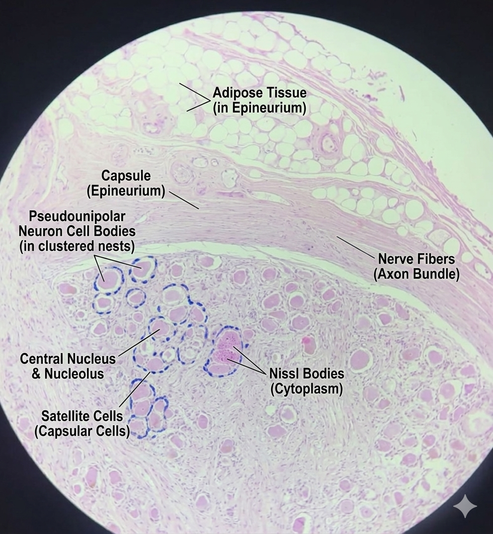
Ganglion spinale - HE

1. The Capsule (The Outer Shell)

The entire ganglion is enveloped in a thick layer of dense irregular connective tissue called the epineurium (or simply the capsule). You will often see small blood vessels and adipose tissue surrounding it.

2. Pseudounipolar Neurons (aka ganglion cells)

The most striking feature of a spinal ganglion slide is the collection of pseudounipolar sensory neurons.

  • Location: They are typically arranged in dense clusters or "nests" toward the periphery of the ganglion.

  • Appearance: These cells are large, spherical, and have a centrally located, pale nucleus with a very prominent, dark "owl’s eye" nucleolus.

  • Nissl Bodies: In H&E stains, the cytoplasm is granular and basophilic (purplish) due to the high density of RER.

3. Satellite Cells (The "Ring")

Each large neuron is surrounded by a neat ring of small, flat cells called satellite cells (or capsular cells).

  • Function: They provide structural and metabolic support to the neuron.

  • Identification: Look for a "halo" of tiny, dark nuclei hugging the perimeter of the large neuronal cell body.

4. Nerve Fiber Bundles

In the center of the ganglion, you will see bundles of nerve fibers (axons) passing through.

  • In H&E, these look like wavy, pink longitudinal streaks.

  • In AgNO3, these look like dark brown or black threads.

<p>Ganglion spinale - HE</p><p class="ds-markdown-paragraph"><strong>1. The Capsule (The Outer Shell)</strong></p><p>The entire ganglion is enveloped in a thick layer of dense irregular connective tissue called the <strong>epineurium</strong> (or simply the capsule). You will often see small blood vessels and adipose tissue surrounding it.</p><p><strong>2. Pseudounipolar Neurons (aka ganglion cells)</strong></p><p>The most striking feature of a spinal ganglion slide is the collection of <strong>pseudounipolar sensory neurons</strong>.</p><ul><li><p><strong>Location:</strong> They are typically arranged in dense clusters or "nests" toward the periphery of the ganglion.</p></li><li><p><strong>Appearance:</strong> These cells are large, spherical, and have a centrally located, pale nucleus with a very prominent, dark "owl’s eye" <strong>nucleolus</strong>.</p></li><li><p><strong>Nissl Bodies:</strong> In H&amp;E stains, the cytoplasm is granular and basophilic (purplish) due to the high density of RER.</p></li></ul><p><strong>3. Satellite Cells (The "Ring")</strong></p><p>Each large neuron is surrounded by a neat ring of small, flat cells called <strong>satellite cells</strong> (or capsular cells).</p><ul><li><p><strong>Function:</strong> They provide structural and metabolic support to the neuron.</p></li><li><p><strong>Identification:</strong> Look for a "halo" of tiny, dark nuclei hugging the perimeter of the large neuronal cell body.</p></li></ul><p><strong>4. Nerve Fiber Bundles</strong></p><p>In the center of the ganglion, you will see bundles of nerve fibers (axons) passing through.</p><ul><li><p>In <strong>H&amp;E</strong>, these look like wavy, pink longitudinal streaks.</p></li><li><p>In <strong>AgNO3</strong>, these look like dark brown or black threads.</p></li></ul><p></p>
8
New cards
term image

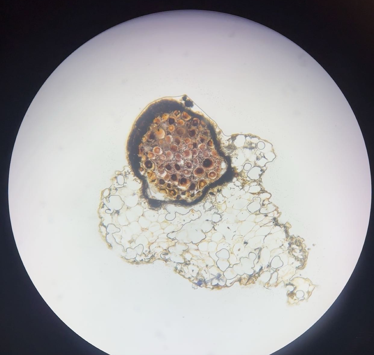
Ganglion spinale - AgNO3

1. The Capsule (The Outer Shell)

The entire ganglion is enveloped in a thick layer of dense irregular connective tissue called the epineurium (or simply the capsule). You will often see small blood vessels and adipose tissue surrounding it.

2. Pseudounipolar Neurons (aka ganglion cells)

The most striking feature of a spinal ganglion slide is the collection of pseudounipolar sensory neurons.

  • Location: They are typically arranged in dense clusters or "nests" toward the periphery of the ganglion.

  • Appearance: These cells are large, spherical, and have a centrally located, pale nucleus with a very prominent, dark "owl’s eye" nucleolus.

  • Nissl Bodies: In H&E stains, the cytoplasm is granular and basophilic (purplish) due to the high density of RER.

3. Satellite Cells (The "Ring")

Each large neuron is surrounded by a neat ring of small, flat cells called satellite cells (or capsular cells).

  • Function: They provide structural and metabolic support to the neuron.

  • Identification: Look for a "halo" of tiny, dark nuclei hugging the perimeter of the large neuronal cell body.

4. Nerve Fiber Bundles

In the center of the ganglion, you will see bundles of nerve fibers (axons) passing through.

  • In H&E, these look like wavy, pink longitudinal streaks.

  • In AgNO3, these look like dark brown or black threads.

9
New cards
term image

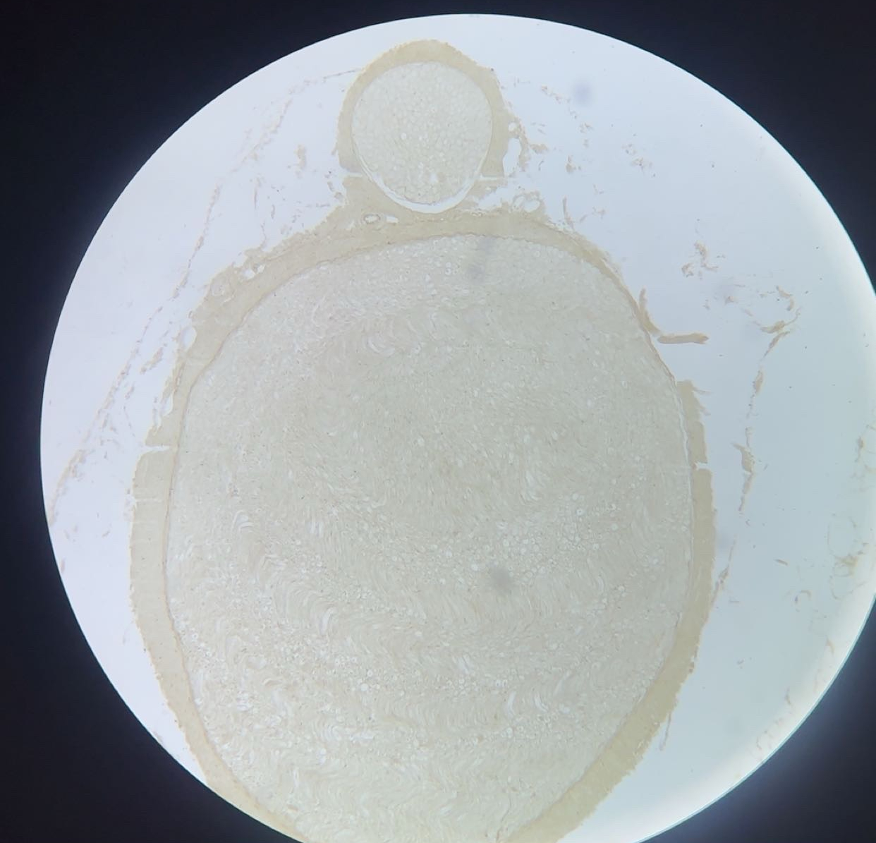
Peripheral nerve - OsO4

The nerve is organized into three distinct layers of connective tissue (from largest to smallest):

  • Epineurium: The outermost, thick layer of dense irregular connective tissue. It holds multiple fascicles together and often contains adipose tissue and vasa nervorum (blood vessels).

  • Perineurium: A specialized, denser layer that surrounds each individual fascicle (bundle of axons). In a slide, it looks like a distinct "ring" or "sleeve" around the bundle. It acts as a blood-nerve barrier.

  • Endoneurium: The delicate, thin layer of loose connective tissue surrounding each individual axon. In a cross-section, it’s the space between the nerve fibers.


Each nerve fiber consists of an axon surrounded by Schwann cells. The fibres may be myelinated or non-myelinated.

Stain: focuses on lipids. Best for seeing myelin sheaths and nodes of ranvier.

10
New cards
term image

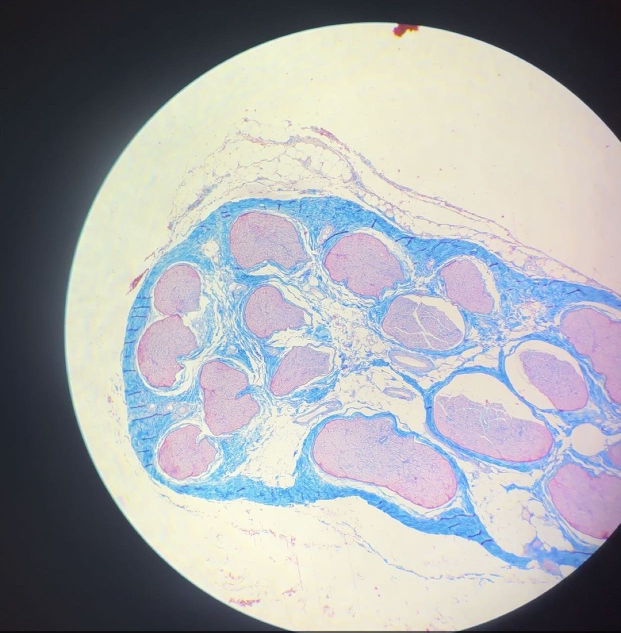
Peripheral nerve - azan

The nerve is organized into three distinct layers of connective tissue (from largest to smallest):

  • Epineurium: The outermost, thick layer of dense irregular connective tissue. It holds multiple fascicles together and often contains adipose tissue and vasa nervorum (blood vessels).

  • Perineurium: A specialized, denser layer that surrounds each individual fascicle (bundle of axons). In a slide, it looks like a distinct "ring" or "sleeve" around the bundle. It acts as a blood-nerve barrier.

  • Endoneurium: The delicate, thin layer of loose connective tissue surrounding each individual axon. In a cross-section, it’s the space between the nerve fibers.


Each nerve fiber consists of an axon surrounded by Schwann cells. The fibres may be myelinated or non-myelinated.


  • Key Feature:

    • Azan highlights collagen (blue) vs. neural elements (pink/red)

11
New cards
term image

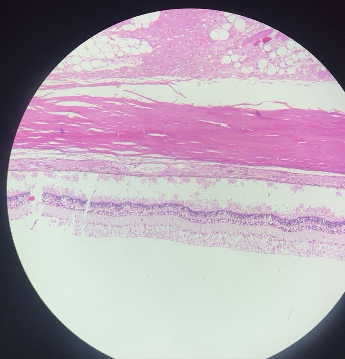
Eye - HE


3 tunics:

  • fibrous layer - sclera, cornea

  • vascular layer - choroid, ciliary body and iris

  • retina


sclera: white of eye. it is opaque, protective layer

1) conjunctival epithelium - str. sq. non-keratinized epithelium

2) episclera

3) stroma

4) l . fusca

5) endothelium


cornea:

1) corneal epithelium - str. sq. non-keratinized epithelium

2) ant. limiting membrane

3) stroma

4) post. limiting lamina

5) endothelium


Retina’s 10 layers:

  • Pigment Epithelium: A single layer of cuboidal cells filled with dark melanin.

  • Photoreceptor Layer: The outer and inner segments of Rods and Cones.

  • External Limiting Membrane: A thin line of junctions between photoreceptors and Müller cells.

  • Outer Nuclear Layer: Dense, dark-blue nuclei of the Rods and Cones.

  • Outer Plexiform Layer: A pale pink layer where photoreceptors synapse with bipolar cells.

  • Inner Nuclear Layer: Nuclei of Bipolar, Amacrine, and Horizontal cells.

  • Inner Plexiform Layer: A pale pink synaptic layer.

  • Ganglion Cell Layer: A sparse layer of very large, multipolar neurons.

  • Nerve Fiber Layer: Axons that travel toward the optic disc (forming the Optic Nerve).

  • Internal Limiting Membrane: The innermost boundary.

mnemonic: Peter Pan Only Offered Orange Ice In Geoffrey’s New Island

12
New cards
term image

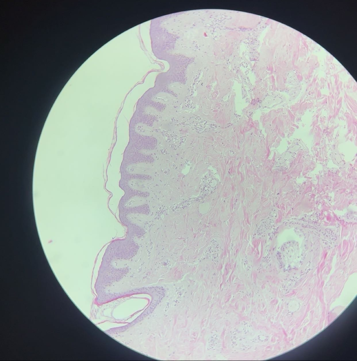
Skin (cutis) - HE

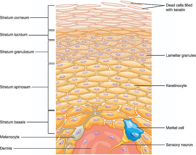
1) Epidermis-stratified squamous keratinized epithelium

  • str. basale - has stem cells

  • st. spinosum - desmosomes

  • st. granulosum - 3-5 dead flattened cells

  • str. lucidum - clear line of dead cells

  • str. corneum - outermost layer, with flattened, dead, keratin-filled cells

2) Derma - thick irregular CT

  • papillary layer - forms ‘humps’ aka dermal papillae that interlock with epidermis

  • reticular layer - layer of thick dense irregular CT

3) Hypodermis- subcutaneous tissue

  • fat is the major component

  • also contains fibroblasts, macrophages. etc.

  • pacinian corpuscle, sebaceous gland, hair follicles and sweat glands

<p>Skin (cutis) - HE</p><p class="ds-markdown-paragraph">1) <strong>Epidermis</strong>-stratified squamous keratinized epithelium</p><ul><li><p class="ds-markdown-paragraph">str. basale - has stem cells</p></li><li><p class="ds-markdown-paragraph">st. spinosum - desmosomes</p></li><li><p class="ds-markdown-paragraph">st. granulosum - 3-5 dead flattened cells</p></li><li><p class="ds-markdown-paragraph">str. lucidum - clear line of dead cells</p></li><li><p class="ds-markdown-paragraph">str. corneum - outermost layer, with flattened, dead, keratin-filled cells</p></li></ul><p>2) <strong>Derma - thick irregular CT</strong></p><ul><li><p>papillary layer - forms ‘humps’ aka dermal papillae that interlock with epidermis</p></li><li><p>reticular layer - layer of thick dense irregular CT</p></li></ul><p>3) <strong>Hypodermis</strong>- subcutaneous tissue</p><ul><li><p>fat is the major component</p></li><li><p>also contains fibroblasts, macrophages. etc. </p></li><li><p>pacinian corpuscle, sebaceous gland, hair follicles and sweat glands</p></li></ul><p></p>