Cardiovascular System Lecture 1 and 2
1/206
There's no tags or description
Looks like no tags are added yet.
Name | Mastery | Learn | Test | Matching | Spaced | Call with Kai | Chat |
|---|
No analytics yet
Send a link to your students to track their progress
207 Terms
Components of the circulatory system
Pump (heart)
Liquid (blood)
Container (vasculature)
Control system
Add adaptibility notes and image
yes
Add circulatory system image
yes
Pump
A pump is a device that moved fluids by mechanical action. Pumps consume energy to perform mechanical work.
Herat anatomy overview
4 chambers
4 valves
4 chambers of heart
Right atrium
Right ventricle
Left atrium
Left ventricle
4 valves of heart
Right atrioventricular valve
Pulmonic valve
Left atrioventricular valve
Aortic valve
Add image of of heart
yes
Right atrioventricular valve
Also known as the tricuspid valve, it is located between the right atrium and right ventricle, preventing backflow of blood into the atrium during ventricular contraction.
Pulmonic valve
Also known as the pulmonary valve, it is situated between the right ventricle and the pulmonary artery, allowing blood to flow from the heart to the lungs for oxygenation.
Left atrioventricular valve
Also known as the mitral valve, it is located between the left atrium and left ventricle, preventing backflow of blood into the atrium during ventricular contraction.
Aortic valve
Also known as the aortic semilunar valve, it is positioned between the left ventricle and the aorta, allowing blood to exit the heart and enter the systemic circulation.
Add heart gross anatomy image
yes
Add heart cross section image
yes
Blood flow through heart
Right atriu
Right ventricle
Pulmonary artery
Lungs
Pulmonary veins
Left atrium
Left ventricle
Aorta
Body
Vena cava
Add blood flow through heart image of stuff on side
yes
Add vascular network iamge
yes
Components of the heart
Myocardium
Valves
Conduction system
Coronary vasculature
Myocardium
The myocardium is the muscular layer of the heart responsible for contracting and pumping blood throughout the body. It is composed of cardiac muscle tissue and forms the bulk of the heart wall.
Conduction system
The conduction system of the heart is a group of specialized cardiac muscle cells that generate and propagate electrical impulses, coordinating the heart's rhythm and ensuring efficient pumping of blood.
Coronary vasculature
The coronary vasculature refers to the network of blood vessels that supply blood to the heart muscle itself. This includes coronary arteries and veins that ensure sufficient oxygen and nutrient delivery to the myocardium.
Add left AV valve appartud image
yes
What cells reside in myocardium
Myocytes
Specialized conducting tissue (modified myocytes)
Fibroblasts
Endothelial cells
Nervous tissue
Myocytes
Muscle cells responsible for contraction of the heart, facilitating pumping action.
Myocyte examples
Cardiac muscle cells and Sinoatrial node cells
Add images of myocytes
yes
Specialized conducting tissue (modified myocytes)
includes the bundle of His and Purkinje fibers, which facilitate electrical conduction in the heart.
Specialized conducting tissue (modified myocytes) examples
include bundle branches and atrioventricular node.
Add Specialized conducting tissue (modified myocytes) imahes
yes
Fibroblasts
are a type of cell in connective tissue that produce collagen and other fibers. They play a critical role in wound healing and tissue repair.
Fibroblasts examples
include myofibroblasts and adipocytes.
Endothelial cells
are specialized cells that line the blood vessels and lymphatic vessels, playing a key role in vascular function and regulation.
Endothelial cell examples
include capillary endothelial cells and arterial endothelial cells.
Add iaes of fibroblasts and endothelial cells
yes
Nervous tissue
is a type of tissue that is responsible for transmitting signals throughout the body. It is composed of neurons and glial cells, playing a crucial role in communication and coordination of bodily functions.
Nervous tissue examples
include neurons and glial cells.
Add nervous tissue image
yes
Add myocardium image
yes
Add cardiac muscle image
yes
Add cardiac mopnocute image
ye
Add cardiac mucle v. sksletal musle image
yes
Add images of change in pressuie that are diving blood flow in heart
ye
What is the driving force for the movement of blood through vasculature?
The pressure gradient created by the contraction of the heart, particularly during systole. This pressure pushes blood from areas of higher pressure to areas of lower pressure, facilitating circulation.
Add chnages in pressure taht drive blood flow image
d
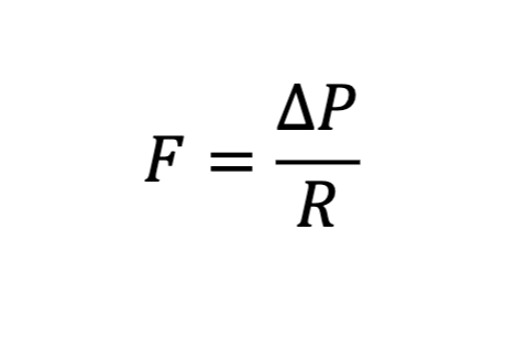
What does this show?
Pressure gradients drive the movement of blood through the circulatory system

What does each variable mean?
(Flow Rate): The volume of fluid moving through the system per unit of time (e.g., liters per minute).
(Pressure Gradient/Change in Pressure): The difference in pressure between the start and the end of the vessel. Pressure drives the fluid forward, so a higher difference creates more flow.
(Resistance): The friction or obstruction that the fluid encounters as it moves through the pipe or vessel. Higher resistance slows down the flow
Add pressures i the vasicular circuit image
d
Add transition form diastole yo systole image and othe rimage
yes
Diastole
The phase of the cardiac cycle when the heart muscle relaxes and the chambers fill with blood. It occurs before systole, which is the contraction phase.
Systole
The phase of the cardiac cycle where the heart muscles contract, pumping blood out of the chambers and into the arteries. It follows diastole and is essential for circulation.
Add conducting system of heart image
yes
Add wiggers diagram of cardiac cycle mage
yea
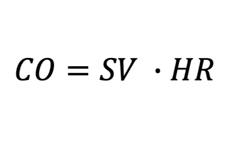
What does this mean?
Cardiac output

What do each of these variables mean?
CO (Cardiac Output): The volume of blood pumped by the heart per minute, usually expressed in liters per minute (L/min) . A normal resting adult has a CO of about 5 L/min.
SV (Stroke Volume): The volume of blood ejected by the left ventricle with each single heartbeat, typically measured in milliliters per beat (mL/beat). The average resting SV is about 70 mL .
HR (Heart Rate): The number of times the heart beats per minute (bpm). A normal resting heart rate typically ranges from 60 to 100 bpm
What happens when something goes wrong with the heart? (2)
Heart disease
Heart failure
Heart disease
Something is wrong with the heart
Types of heart disease
can include coronary artery disease, arrhythmias, and valvular heart disease.
Heart failure
Heart is unable to deliver sufficient blood to the tissues
Heart failure types
include systolic and diastolic heart failure.
4 chambers of heart
Right atrium (RA)
Right ventricle (RV)
Left atrium (LA)
Left ventricle (LV)
4 valves of heart
2 Atrioventricular (AV) valves
Right AV valve
Left AV valve
Pulmonic valve
Aortic valve
Major blood vessels in heart
Cranial and caudal vena cava
Pulmonary trunk
Pulmonary veins
Aorta
Cranial and caudal vena cava
Major veins that drain into RA
Pulmonary trunk
From RV to lungs
Splits into left and right pulmonary arteries
Deoxygenated blood
Pulmonary veins
From lungs to LA
Oxygenated blood
Aorta
From LV to systemic circulation
Add blood vessel image
d
Heart histology
heart is composed of 3 layers
3 layers of heart
Endocardium
Myocardium
Epicardium
Endocardium
Lines the atria and ventrciles
Myocardium
Cardiac muscle
Contractile units = sarcomeres
Sarcomeres
The basic contractile units of cardiac muscle, composed of actin and myosin filaments that enable muscle contraction.
Contractile units
Composed of actin/myosin filaments
Contraction requires ATP and Ca2+
Epicardium (Visceral Pericardium)
Outer layer of simple squamous cells
Lines pericardial cavity
Intercalated disks
In cardiac muscle; anchoring structures containing gap junctions
Cardiac muscle cells
Faintly striated, branching, mononucleated cells, which connect by means of intercalated disks to form a functional network
The action potential
Travels through all cells connected together forming a functional syncytium in which cells function as a unit
Syncytium
A multinucleated cell formed by the fusion of individual cells, allowing coordinated contraction in cardiac muscle tissue.
Add cardiac msucle structure image
d
Add heart image d
d
Add blood flow through heart chart
d
Problem List:
Grade V/VI basilar systolic murmur
Weak synchronous femoral pulses
More diagnostics are necessary - WHICH?
Electrical analysis of heart
Functional analysis of heart
START WITH THE HEART
Pulmonary circulation
To lungs from RV
Arteries deoxygenated
Veins oxygenated
Systemic circulation
To body from LV
Arteries oxygenated
Veins deoxygenated
Add overview of circulation image
d
Cardiovascular Phyiology Goal
Circulation of blood
Delivers oxygen and metabolic substrates to tissues
Removes carbon dioxide and metabolic byproducts
In Cardiovascular Physiology, blood flow is driven by pressure gradients
Driving force is rhythmic pumping of heart
Modulated by constricting or dilating blood vessle sto change their resistance to flow
Phases of Cardiac Cycle
Phase 1: Diastole
Phase 2: Systole
Phase 3: Systole
Phase 4: Diastole
Phase 1
Diastole
Opening of AV valves
Ventricular filling and atrial contraction
Phase 2
Systole
Closing of AV valves
Isovolumetric ventricular contraction
All valves closed
Phase 3
Systole
Opening of pulmonic and aortic valves
Ventricular ejection
Phase 4
Diastole
Closing of pulmonic and aortic valves
Isovolumetric ventricular relaxation (all valves closed)
Add cardiac cycle image
d
dd cardiac cycle graoh image
d
3 equations
Blood pressure (BP)
Cardiac Output (CO)
Stroke Volume
Blood Pressure (BP)
Cardiac Output (CO) x Total Peripheral Resistance (TPR)
Cardiac Output (CO)
Heart Rate (HR) x Stroke Volume (SV)
Stroke Volume
Preload Contractility - Afterload
Preload
The load imposed on the heart prior to systole (end dystolic volume)
Contractility
Number of actin-myosin interactions during systole (intracellular calcium)