Metabolism - Basic Science Core
 Call Kai
 Learn
Learn Practice Test
 Spaced Repetition
 Match
Match Flashcards
 Knowt Play
1/92

Earn XP
Description and Tags
Metabolism/Phyisology Test
Name | Mastery | Learn | Test | Matching | Spaced | Call with Kai | Chat |
|---|
No analytics yet
Send a link to your students to track their progress
93 Terms
Gross Anatomy of the Small Intestine (3 Parts)
1) the duodenum
2) the jejunum
3) the ileum
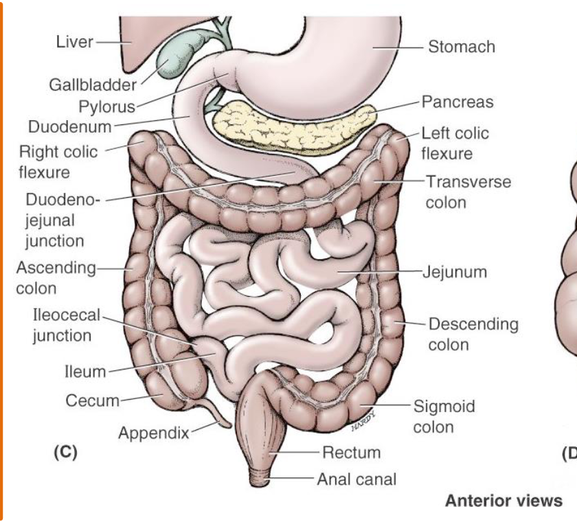
duodenum
The duodenum constitutes the first 10-12 inches. It begins at the pyloric valve, arcs around the head of the pancreas and passes to the left, and ends at a sharp bend called the duodenojejunal flexure
_
The duodenum receives the stomach contents, pancreatic juice and bile.
Duodenojejunal Flexure Junction
Arcs around the head of the pancreas and passes to the left, and ends at a sharp bend called the duodenojejunal flexure
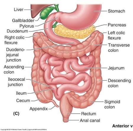
jejunum
The jejunum is about 8 feet long and extends from the duodenum to the ileum.
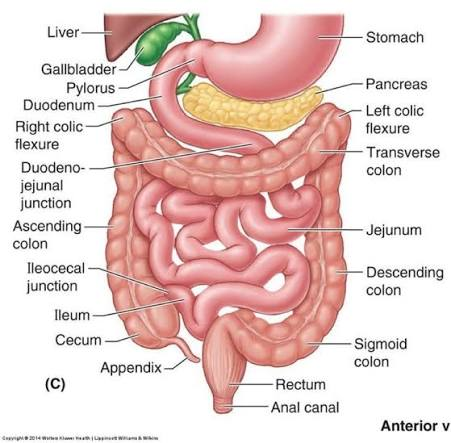
ileum
The ileum is about 12 feet in length and joins the large
intestine at the ileocecal valve.
Ileocecal valve
The end of the small intestine feeding into the ascending colon (start of the large intestine)
Surface of the Small Intestine
In order for absorption of nutrients, electrolytes, and water in the small intestine to occur,
There needs to be a maximal surface area - this is accomplished through:
Plicae circulares
Villi and microvilli
Plicae Circulares
Large, permanent transverse folds of the mucosa and submucosa that project into the lumen of the small intestine.
Help the body absorb nutrients
villi
Small, finger-like projections made of multiple cells protruding from the mucosa. Range from 0.5 mm to 1.5 mm long
Microvilli
Extremely tiny, microscopic folds of the plasma membrane on the apical surface of individual epithelial cells.
Form a dense "brush border"
Carbohydrates (How we absorb them in the human body)
Carbohydrates are widely distributed the human body they have important structural and metabolic roles
_
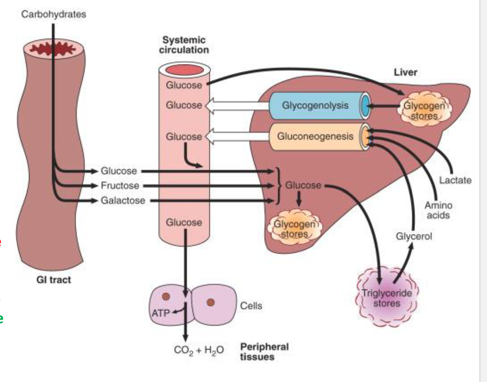
The most important carbohydrate; most dietary carbohydrate is absorbed into the bloodstream as glucose formed by hydrolysis of dietary starch and disaccharides, and other sugars are converted to glucose in the liver (more on this in just a bit)
Glucose
Glucose is the major metabolic fuel of humans and a universal fuel of the fetus
_
Glucose is the precursor for synthesis of all the other carbohydrates in the body, including:
glycogen for storage,
ribose and deoxyribose in nucleic acids,
galactose for synthesis of lactose in milk, in glycolipids, and in combination with protein to form glycoproteins
Major Dietary Carbohydrates Types (3)
Monosaccharides
Disaccharides
Polysaccharides
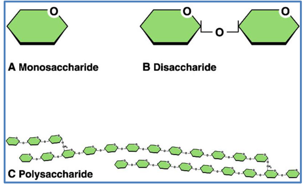
Monosaccharides (3 Dietary Carbohydrates)
Glucose
Fructose
Galactose
Disaccharides (3 Dietary Carbohydrates)
Sucrose (Table Sugar)
Lactose (Milk Sugar) → a galactose and Glucose
Maltose (Sugar from Malt grain)
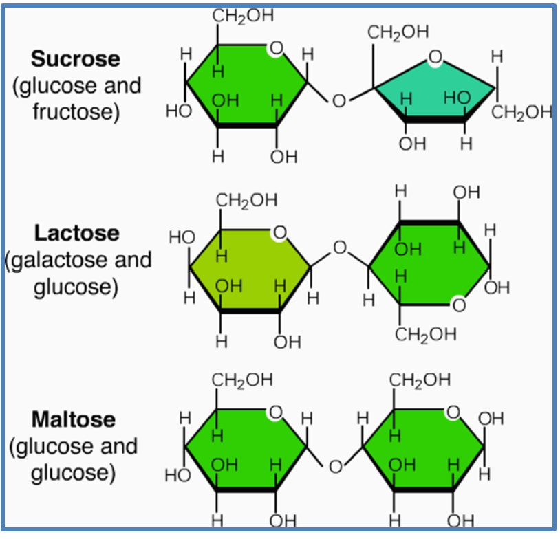
Polysaccharides (3 Dietary Carbohydrates)
Glycogen (We eat from animals)
Starch (We eat from plants)
“Fiber” (cellulose, others) → Humans cannot digest this
Digestion of Carbohydrates
To absorb carbohydrates it is essential to break them down into there smallest components
_
Amylase → Means to digest Carbohydrates
Two Digestive Enzymes we go over:
1) Salivary amylase and pancreatic amylase:
Degrades polysaccharides into disaccharides.
_
2) Disaccharidases:
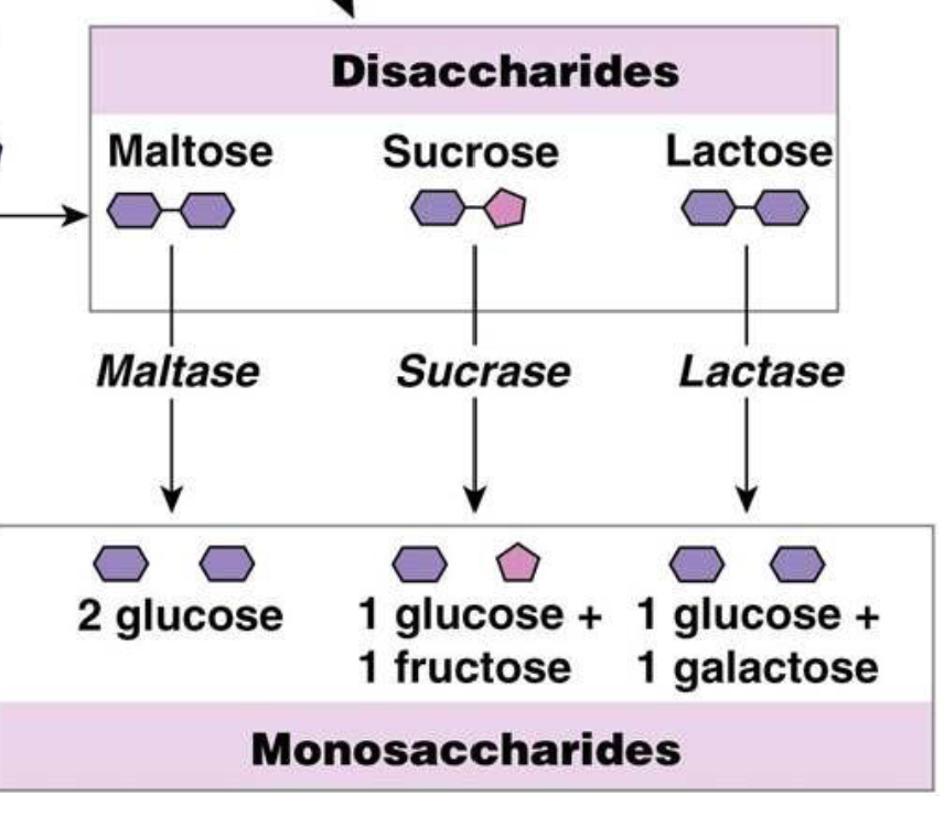
Degrades disaccharides into monosaccharides
Located in the small intestine
Disaccharidases
Disaccharides are degraded in the small intestine by a group of brush border enzymes called disaccharidases
Named after the disaccharide they breakdown
Absorption of Glucose and galactose
Glucose and galactose are absorbed (via secondary active transport) through a Na+-dependent cotransporter known as SGLT1.
_
A high luminal concentration of Na+ facilitates absorption of these 2 sugars against their gradients.
Absorption of Fructose
Fructose enters by facilitated diffusion via glucose transporter 5 (GLUT5)
GLUT5 does not require Na+
How are monosaccharides are transported out of the enterocytes
Transported into the capillaries by GLUT2
Facilitated Diffusion (How this is done)
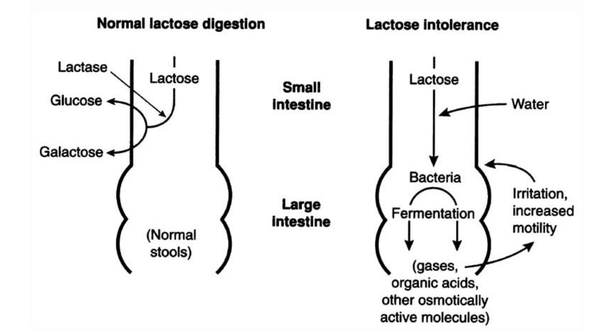
Lactose Intolerance
A Intolerance makes you fart a lot, you can just avoid lactose or take medication
Normally lactose is broken down into Glucose & Galactose in the small intestine
_
Lactose Intolerent people, cannot breakdown lactose and it ferments in the large intestine by bacteria
Every Carbohydrate in the body eventually turn into …
Glucose
Metabolic fates of glucose: (4)
Stored as glycogen in the liver and skeletal muscle
_
Stored as triglyceride in adipose tissue
_
Metabolized into pyruvate or lactate via glycolysis
_
Metabolized in the pentose phosphate pathway
Glycolysis Facts
Involves 10 enzymatic reactions
Occurs in the CYTOSOL of cells
Does NOT require oxygen (i.e., glycolysis is ANAEROBIC)
Results in the formation of:
2 ATP molecules (a very small amount of energy)
2 NADH (nicotinamide adenine dinucleotide + hydrogen) molecules, which if oxygen is present, can produce 5 ATP later on in the ETC
Pyruvate (Glycolysis)
End product pf Glycolysis is PYRUVATE
Most of the pyruvate undergoes OXIDATIVE degradation in the mitochondria if AEROBIC conditions exist
If no Oxygen
Conversion of pyruvate into LACTATE occurs under ANAEROBIC conditions or in those cells (RBCs, platelets) that have no mitochondria and therefore lack the ability to oxidatively degrade pyruvate altogether
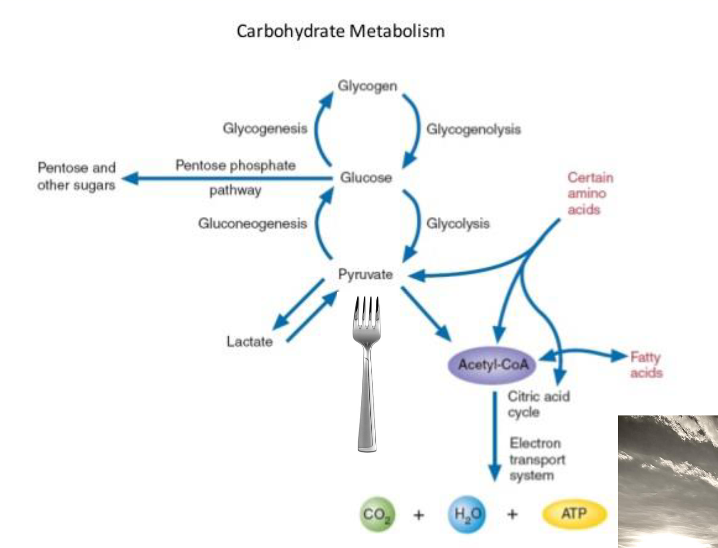
For every 1 molecule of glucose … (Glycolysis)
2 molecules of pyruvate are produced
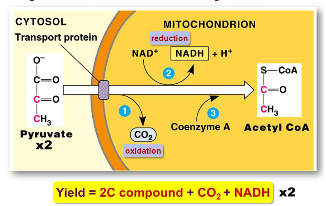
Pyruvate to Acetyl CoA
Catalyzed by pyruvate dehydrogenase in the mitochondrial membrane
Krebs (Citric Acid, TCA) Cycle
Occurs in the MITOCHONDRIA of cells
Does not USE oxygen…but will STOP in its
absence
Results in the formation of:
• 6 molecules of NADH
• 2 molecules of FADH2 (flavin adenine
dinucleotine + 2 hydrogen)
• 2 molecules of GTP
Generates CO2 as a waste product
Four VITAMINS are essential for the Krebs cycle:
Thiamine (vitamin B1)
Riboflavin (vitamin B2)
Niacin (vitamin B3)
Pantothenic acid (vitamin B5)
_
If you have vitamin deficiencies of any of these the Krebs cycle may not be as efficient
Respiratory (Electron Transport) Chain
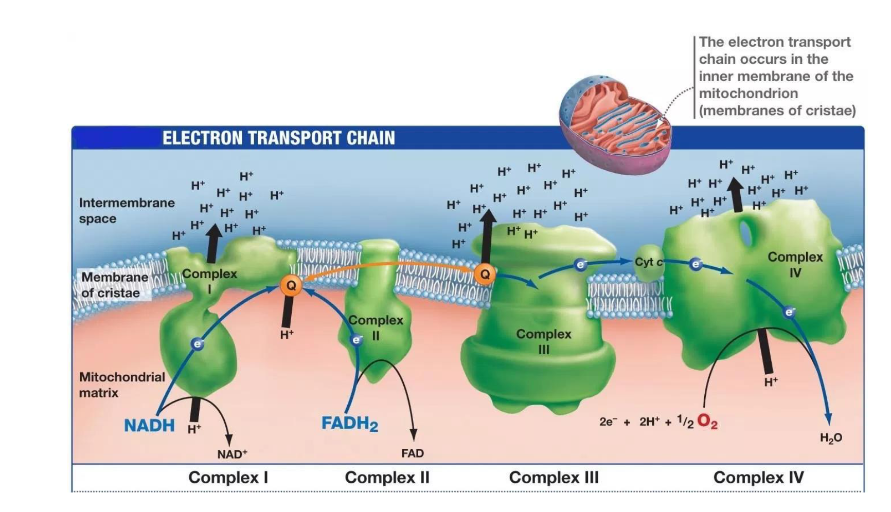
Found in the MITOCHONDRIA of cells (specifically the inner mitochondrial membrane)
Consists of 4 protein complexes (called I-IV)
Respiratory (Electron Transport) Chain
ABSOLUTELY requires oxygen
Converts NADH and FADH2 that
was created in the TCA cycle to
NAD+ and FAD by “stripping
away” hydrogen (H+) molecules
ATP SYNTHASE is the key
enzyme associated with the ETC
that creates ATP from the H+
gradient
Ultimately:
• Each NADH to NAD+
generates 2.5 ATP from ADP
• Each FADH2 to FAD generates
1.5 ATP from ADP
ATP SYNTHASE
ATP SYNTHASE is the key
enzyme associated with the ETC
that creates ATP from the H+
gradient
Final Reaction Tally of Complete oxidation of Glucose
Every molecule of Glucose
Net Yield of ATP is 32
NEED OXYGEN
Hexose Monophosphate Shunt (HMP; Pentose
Phosphate Pathway)
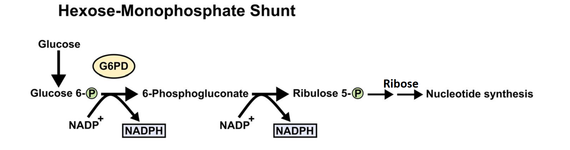
Is an alternate route for the metabolism of glucose
Has nothing to do with creating energy (ATP)…..instead the pathway generates
NADPH (nicotinamide adenine dinucleotide phosphate) and 5-carbon sugars called
PENTOSES (the most important being RIBOSE)
Occurs in the cytosol, no organelles are needed, so this pathway can happen in all
cells
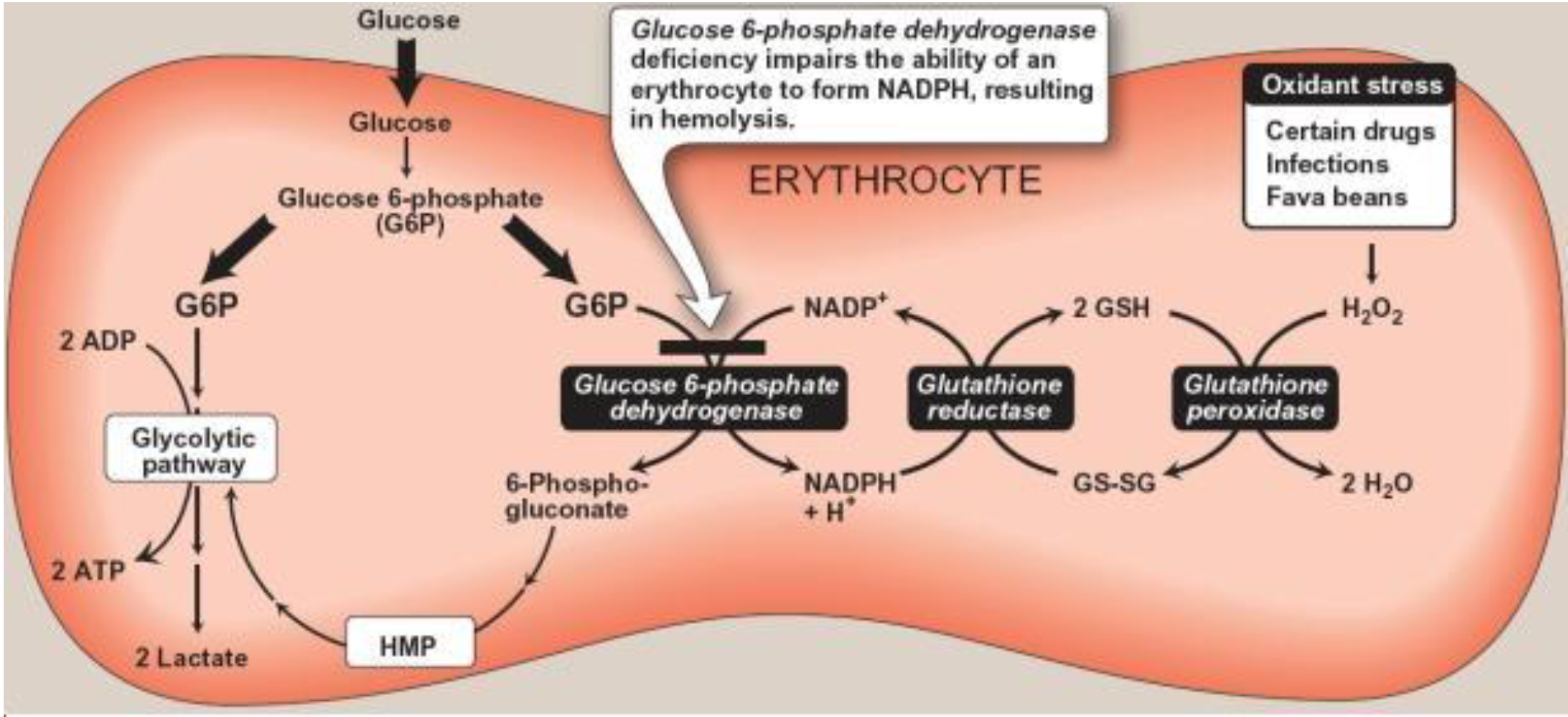
The key enzyme in the pathway is glucose-6-phosphate-dehydrogenase (G6DP)
So…..what is RIBOSE used for in the Hexose Monophosphate Shunt
Basically for synthesizing the
nucleotides found in RNA and
DNA, especially in cells that are
rapidly dividing (both normal and
cancerous
So…..what is NADPH used for?
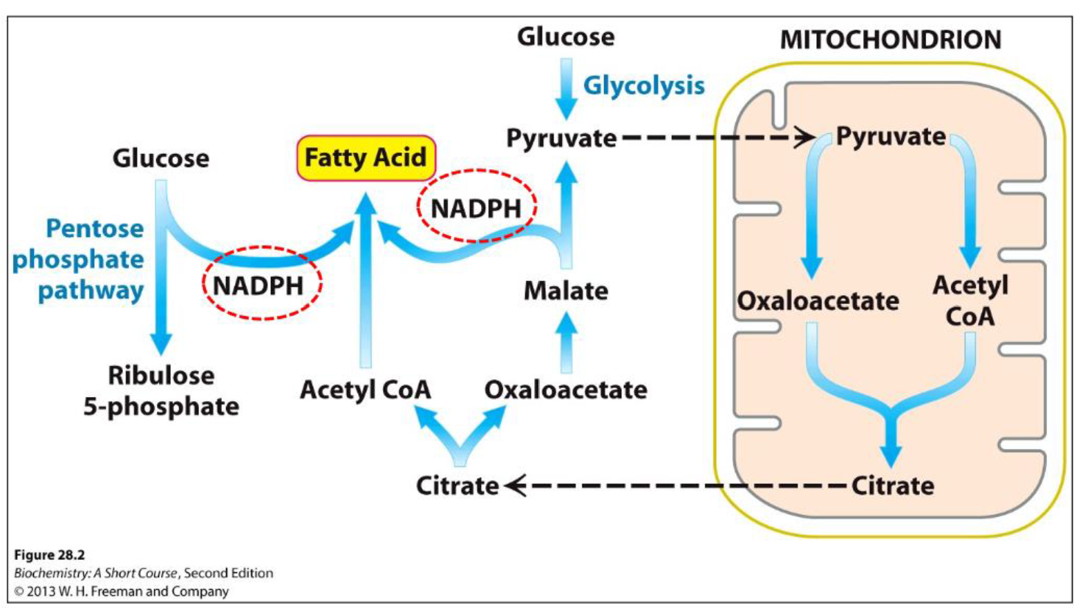
1) For synthesizing fatty acids
2) For indirectly eliminating
toxic oxygen “radicals” (e.g.,
HYDROGEN PEROXIDE)
For synthesizing fatty acids
or synthesizing fatty acids
& cholesterol in liver,
adipose tissue &
mammary glands and for
synthesizing steroid
hormones in certain
endocrine glands (ovaries,
testes, adrenal glands,
placenta)
Basically, NADPH donates
electrons (i.e., it is a REDUCING
AGENT) in the anabolic reactions
used to make fatty acids,
cholesterol and steroid hormones
For indirectly eliminating
toxic oxygen “radicals” (e.g.,
HYDROGEN PEROXIDE)—
highly reactive oxygen
molecules produced by
some reactions in the body,
which can cause significant
damage to cells
So………which cells produce
lots of hydrogen peroxide?
RBCs (erythrocytes)
RBCs generating hydrogen peroxide (
ll day long………and can be
induced to make more by certain medications, infections and fava
beans…..yes, fava beans…
RBCs possess a very important
chemical called GLUTATHIONE, which
can exist in both a reduced state (GSH)
and an oxidized state (GSSG)
It is critical for glutathione to be in the
REDUCED STATE (GSH)….or if you
prefer, we want glutathione to be an
ANTI-OXIDANT
GLUTATHIONE
RBCs possess a very important
chemical called GLUTATHIONE, which
can exist in both a reduced state (GSH)
and an oxidized state (GSSG)
It is critical for glutathione to be in the
REDUCED STATE (GSH)….or if you
prefer, we want glutathione to be an
ANTI-OXIDAN
What keeps glutathione to be in the
REDUCED STATE
NADPH
Glutathione and RBCs
Glutathione is THE major endogenous antioxidant produced by all cells, participating directly
in the neutralization of free radicals and reactive oxygen compounds, as well as maintaining
exogenous antioxidants such as vitamins C and E in their reduced (active) forms
-Here’s where it all comes together to prevent HEMOLYTIC ANEMIA
Glycogenesis
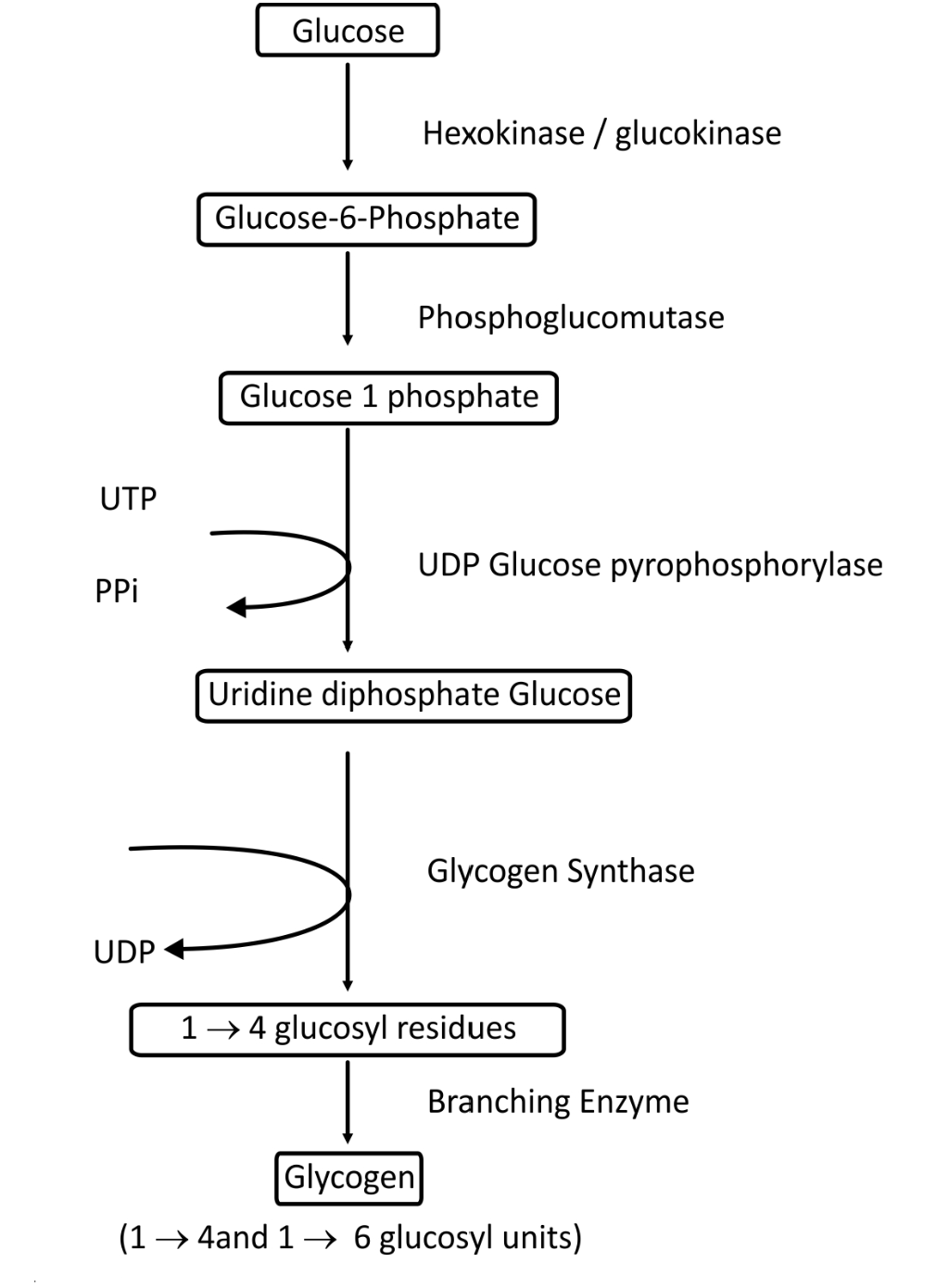
Occurs in fed state in liver and skeletal muscle
Helps prevent hyperglycemia because it allows
sequestration of blood glucose is the storage
form of GLYCOGEN
Glucokinase is found in liver hepatocytes,
whereas hexokinase is found in skeletal muscle
cells
So what happens if there is still a lot of blood
glucose and the liver and skeletal muscle hang
there “No Vacancy” signs…………?
Insulin function
Key enzyme in both tissues is GLYCOGEN
SYNTHASE which is stimulated by INSULIN
Skeletal muscle can store 350-500 grams
(provides about 1400-2000 calories) and liver
can store about 100 grams (provides about 400
calories)
Insulin Stores glucose a glycogen (in the liver)
If you eat too much carbs the glucose is used as triglyceride stores
Process of Glycogenesis
Fatty Acid Synthesis
Fatty acids are synthesized from ACETYL-CoA
Occurs mainly in adipose tissue and liver
A very round- about set of pathways between the cytosol and mitochondria….
WHAT’S HAPPENS WHEN WE
NEED TO “MAKE” GLUCOSE?
1. GLYCOGENOLYSIS
2. GLUCONEOGENESIS
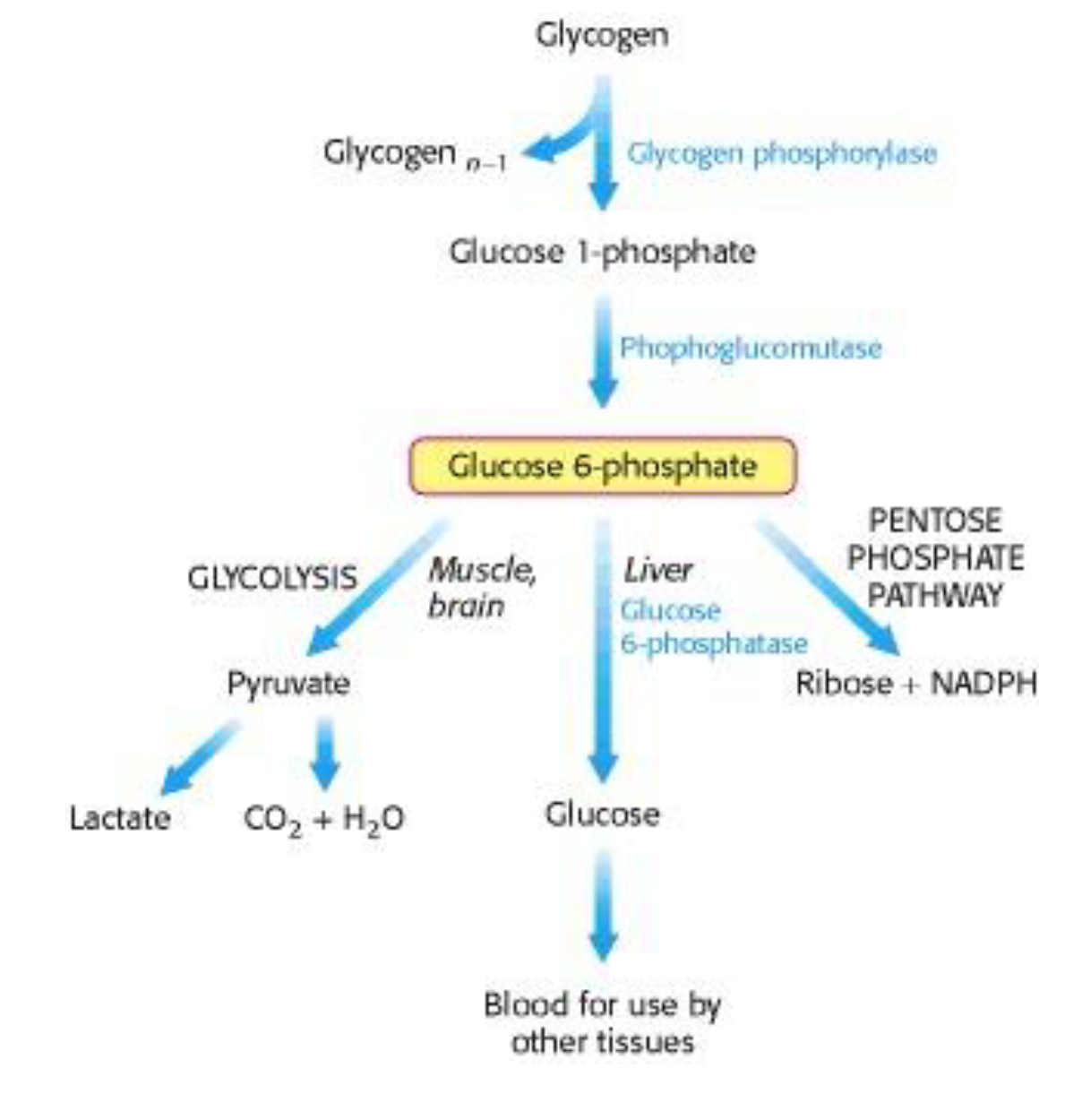
Glycogenolysis
Occurs in fasting state in liver and skeletal muscle…….BUT the pathways are slightly different between the two tissues
In the liver, glycogenolysis helps prevent hypoglycemia because it allows for the breakdown of glycogen into glucose
In the skeletal muscle, glycogenolysis provides a locally available source of glucose-6- phosphate that can then enter the glycolysis pathway
Gluconeogenesis
Synthesizes glucose from non-
carbohydrate sources, namely:
• amino acids (except leucine and
lysine)
• lactate
• glycerol (from triglycerides)
Occurs primarily in liver hepatocytes,
and to a minor extent, in epithelial cells
of the kidneys and small intestine
Gluconeogenesis occurs in 11 steps and
is NOT simply a reversal of glycolysis
THIS IS MAKING GLucose without Carbohydrates
There are two important inter-organ cycles Involving glycolysis in
muscle and gluconeogenesis in liver
1) Cori Cycle
2) Alanine Cycle
Cori Cycle
There are two important inter-organ cycles involving glycolysis in muscle and gluconeogenesis in liver.
In the CORI CYCLE, glucose is metabolized to pyruvate and then to lactate in muscle, the lactate is released into the blood and carried to the liver, where it is reconverted to pyruvate and used for gluconeogenesis, and the resulting glucose is released and travels back to muscle
Alanine Cycle
The ALANINE CYCLE is similar to the Cori cycle, except that muscle pyruvate is converted to the amino acid alanine rather than lactate by transamination. In the liver, alanine is transaminated back to regenerate pyruvate, and the excess amino groups are disposed of as UREA
Types of Lipids (3)
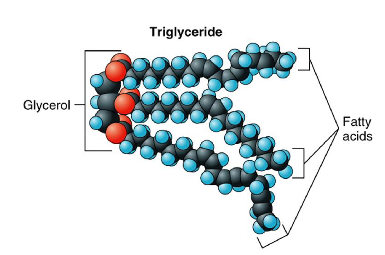
1) Triglycerides (triacylglycerols)
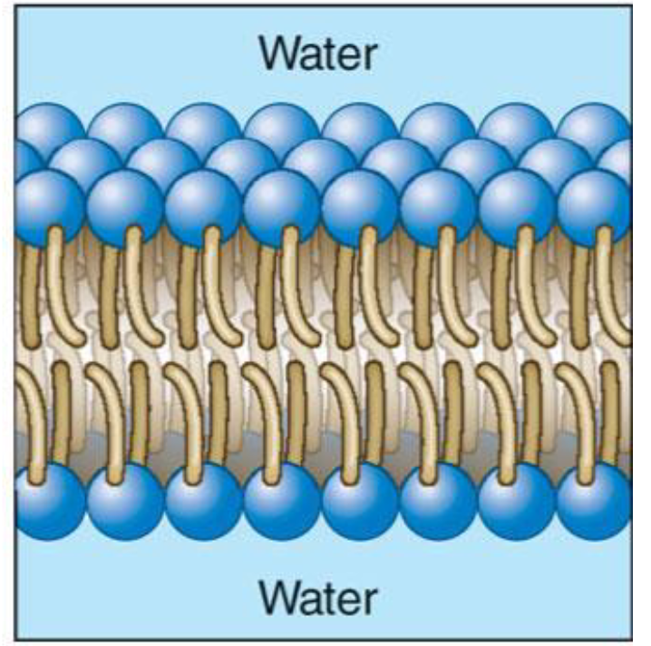
2) Phospholipids
3) Cholesterol
Triglycerides
Fats → saturated (animal) and unsaturated (oils)
also known as triacylglycerols
are mainly used by cells for energy
Phospholipids
Phospholipids are mainly used by cells for building cell (plasma) membranes.
Cholesterol
Cholesterol is also used by cells for building cell (plasma)
membranes and is also the building block for steroid hormones
(e.g. – testosterone, estrogen, progesterone, cortisol and
aldosterone). When cholesterol travels in the blood, it does so
mainly as LDL-C (low density lipoproteins) & HDL-C (high density
lipoprotein) more on this later………
Two Types of Cholesterol
1) LDL-C (low density lipoproteins)
2) HDL-C (high density lipoprotein
Overview – Digestion & Absorption of Lipids
emulsification
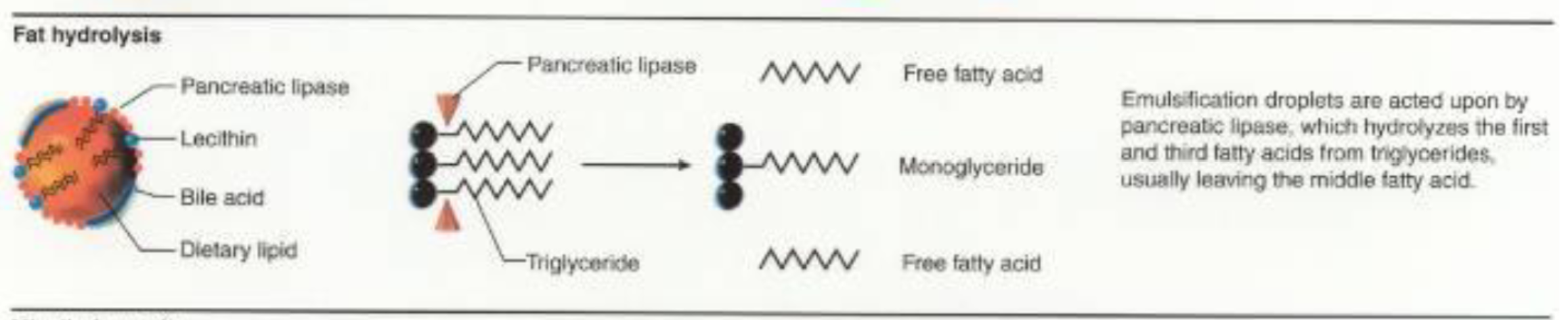
• pancreatic lipase
• colipase
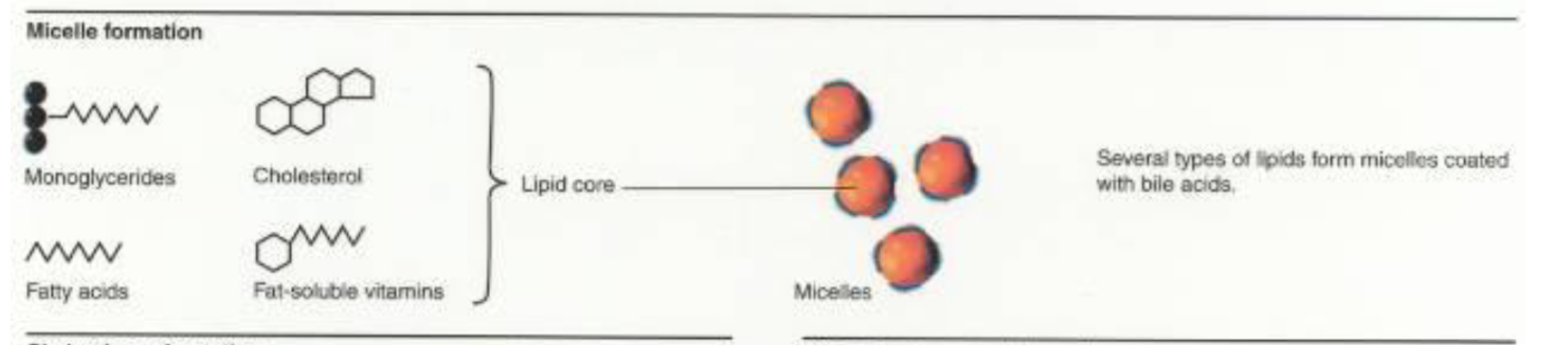
• micelles
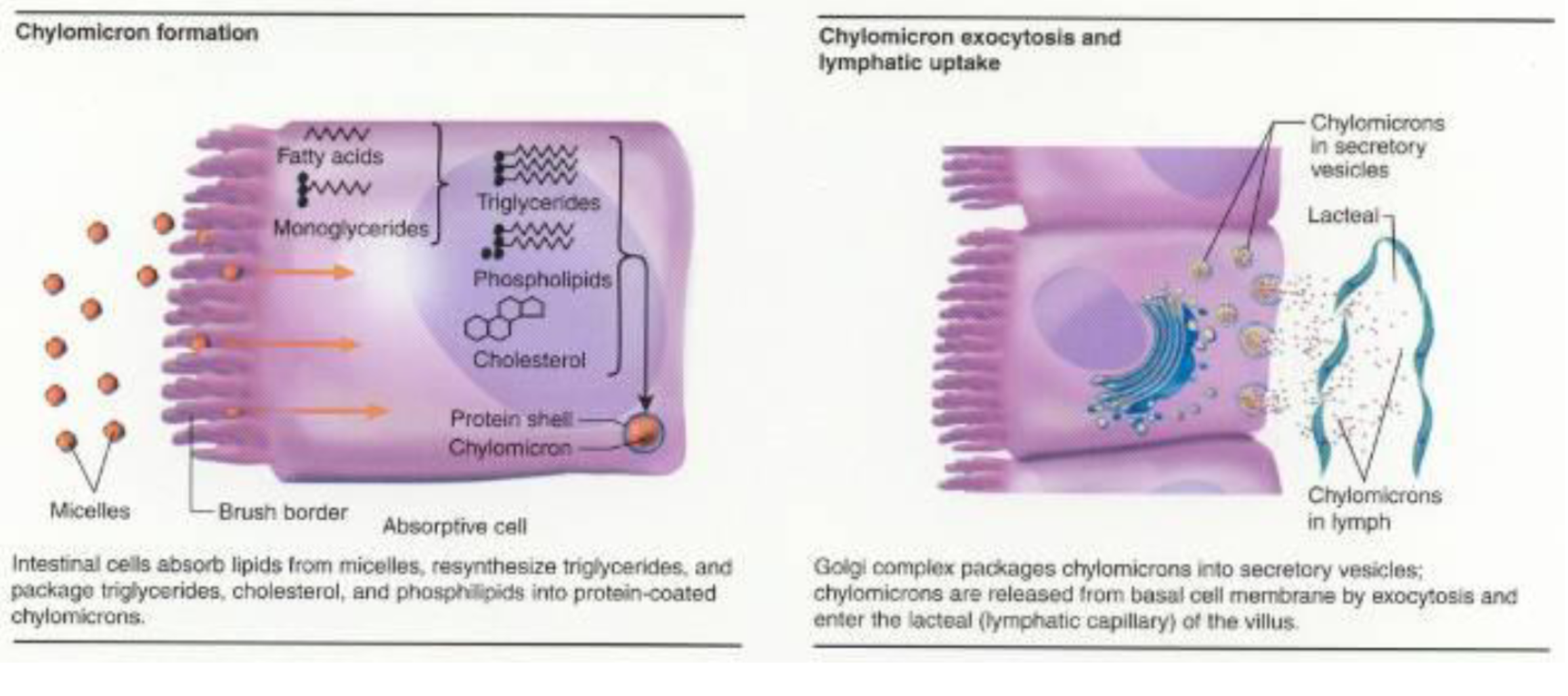
• chylomicrons
• lacteals
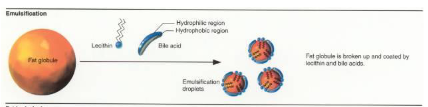
emulsification
Requires bile → make a fat molecule into droplets, this allows the surface area to increase
Transport of Chylomicrons
Once the
chylomicrons enter
the lymphatic
circulation, they are
transported upward
through the thoracic
duct and emptied
into the venous
circulation at the
juncture of the
jugular and
subclavian veins.
Lipemia
usually occurs within an hour after eating a fatty meal.
Chylomicrons have a short half-life (< 60 min), so the plasma usually
becomes clear within a few hours.
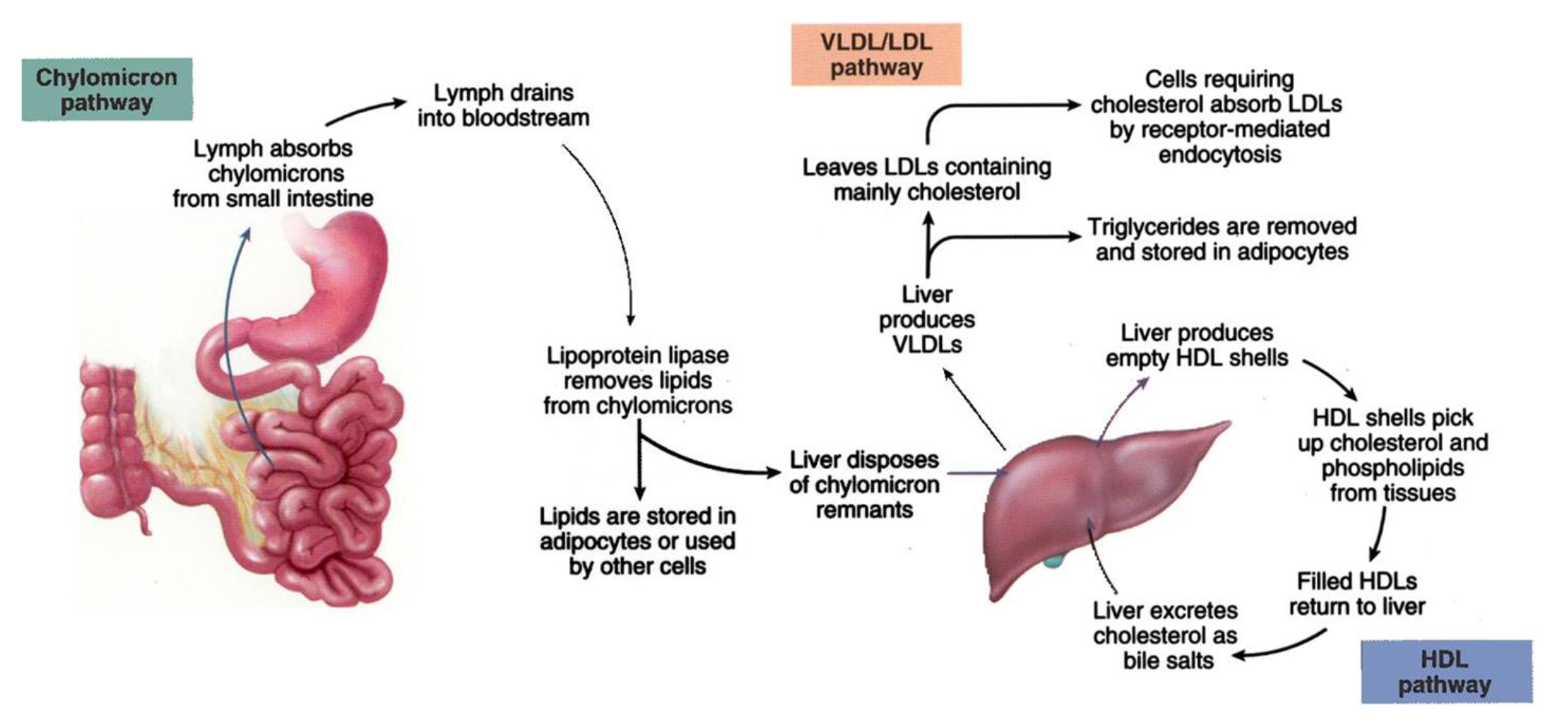
Fate of Chylomicrons
Chylomicrons are removed from the blood as they pass through the capillaries of
adipose tissue and the liver. Both contain large quantities of lipoprotein lipase.
This enzyme is especially active in the capillary endothelium, where it hydrolyzes
the TGs of chylomicrons into fatty acids & glycerol, which diffuse into adipocytes
(storage) & hepatocytes (re-packaged into lipoproteins & exported to blood). It is
also possible for other tissues to use the contents of chylomicrons.
Types of Lipoproteins (5)
Allows Fat to transport in the blood
_
There are generally 5 classes of lipoproteins:
Chylomicrons
Very low density lipoproteins (VLDL)
Intermediate density lipoproteins (IDL)
Low density lipoproteins (LDL)
High density lipoproteins (HDL)
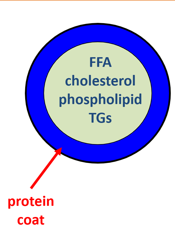
Very low density lipoproteins (VLDL)
high TGs / moderate cholesterol & phospholipids
Intermediate density lipoproteins (IDL)
moderate TGs, cholesterol & phospholipids
Low density lipoproteins (LDL)
low TGs / high cholesterol & phospholipids
Bad Cholesterol
High density lipoproteins (HDL)
low TGs, cholesterol & phospholipids
(good Cholesteerol)
Clinically which Lipids are ussally tested
1) Clinically, LDL-C & HDL-C levels are the only lipoproteins routinely monitored and used to screen for atherosclerosis, MI & stroke risk, heart disease, etc.
LDL-C vs. HDL-C
The role of LDLs (bad cholesterol) is to transport cholesterol and other lipids from
the liver to the peripheral tissues, making them available (via receptor mediated
endocytosis) for membrane or steroid hormone synthesis. The function of HDLs
(good cholesterol) is to transport excess cholesterol from peripheral tissues to the
liver, where it is broken down and becomes part of the bile
LDL Receptor Mediated Endocytosis
After a while the body refuses to take this fat, and leaves it in the blood
Because it leaves you in the blood eventually Atherosclerotic plaque is formed
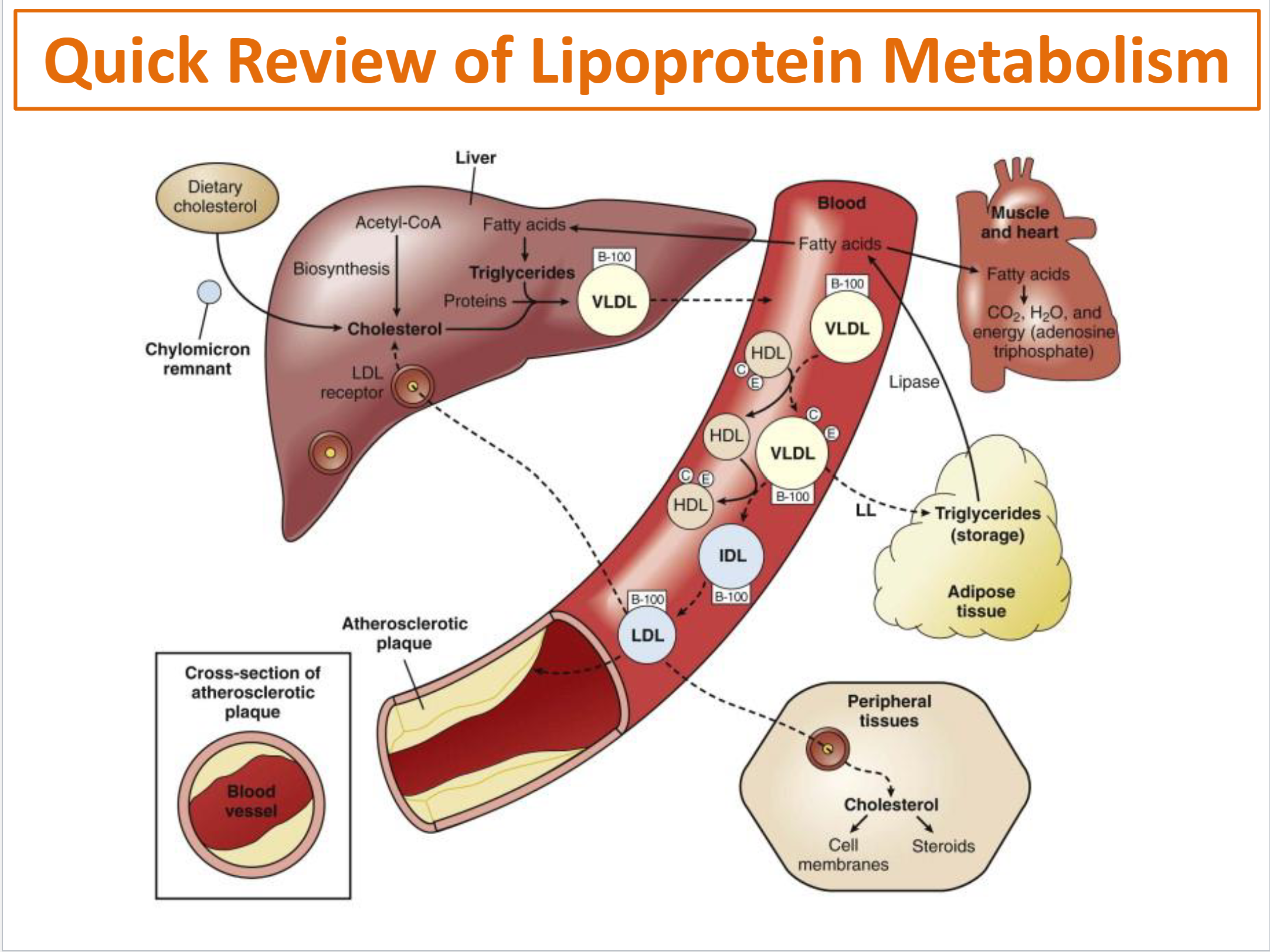
There are 2 main sources of cholesterol in the blood:
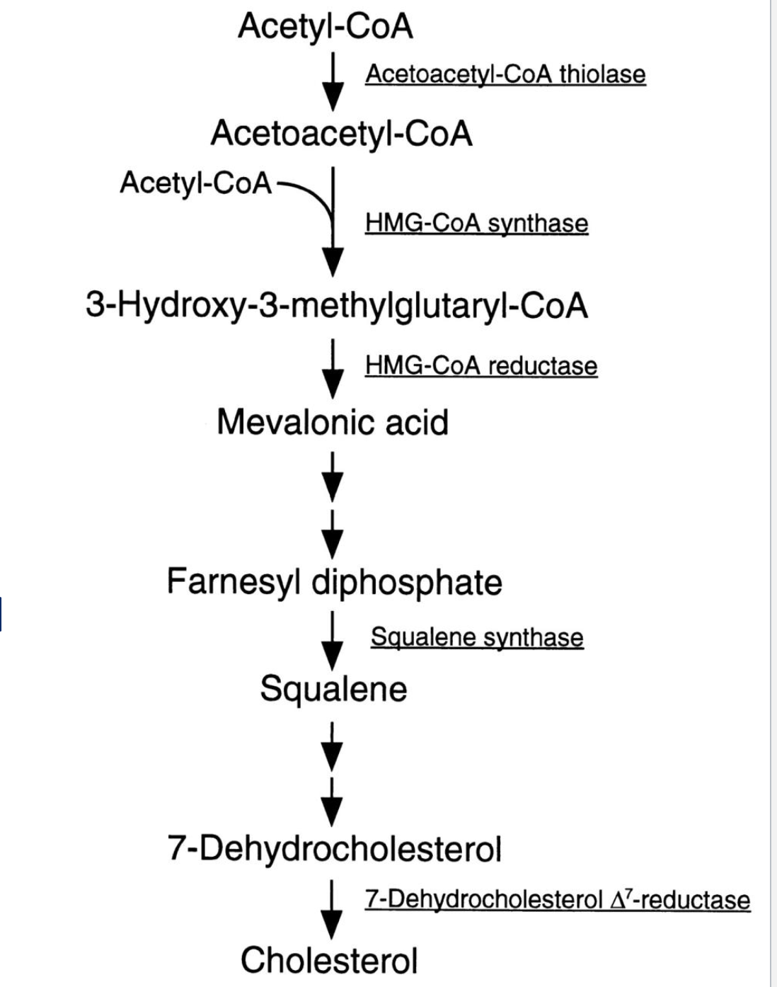
Cholesterol De Novo Synthesis
Using acet-l COA to create cholesterol by liver
hepatocytes
The amount this is used is genetic
Most important emnzynme is the HMG-CoA Reductase (what is used to stop this
HMG-CoA Reductase Inhibitors
- Statins
Statins are the most effective drugs currently in use for
treating elevated LDL-C – the major ones on the USA
market include:
Atorvastatin (Lipitor)
Fluvastatin (Lescol)
Lovastatin (Altoprev, Mevacor)
Pravastatin (Pravachol)
Simvastatin (Zocor)
Rosuvastatin (Crestor)
Pitavastatin (Livalo)
Lipolysis and Fatty Acid (Beta) Oxidation
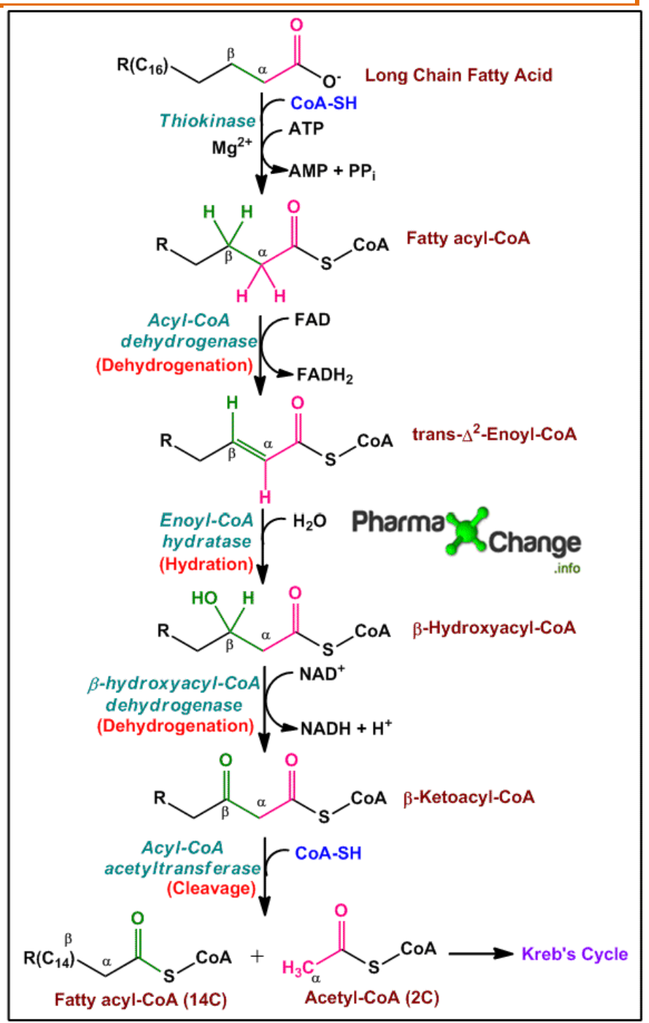
FATTY ACID (BETA) OXIDATION is the mitochondrial aerobic process of breaking down a fatty acid into acetyl-CoA units (each containing 2 carbons).
_
Acetyl-CoA is then “fed” into the Krebs cycle, where NADH, FADH2 and GTP are generated…..and then NADH and FADH2 are used by the ETC to make ATP
_
Occurs mainly in the liver, skeletal muscles and heart (Good At breaking down fat)
Lipolysis and Fatty Acid (Beta) Oxidation Diagram
Ketone Metabolism Occurs?
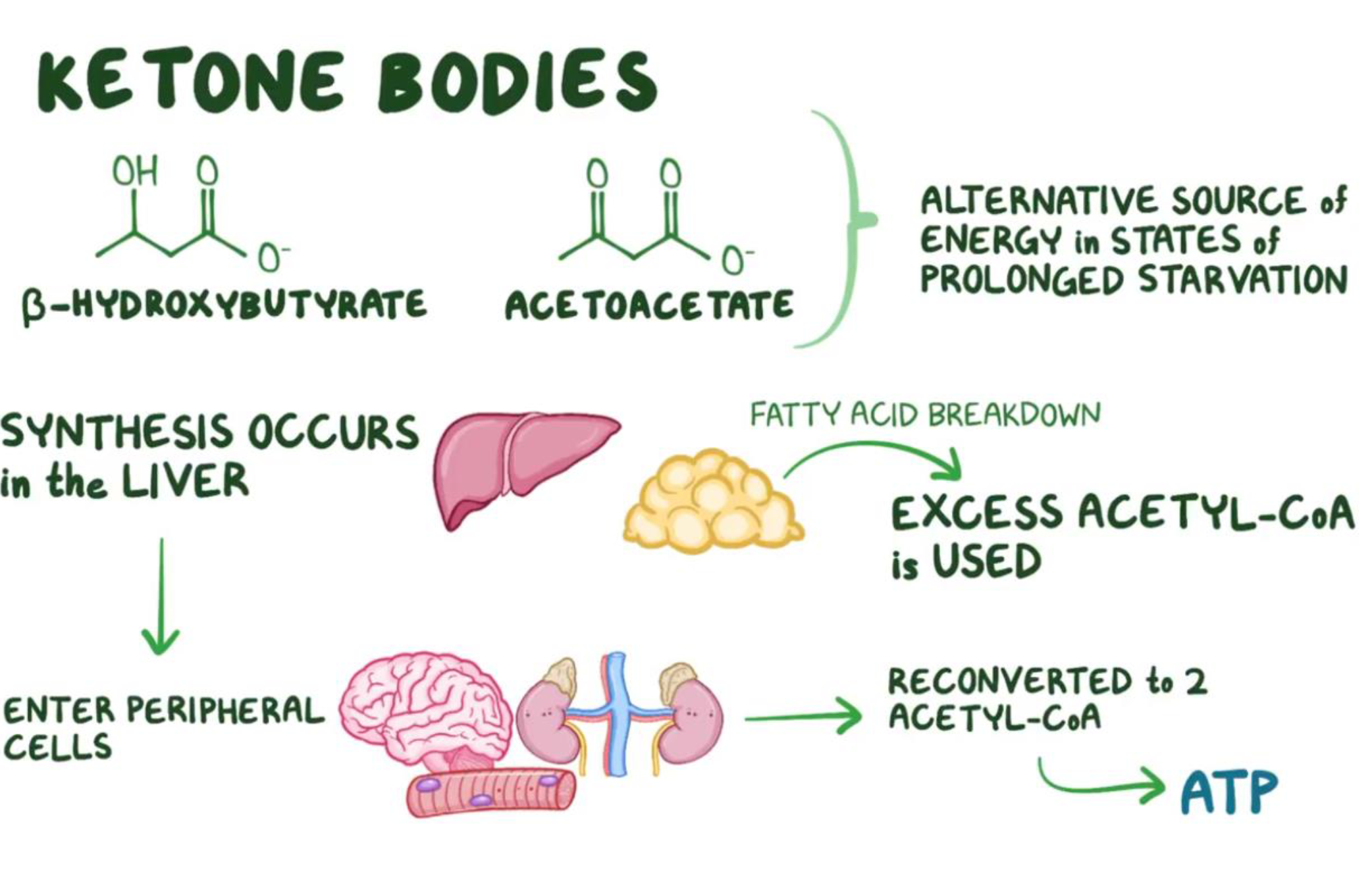
The synthesis of ketones (or ketone bodies) occurs in the mitochondria of liver hepatocytes
Made only in the liver
Ketones
Fatty product
They are Acidic (Can lower the ph of the blood to be dangerous)
Can be used to make atp
Become a problem if there is too many of them
Ketone Metabolism
During prolonged starvation or fasting, oxaloacetate can eventually become depleted in the liver due to extensive gluconeogenesis → this impedes the entry of Acetyl-CoA into the Krebs’ cycle
_
All of this excess acetyl-CoA has no where to go, so it accumulates………
The liver then converts the excess acetyl-CoA into ketone bodies (3 types), which can then enter peripheral tissues (e.g., brain, skeletal muscle, kidneys) and be reconverted back to acetyl-CoA to make ATP via the Krebs’ cycle and ETC
Ketone Bodies
Synthesis of amino acids
is of paramount importance to the human body.
Once synthesized or ingested, amino acids are used as BUILDING BLOCKS not only for proteins but also for several other critical biological molecules such as:
nucleic acids (both purines and pyrimidines),
hormones,
neurotransmitters,
anti-oxidants
various signaling molecules.
Digestion of Proteins & Absorption of Amino Acids
The stomach does a good job of denaturing proteins, but it is not as effective cleaving off individual amino acids (pepsin does some of this).
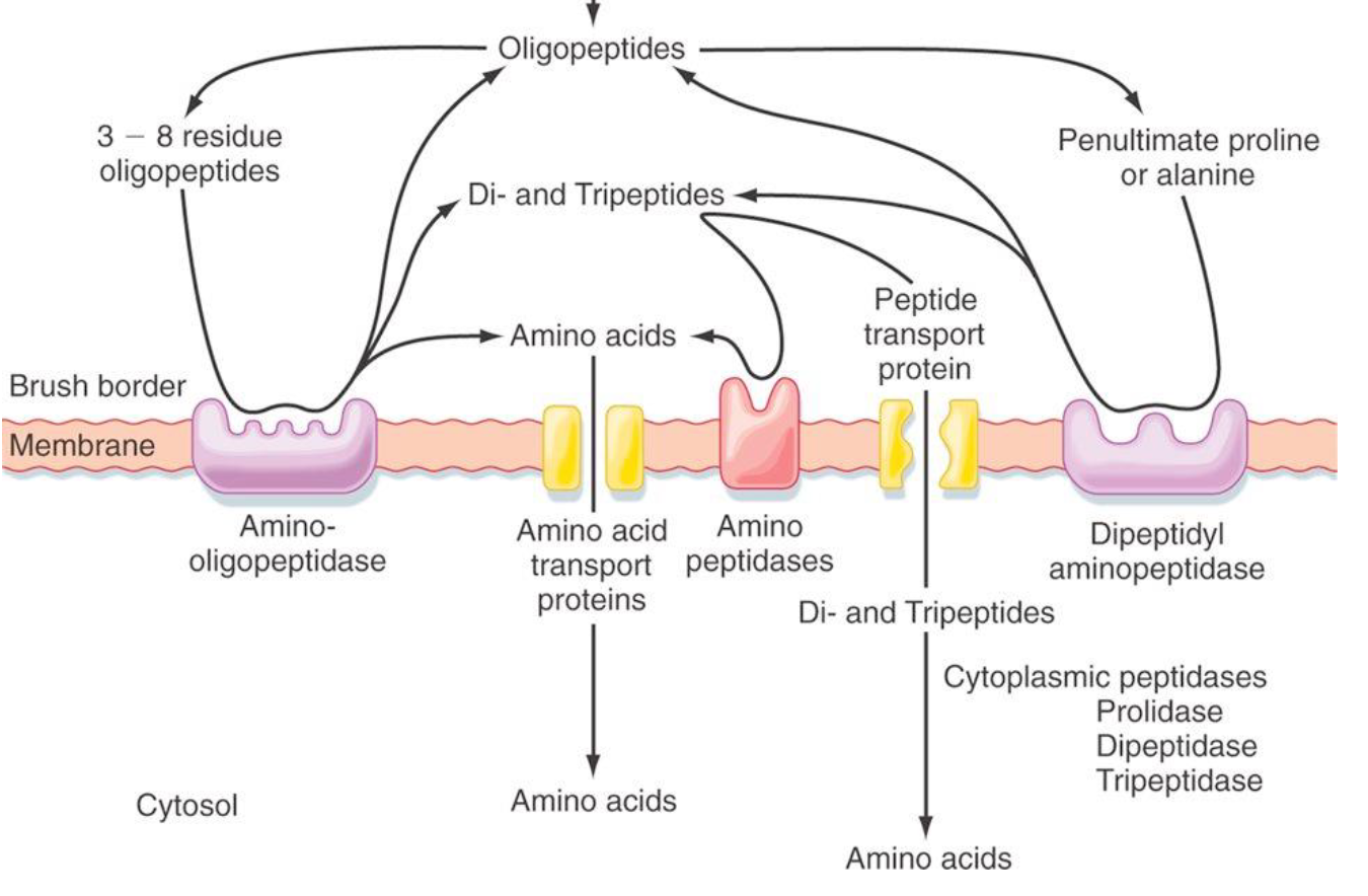
Cleaving off amino acids occurs mainly at the brush border with
aminopeptidases and with the active pancreatic enzymes (proteases).
Absorption
of Amino Acids
Once the digestion of
proteins is complete, most
amino acids or small
peptides are transported
via the same co-transport
mechanism utilized by
glucose -- some amino acids
do not require this sodium
co-transport mechanism
but instead are transported
in the same way that
fructose is moved, i.e. by
facilitated diffusion
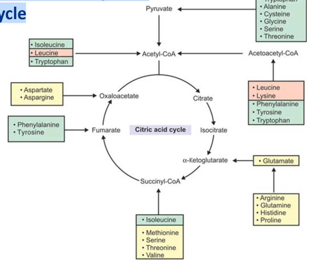
Can Amino Acids Be Used For Energy?
Yes!
1. Gluconeogenesis 18/20 (except leucine and lysine)
2. Ketone body formation (leucine and lysine)
3. Directly fed into Krebs cycle
Can Amino Acids Be Broken Down Into Waste?
Yes!
Ultimately, the amine groups cleaved from the
amino acids are converted into ammonia (NH3)
or ammonium ions (NH4+), which are then
converted into urea.
urea
Essentially all urea formed in the human body is synthesized in the liver
After its formation, urea diffuses from liver
hepatocytes into the body fluids and is excreted
by the kidneys.
hepatic coma or hepatic encephalopathy
In serious liver disease, ammonia can often accumulate in the blood and lead to a state
azotemia or uremia
A build-up of urea in the blood can lead to a state of azotemia or uremia, in which high levels of urea become toxic to a variety of tissues.
This is often a symptom of renal disease or failure.