RECEPTORS
1/17
There's no tags or description
Looks like no tags are added yet.
Name | Mastery | Learn | Test | Matching | Spaced | Call with Kai |
|---|
No analytics yet
Send a link to your students to track their progress
18 Terms
Receptors
Structures, detect change in body's internal environment. Grouped in sense organs ce.g.ear or eye or specialized nerveendings inbodyparts e.g. skin, different receptors are able to respond to different types of stimuli
Thermoreceptors
Respond to heat and cold, different receptors required for each
External temp- detected by peripheral thermoreceptors in skin, send info to hypothalamus (allows conscious awareness of temp)
Internal temp- monitored by thermoreceptors in hypothalamus, detect temp of blood flowing through the brain (core temp)
The hypothalamus can use to regulate body temp
Chemoreceptors
stimulated by particular chemicals
Found in mouth and nose, give sensitivity to taste and smell
Some internal chemoreceptors are sensitive to composition of body fluids
E.g. in blood vessels- sensitive to ph and co2 and 02 concentrations, involved in regulating heartrate and breathing
Mechanoreceptors
(Touch receptors), found mainly in skin and detect touch, those close to surface are sensitive to light touch
More numerous in sensitive areas (e.g. lips, fingertips)
Receptors deeper in skin are sensitive to pressure + vibrations
Nociceptors
(Pain), stimulated by damage to tissues such as: a cut, heavy bump, poor blood flow to tissue, excessive stimulation from stimuli such as heat or chemicals, concentrated in membranes, epidermis, and mucus membranes (lining of internal structures), not present in brain
Don’t adapt quickly, pain continues and worsens, while stimuli is present (protective function)
Osmoreceptors
Located in hypothalamus, sensitive to osmotic pressure and respond to very small changes
Osmotic pressure, determined by concentration of substances, dissolved in blood plasma
(High solute conc= high osmotic pressure)
Stimulate hypothalamus so body’s water content maintained within narrow limits
four properties all reflexes have
1. Stimulus- requires a trigger
2. Involuntary – occurs without conscious thought
3. Rapid- small number of neurons involved
4. Stereotyped – occurs the same way every time
what is a reflex
rapid, automatic response to a change in the external or internal environment
Most reflexes coordinated by the spinal cord rather than brain as mostly protected and need to be carried out automatically (spinal reflex) some reflexes involve the unconscious part of the brain.
Pathway nerve impulse follows known as the reflex arc
example of a reflex
e.g. protecting the body from injury- blinking, coughing, sneezing, constriction of the pupil
also- secretion of saliva, gag reflex, knee reflex
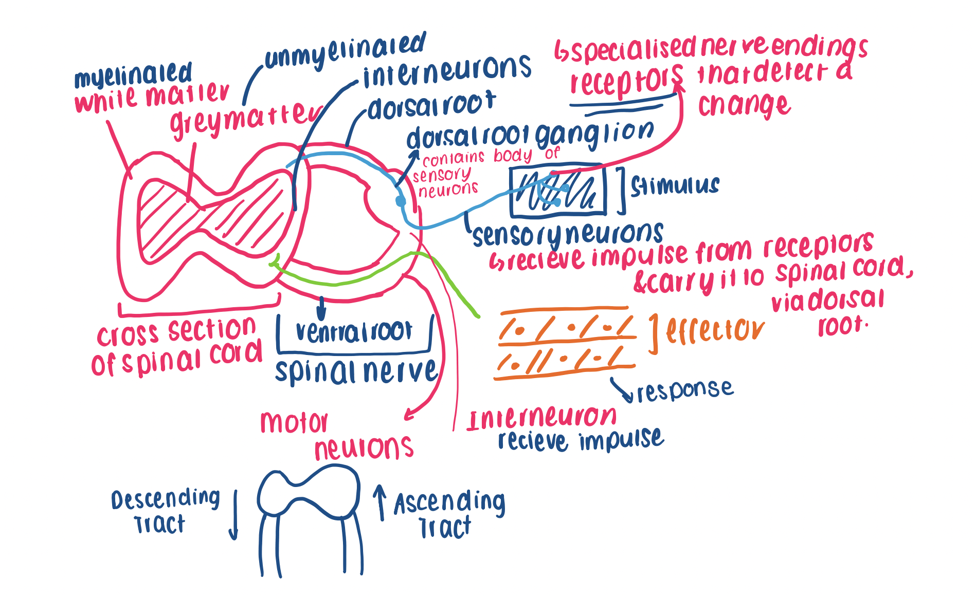
components of a reflex arc
resting membrane potential
Difference in charge between extracellular env + the intracellular env, caused by a difference in ion conc, when a cell is at rest.
Nerve impulse made up of multiple action potentials
Ions contributing to RPM
- K+, Na+, anions, Cl-
what is the value of the RMP
determine that the RMP indicates the intracellular environment is more negative than the extracellular environment
Value= -70mv (millivolts)
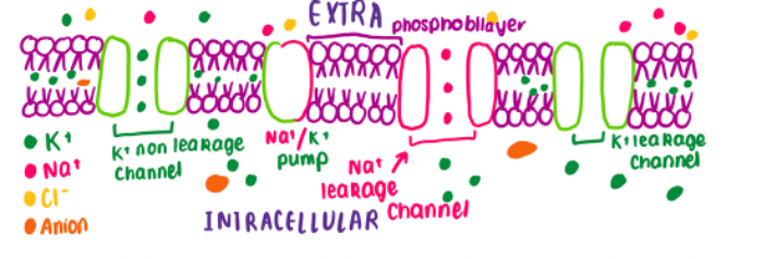
important points about resting membrane potential
- Embedded through the membrane are channel proteins called leakage channels, which remain open + passively move ions along concentration gradient
- The membrane contains more K+ leakage channels than Na+ leakage channels, meaning it is more permeable to K+
- The anions and chlorine do not move, as the membrane is not permeable
- The Na+/ K+ pump is a carrier protein that actively pumps these ions against their conc gradient, to maintain RMP of -70mv
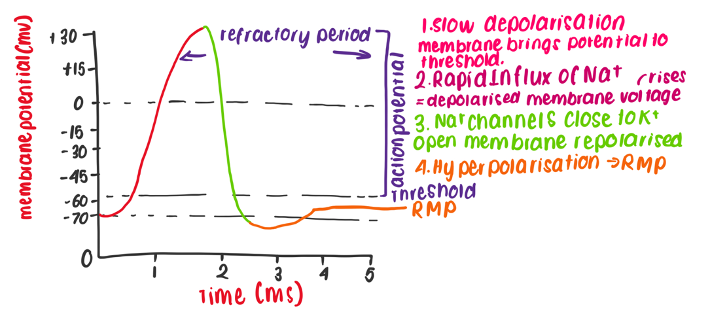
phases of the action potential
1.Depolarisation, NA+ MOVEMENT
2.Intracellular env becomes more positive than extracellular env
3.Repolarisation
4.Hyperpolarisation
overview of the phases of action potential
Depolarisation:
1.Sudden increase in membrane potential (cell is becoming more positive)
Caused by stimulation that exceeds 15mv “threshold” of cell
Na+ ligand channels open in response to stimulation via neurotransmitter/ sensory receptor (slow depolarisation)
2.This causes Na+ to move into the cell + the intracellular environment becomes more positive then the extracellular environment (voltage gated na+ channels open)
Original polarity of membrane reaches approx. +40mv= depolarised
3.Repolarisation- Na+ channels will close, preventing the influx of Na+ ions
Voltage gated K+ channels open, increasing the flow of K+ out of the cll
The intracellular environment becomes more neg than the outside= decrease in membrane potential
Membrane is now polarised
4.Hyperpolarisation- Membrane can now become hyperpolarised if K+ channels remain open longer than needed, Membrane potential decreases lower than RMP
steps to establishing RMP (refer to images on notes)
Steps to establishing RMP
1. Higher conc of K+ INTRA than
EXRTRACELLULAR
Results in K+ diffusing across leakage channel to extracellular env along conc gradient
2. Na+ is in higher conc in EXTRA than INTRA
Results in Na+ diffusing across the leakage channel
Much less Na+ will diffuse when compares to the amount of K+ (due to less leakage channels) resulting in the membrane potential becoming more negative
The RMP is -70 mv
3. Naturally K+ will passively diffuse back through leakage channels along its conc gradient
To ensure RMP of -70mv is maintained, and the cell is polarised (negative), the sodium/ potassium pump will actively pump 3Na+ out of the cell and 2K+ into the cell
Ensures conc gradient of both K+ and Na+ are kept constant
The carrier proteins bind with ATP to actively transport ions
action potential diagram
what is the refractory period
approx. 2-3 milliseconds after an action potential, where axon can’t conduct another axon potential
Ions return to original concentrations inside + outside of the cell
Period prevents- nerve impulse travelling backwards
Membrane reaches threshold of -55mv until it returns to RMP of -70mv
Requires considerable energy