Biosci 221 Cumulative Lab Final
0.0(0)
0.0(0)
Card Sorting
1/122
Earn XP
Description and Tags
Safety Lab, Metric Info, Aseptic Techniques, Microscope Lab, Protists, Fungi, Helminths, Microbial Growth, Control of Microbial Growth, Isolation Methods, Populations, Dilutions, Water/Food/Milk Lab, Transformation Lab, Staining Lab, Physiological Testing, & Clinical and Immunological Testing
Study Analytics
Name | Mastery | Learn | Test | Matching | Spaced | Call with Kai |
|---|
No study sessions yet.
123 Terms
1
New cards
What are the BioSafety Levels?
**BSL-1**: not known to cause disease in healthy adults
**BSL-2:** associated with disease
**BSL-3:** indigenous or exotic agent disease-causing agents
**BSL-4:** dangerous or exotic agents, highest risk, and no treatments
**BSL-2:** associated with disease
**BSL-3:** indigenous or exotic agent disease-causing agents
**BSL-4:** dangerous or exotic agents, highest risk, and no treatments
2
New cards
What kind of microbiology lab do we have at COC?
**BSL 2** b/c we work with *Staphylococcus aureus*, perform environmental sampling, and some individuals may be immunocompromised
3
New cards
What are the lab requirements for BSL 1 and 2?
BSL 1: lab coats, eyewear, and gloves, as needed
**BSL 2:** lab coats, eyewear, gloves, no eating/drinking, decontaminate surfaces, washing hands before and after, closed-toed shoes, no touching face, clean spills properly, keep hair back
**BSL 2:** lab coats, eyewear, gloves, no eating/drinking, decontaminate surfaces, washing hands before and after, closed-toed shoes, no touching face, clean spills properly, keep hair back
4
New cards
How would we handle a spill in lab?
* Stand still and assess the situation
* alert people around you and don’t put the tube down
* someone will bring you the biohazard rack for the tube
* then someone will spray the spill with sanisol and leave it for ten minutes
* then you can walk carefully over to wash your hands!
* alert people around you and don’t put the tube down
* someone will bring you the biohazard rack for the tube
* then someone will spray the spill with sanisol and leave it for ten minutes
* then you can walk carefully over to wash your hands!
5
New cards
Other safety info from the LS Safety lab?
* Put items in the cabinet, wash hands, disinfect station with Sanisol, and iPad with Sanisol
* Proper removal of gloves into a hazardous waste bin; must wear them when handling BSL 2 microorganisms or stains
* gloves should fit snugly, use caution when near a heat source as gloves can melt, and put on a new pair of gloves if they are contaminated or have a hole in them
* Proper removal of gloves into a hazardous waste bin; must wear them when handling BSL 2 microorganisms or stains
* gloves should fit snugly, use caution when near a heat source as gloves can melt, and put on a new pair of gloves if they are contaminated or have a hole in them
6
New cards
Conversions between ^^ul/um^^ and um/mm
* A micrometer is 1000 times smaller than a millimeter. 1mm = 1000um
* 1ul = 10^9 cubic um?
* 1ul = 10^9 cubic um?
7
New cards
Pipets: what types and when do we use each?
* **Serological** = for measuring and transferring milliliter volumes of liquid
* **Micropipette** = a very fine pipette for accurately and precisely measuring, transferring, or injecting very small quantities of liquid.
* **Transfer** = disposable and plastic, most basic, used for rough measurements only
* **Micropipette** = a very fine pipette for accurately and precisely measuring, transferring, or injecting very small quantities of liquid.
* **Transfer** = disposable and plastic, most basic, used for rough measurements only
8
New cards
Graduated cylinders, when do we use?
Graduated cylinders are for precise measurements; reading a liquid’s volume
9
New cards
Temps - room, body, refrigerator, freezer
* Freezer = 32°F (0°C)
* Refrigerator = 39°F (4°C)
* Room = 70°F (21°C)
* Body = 98.6° F (37° C)
* Refrigerator = 39°F (4°C)
* Room = 70°F (21°C)
* Body = 98.6° F (37° C)
10
New cards
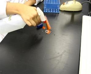
Know how to transfer bacteria from tube to tube or tube to plate, etc.
(photo)
(note: open image in new tab, difficult to see)
(note: open image in new tab, difficult to see)
11
New cards
How to prevent contamination
Always use proper aseptic technique
12
New cards
How to use the loop, needle or swab
Flaming of the loops and needles, until they become red hot, facilitates their sterilization such that it prevents cross-contamination of samples.
Following this, when they are cooled, they are ready to pick up, transfer and inoculate the microbial sample into the media or prepare a smear.
A cotton swab is usually used to obtain a sample from a patient or an environmental source; it can also be used to inoculate agar plates.
Following this, when they are cooled, they are ready to pick up, transfer and inoculate the microbial sample into the media or prepare a smear.
A cotton swab is usually used to obtain a sample from a patient or an environmental source; it can also be used to inoculate agar plates.
13
New cards
How to use the bacti-incinerator
When plugged in and turned on, the bacti-incinerator can reach high temperatures more than sufficient to incinerate any organic material on an inoculation loop.
To use, simply insert loop or needle into the tunnel for sterilization.
Can also be used to heat fix bacterial smears onto microscope slides.
To use, simply insert loop or needle into the tunnel for sterilization.
Can also be used to heat fix bacterial smears onto microscope slides.
14
New cards
How to identify contamination
Bacterial contamination is easily detected by __visual inspection__ of the culture within a few days of it becoming infected
15
New cards
What is the difference between brightfield, darkfield and phase contrast
* **Bright-field** (BF) microscopy is when light passes through the specimen and is collected in the objectives. The stained specimens appear dark against a “bright” background.
* In **Dark-field** (DF) microscopy, some of the rays of light are blocked as they pass through the condenser. It shows the specimens bright on a dark background.
* **Phase Contrast** microscopy is a technique for viewing live specimens, which enables one to see the details and finer structures of cells. The thickness of different parts of the specimen will refract (bend) the light to different degrees; enhances contrast.
16
New cards
How do you use phase contrast
Depending on the lens, switch to PH1, PH2, or PH3
You may use phase contrast when you have difficulty finding your specimen or when you are looking at __living, unstained cells.__
You may use phase contrast when you have difficulty finding your specimen or when you are looking at __living, unstained cells.__
17
New cards
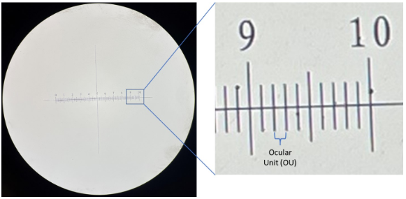
How do you measure organisms
using the **ocular micrometer**
18
New cards
What are the calibrations of the ocular micrometer for each of the objective lens
(photo)
19
New cards
How do you determine the total magnification
**total magnification** = (ocular lens magnification) x (objective lens magnification)
the ocular lens magnification on the compound microscope is 10x
the ocular lens magnification on the compound microscope is 10x
20
New cards
What does oil do in oil immersion
light passes from glass to oil to glass which __increases resolution__
21
New cards
How do you adjust the amount of light
adjust the **occulting disk**
22
New cards
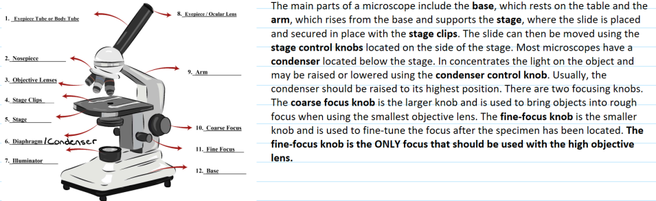
What are the parts of the microscope
(photo)
23
New cards
How do you use the microscope to observe bacteria
Using an oil-immersion objective lens on a microscope, a magnification of 1000× can be achieved to view bacteria → can determine shape, arrangement, and size of bacteria
24
New cards
How do you put your microscope away
using both hands; one holding the handle on the arm of the microscope, the other holding the bottom of the microscope on the other side
25
New cards
What are the general characteristics of the protists?
eukaryotic and unicellular
26
New cards
Compare and contrast algae versus protozoa
* **Protozoa**: most are harmless free-living in a moist habitat but some are animal parasites; use locomotion; chemoheterotrophic; no cell wall
* **Algae**: single-cells or colonial; photoautotroph; photosynthetic; cell wall; causes Harmful Algal Bloom and Paralytic Shellfish Poisoning
* **Algae**: single-cells or colonial; photoautotroph; photosynthetic; cell wall; causes Harmful Algal Bloom and Paralytic Shellfish Poisoning
27
New cards
What is the difference between a trophozoite and a cyst?
* **Cyst** = the dormant stage; very resistant structures that allow protozoa to survive in adverse environments
* **Trophozoite** = active feeding stage; absorbing nutrients from the host
* **Trophozoite** = active feeding stage; absorbing nutrients from the host
28
New cards
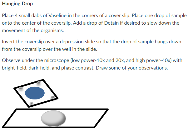
Why would we use a hanging drop slide?
preserves cell shape and arrangement
the Vaseline-sealed depression also slows down the drying-out process, so the organisms can be observed for longer periods.
can see motility
the Vaseline-sealed depression also slows down the drying-out process, so the organisms can be observed for longer periods.
can see motility
29
New cards
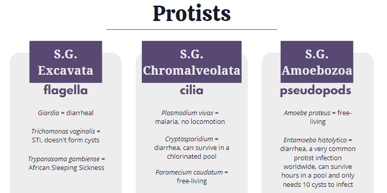
What are the important protists that were studied: Why were these important? Know the important information about them.
(photo)
30
New cards
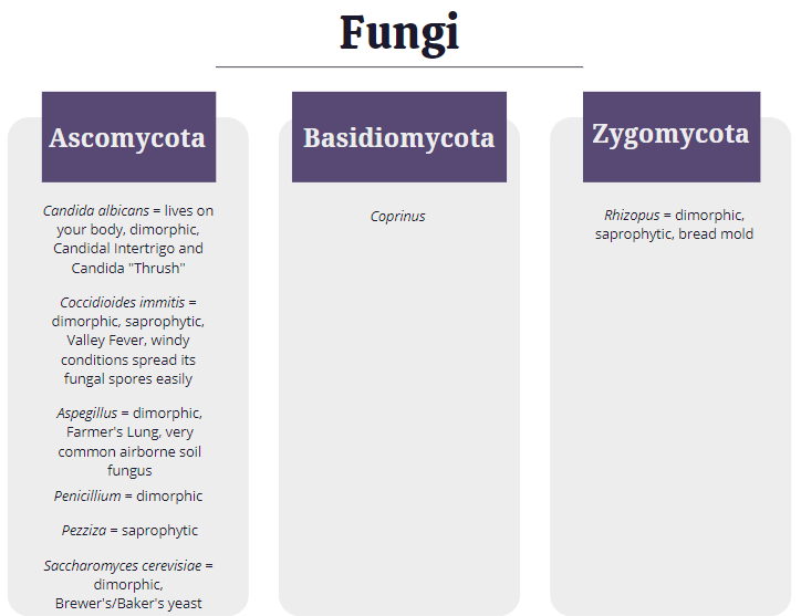
What are the major characteristics of fungi?
chemoheterotrophic organisms that are usually nonmotile and grow by absorbing nutrients from their surroundings; their cells walls are made of chitin
31
New cards
Descriptions of molds versus yeasts
**Yeasts** are single-celled while **molds** are multi-celled (made up of hyphae)
32
New cards
Hyphae and mycelia
**hyphae** = long, threadlike filaments
**mycelia** = mass of hyphae
**mycelia** = mass of hyphae
33
New cards
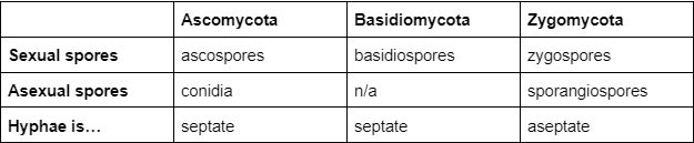
Types of sexual spores - what are the differences?
(photo)
34
New cards
Types of asexual spores - what are the differences? (sporangiospores, conidia)
**conidia** = not in a sac, extend from the hyphae
**sporangiospores** = enclosed in a sac
**sporangiospores** = enclosed in a sac
35
New cards
What are the important fungi that were studied: Why were these important? Know the important information about them.
(photo)
36
New cards
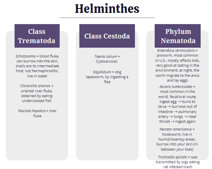
General characteristics of the helminthes
They’re parasitic worms that are multicellular, eukaryotic, invertebrate animals, most are macroscopic but have microscopic life stages
* Phylum Platyhelmintes
* **Class Trematoda**
* flukes, leaf-shaped, intermediate hosts are mollusks, definitive hosts are vertebrae, digestive system
* **Class Cestoda**
* tapeworms, long/ribbon-like bodies, scolex for attachment, two or more hosts, no digestive system
* **Phylum Nematoda**
* roundworms, cylindrical, one host, complete digestive system, diverse and numerous
37
New cards
What is a scolex and a proglottid?
The **scolex** is essentially the head, which contains organs which facilitate attachment to the host tissue
**Proglottids** are segments of the worm’s “neck” which contain eggs
**Proglottids** are segments of the worm’s “neck” which contain eggs
38
New cards
What are the important helminths that were studied: Why were these important? Know the important information about them.
(photo)
39
New cards
Acidophile
prefers acidic conditions (pH 0-5.5)
40
New cards
Neutrophile
prefers neutral conditions (pH 5.5-8.0)
41
New cards
Alkaliphile
prefers basic conditions (pH 8.0-11.5)
42
New cards
How do we use pH to control the growth of bacteria?
__pickling__ is a common method of food preservation and compounds like citric acid are often added to food products to lower the pH and prevent spoilage or toxin formation
43
New cards
Osmotolerant
The ability to grow in an environment with a high osmotic pressure
44
New cards
Osmophiles
Adapted to environments with high osmotic pressures (high salt concentrations)
45
New cards
Plasmolysis
cell shrinkage
46
New cards
Hypertonic
more solutes outside of the cell, higher osmotic pressure; there will be a net movement of water out of the cell
47
New cards
Isotonic
equal amount of solute inside and outside the cell
48
New cards
Hypotonic
more solutes inside of the cell relative to the solution and the net movement of water will be into the cell, which may result in lysis of the cell.
49
New cards
How do we use osmotic pressure to control the growth of bacteria?
__Meat curing:__ removal of water and addition of salt to meat creates a solute-rich environment where osmotic pressure draws water out of microorganisms, thereby retarding their growth
50
New cards
what protects bacteria in a hypotonic environment?
the bacterial cell wall
51
New cards
psychrophile
prefer/capable of growth and reproduction in low temperatures
52
New cards
psychrotroph
cold-tolerant
53
New cards
mesophile
moderate temperatures
54
New cards
thermophile
relatively high temperatures
55
New cards
hyperthermophile
extremely hot environments
56
New cards
cardinal temperatures
the range that a particular bacterium grows in
57
New cards
How do we use temperature to control the growth of bacteria?
Outside of their cardinal temps, the bacteria may not survive
they can be denatured by both high and low temperatures
they can be denatured by both high and low temperatures
58
New cards
anaerobe
cannot survive in the presence of oxygen because they do not have the enzymes to break down toxic by-products and will quickly die in the presence of oxygen
59
New cards
microaerophile
prefer a reduced oxygen atmosphere
60
New cards
facultative anaerobe
have the necessary enzymes and can grow with or without oxygen
61
New cards
strict/obligate anaerobe
tolerate the presence of oxygen but can only grow in the absence of oxygen
62
New cards
aerotolerant anaerobe
can grow in the presence of oxygen but do not use oxygen for growth
63
New cards
Fluid Thioglycollate with resazurin
a medium that uses thioglycollate broth to permit the growth of anaerobic bacteria.
* growth patterns can help distinguish the aerotolerance and oxygen requirements of bacteria
* resazurin = reactant/indicator of FTM
* a dye that changes the color to pink when oxygen is present
* growth patterns can help distinguish the aerotolerance and oxygen requirements of bacteria
* resazurin = reactant/indicator of FTM
* a dye that changes the color to pink when oxygen is present
64
New cards
anaerobe box
specialized container used to create an anaerobic environment for the growth of anaerobic bacteria on plates
65
New cards
^^significant results^^ - like where did certain oxygen types grow, regular incubator, anaerobe chamber or both?
…
66
New cards
What are the definitions of disinfectant and antiseptic?
**Disinfectants** should be fast acting, stable, easy to prepare, inexpensive, and easy to use
Ex.:
* Natural disinfectant = vinegar
* Chemical disinfectant = chlorine bleach or products containing chlorine
**Antiseptics** are antimicrobial chemicals safe for use on living skin or tissues
* Ex. hydrogen peroxide and isopropyl alcohol
Ex.:
* Natural disinfectant = vinegar
* Chemical disinfectant = chlorine bleach or products containing chlorine
**Antiseptics** are antimicrobial chemicals safe for use on living skin or tissues
* Ex. hydrogen peroxide and isopropyl alcohol
67
New cards
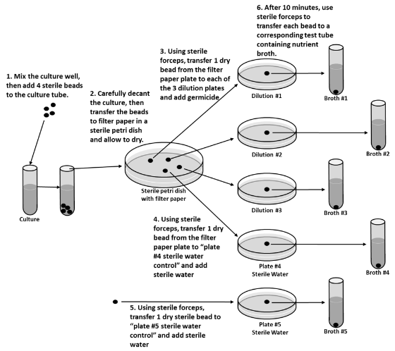
Use dilution test
The use-dilution test is commonly used to determine a chemical’s disinfection effectiveness on an inanimate surface in clinical settings
68
New cards
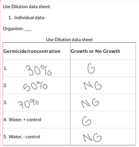
Any significant results for Use dilution?
(photo)
69
New cards
Kirby Bauer Method: What was the medium and was there a reason we used that medium?
Mueller Hinton Agar
* a non-selective and non-differential growth medium
* loose agar meaning when you place antibiotics on the media it's allowed to diffuse or seep in, allowing for antibiotic susceptibility tests to be conducted accurately
* a non-selective and non-differential growth medium
* loose agar meaning when you place antibiotics on the media it's allowed to diffuse or seep in, allowing for antibiotic susceptibility tests to be conducted accurately
70
New cards
What was the point of comparing the turbidity to a McFarland Standard?
to help ensure that a consistent amount of bacteria is plated
71
New cards
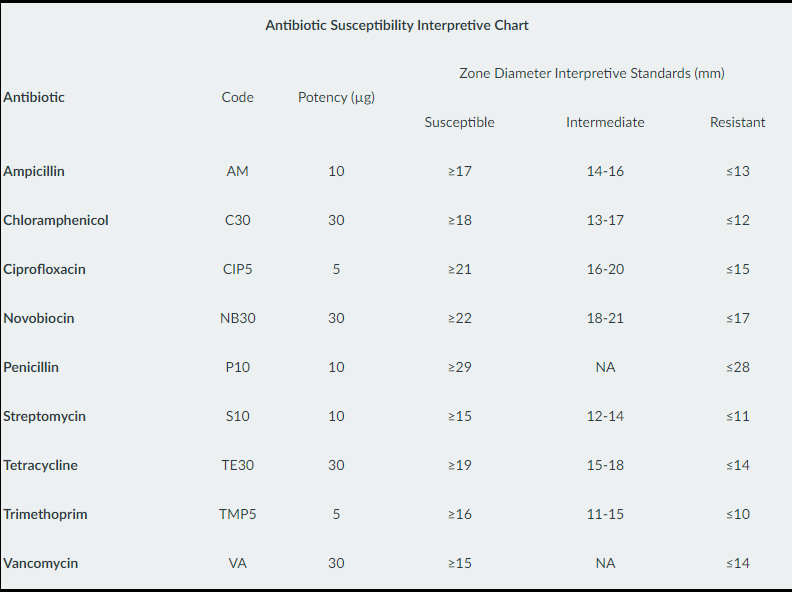
How do you determine if something was Sensitive/Susceptible or Intermediate or Resistant?
The antibiotics are prepared specifically for the antimicrobial sensitivity test by infusing known amounts into standard paper disks. As bacteria begin to grow on a plate, the antibiotics are diffusing out into the agar. If the bacteria are sensitive to the antibiotic, at some point their growth will be inhibited and a zone of no growth (zone of inhibition) will be apparent around the disk. The diameter of the zone is measured and the size of the zone determines whether a bacterium is determined Sensitive (S), Resistant (R), or Intermediate (I) to the antibiotic.
* The size and interpretation of the zone is specific to each bacteria-antibiotic pair and is referenced in a chart.
* The size and interpretation of the zone is specific to each bacteria-antibiotic pair and is referenced in a chart.
72
New cards
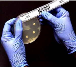
What is the zone of inhibition? How is that determined?
an area of media where bacteria are unable to grow, due to the presence of a drug that impedes their growth
measure the diameter with a ruler
measure the diameter with a ruler
73
New cards
Was there any difference between the results of the GNR verses the GPCs?
Gram-negative bacteria are more resistant to antibiotics than Gram-positive
74
New cards
What were the results of *S. saprophyticus* and *S. epidermidis* on Novobiocin (why was it important to know those results)?
*S. saprophyticus* is resistant
*S. epidermis* is susceptible
Important because it’s how we can differentiate the two
*S. epidermis* is susceptible
Important because it’s how we can differentiate the two
75
New cards
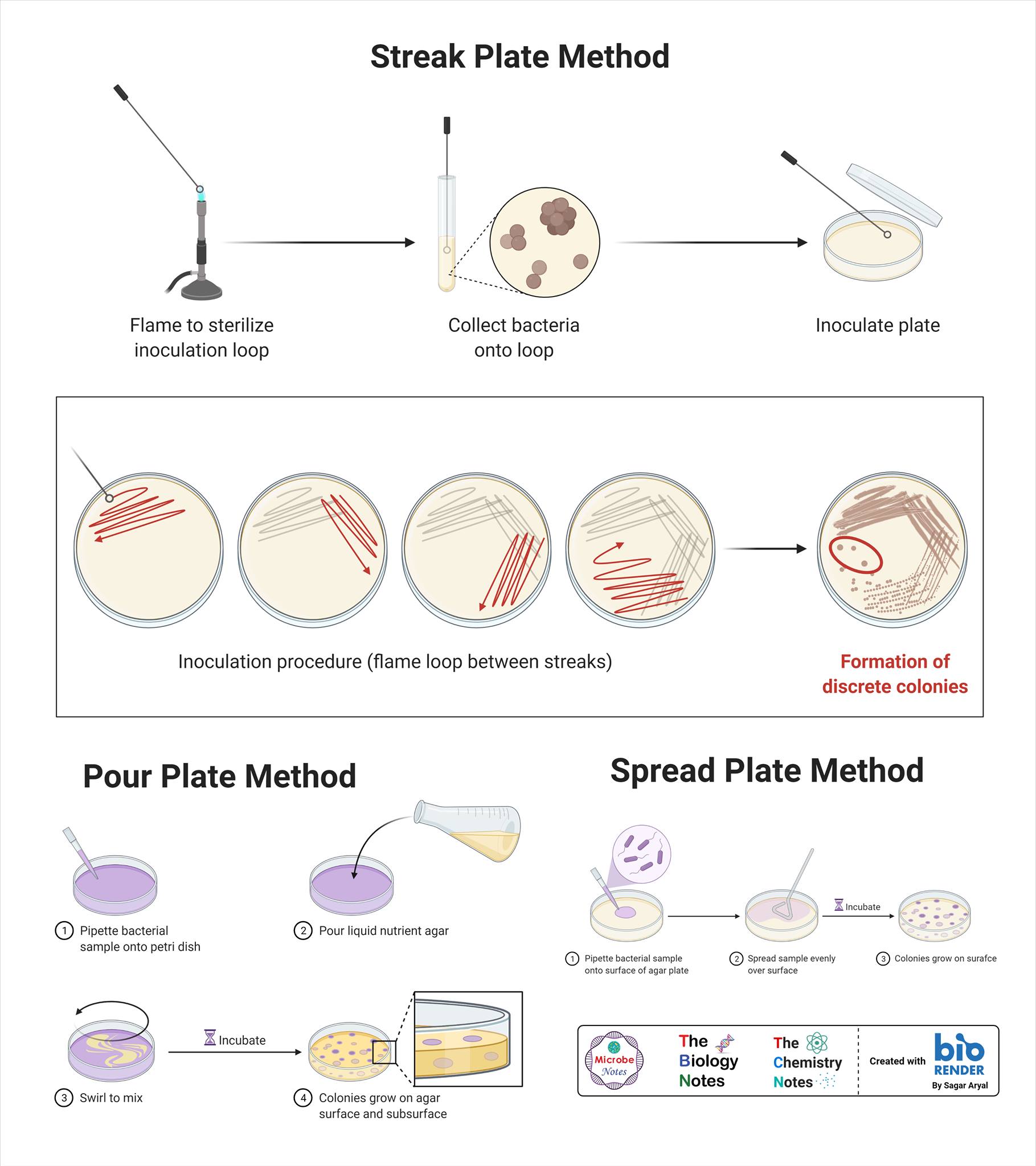
pour vs. streak vs. spread plate
1. **pour plate:** a known volume of the sample is mixed with agar and then poured into a plate
2. **streak plate:** a technique used to have pure colonies
3. **spread plate:** a known volume of the sample is spread on the surface of the agar medium
76
New cards
subculture
**Subculturing** is the transfer of microbes from one growth medium container, such as broth or agar, to another, and allowing the microbes to grow
77
New cards
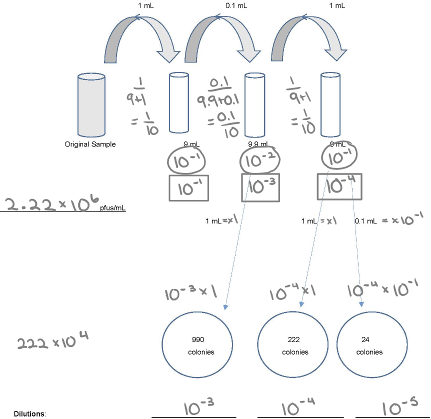
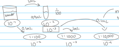
how and why dilutions are needed
Most specimens, based on turbidity, might have millions of bacteria per ml in the sample. If 1.0ml was plated onto agar the number of bacteria would be so many that it would be impossible to count or even discern individual colonies. Therefore, one must perform a series of dilutions with the specimen to obtain a sample that will produce a plate on which colonies can be counted.
78
New cards
optical density and how it relates to population counts
Indirect counts of a sample can be performed by using a spectrophotometer and measuring the turbidity (aka absorbance or Optical Density) of the sample. This is called the Turbidimetric method. A standard curve must be created using the known count of a sample, and then counts of various dilutions can be extrapolated.
79
New cards
direct count
1. Direct microscopic counts of cells can be done, visually, and by various types of electronic particle counters.
2. Direct counts sample small amounts of a larger sample and we can’t distinguish between live cells and dead cells. We could however, see a variety of cell types.
80
New cards
Know how to do dilution
1. Individual dilution
2. Total dilution
3. How much plating: 1.0 ml (x1) or 0.1 ml (x 10^-1)
4. (30-300) x DF
81
New cards
Water Lab
1. HPC - what is it, why do we do it, what do the results show us?
1. **Heterotrophic Plate Count** shows us an estimate of how many (heterotrophic) bacteria might be in the water
2. a standard method __for determining drinking water quality__; a quantitative method used to determine the overall quality of the water
3. shows the effectiveness of disinfectants used in water treatment and differentiating between organisms in the water
2. How would you set up an HPC for tap water, drinking water, or contaminated water?
1. Transfer 1 mL from the original sample to a Petri dish and add TGEA pour
2. Rotate in a figure 8 motion and let harden
3. Incubate at 35 C for 24 hours
4. Count colonies and determine CFU/mL
3. Membrane Filtration - what is the medium, and what are the advantages and disadvantages
1. The **Membrane Filter Technique** utilizes an apparatus containing a filter membrane with a 0.45um pore size that will trap microorganisms larger than that on the surface of the membrane as the water sample passes through the membrane. The membrane is then placed onto a plate of __m-Endo agar__
2. **advantages**: large volumes can be processed at one time; the number of coliforms/mL in the original sample can be calculated; and inexpensive, fast, accurate results
3. **disadvantages**: many organisms dies in the beginning → false negatives
4. Multiple Tube Method for **Most Probable Number** - what media, what is the difference between the 3 parts, the Presumptive, the Confirmed, and the Completed, what are the advantages and disadvantages
1. **media** = lactose broth
2. **Presumptive** → A screening test to sample water for the presence of coliform organisms. If the presumptive test is negative, no further testing is performed, and the water source is considered microbiologically safe. If, however, any tube in the series shows acid and gas, the water is considered unsafe and the confirmed test is performed on the tube displaying a positive reaction.
3. **Confirmed Test** → If a water sample is positive for gas, then it is presumed that the sample contains coliforms. The confirmed test would then be performed via inoculation of a plate of EMB agar from a gas-positive tube.
4. **Completed Test** → Coliform colonies from EMB would be inoculated again into Lactose Broth with a Durham tube and checked for gas, and inoculated on TSA and checked via Gram stain for GNRs. If these tests are positive, it shows that coliforms (not another gas producer) are present and indicates that the water sample is contaminated.
5. **advantages** = cheap and easy
6. **disadvantages** = very time-consuming does not differentiate between coliforms and fecal coliforms
82
New cards
Milk Lab
1. What is the media
1. TGEA pours
2. What is **pasteurization**
1. Milk is heated to a specific temperature for a set period of time to kill harmful bacteria that can lead to diseases
3. What are the concerns about raw milk?
1. It can also be a cause of the transmission of disease to humans.
4. What are we testing for
1. looks for bacteria, not specific to coliforms
2. the bacterial count in milk is the most reliable indication we have of its sanitary quality.
5. What is an APC (SPC)
1. **standard plate count** (or aerobic plate count) estimates the population density of bacteria in a broth by plating a small and dilute portion of the sample and counting the number of bacteria colonies

83
New cards
Food Lab
1. What is the media?
1. Eugon agar pours
2. EMB plate to look for fecal colonies
2. What would be expected from particular foods?
1. high counts for certain types of food (moist, uncooked, lots of handling) and low counts for others (dry, no nutrients, high salt, sugar, high and low pH, etc.)
84
New cards
What is transformation
* Some bacteria may pick up genes from the surrounding environment (from dead bacteria), via “**transformation**.”
* These genes, often on a plasmid, may code for antibiotic resistance mechanisms.
* These genes, often on a plasmid, may code for antibiotic resistance mechanisms.
85
New cards
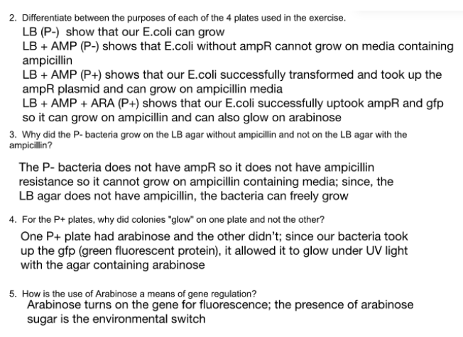
What were the medias used in the transformation exercise and what was their purpose
The PGlo plasmids the bacteria will be uptaking have ampR (ampicillin resistance), gfp (Green Fluorescent Protein) genes, and araC (activator of gfp)
→ Bacteria plated onto media that contains Ampicillin will grow, and if the media also contains arabinose, they will glow
→ Bacteria plated onto media that contains Ampicillin will grow, and if the media also contains arabinose, they will glow
86
New cards
What did the Transformation Efficiency calculation show?
**Transformation efficiency** is the measure of how many cells successfully underwent transformation
TE = (# of transformed colonies/ug DNA) X (final volume at recovery (500ul)/volume plated)
TE = (# of transformed colonies/ug DNA) X (final volume at recovery (500ul)/volume plated)

87
New cards
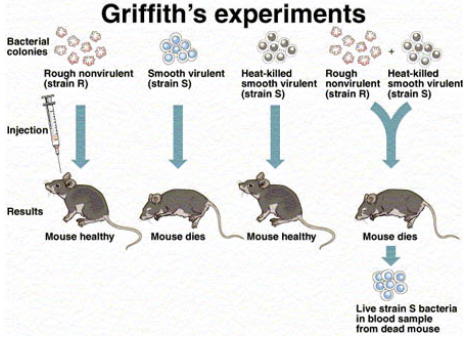
What did Griffith discover in his experiment regarding colony characteristics in relation to bacterial virulence?
During the experiment, Griffith cultured Streptococcus pneumoniae bacteria which showed two patterns of growth. One culture plate consisted of smooth shiny colonies (S) while the other consisted of rough colonies (R).
Griffith injected both S and R strains into mice. The one which was infected with the S strain developed pneumonia and died while that infected with the R strain stayed alive.
In the second stage, Griffith heat-killed the S strain bacteria and injected it into mice, but the mice stayed alive. Then, he mixed the heat-killed S and live R strains. This mixture was injected into mice and they died. In addition, he found living S strain bacteria in dead mice.
IN OTHER WORDS… virulence could be transferred from killed pathogenic bacteria to live non-pathogenic bacteria. He called this process “transformation.”
88
New cards
Negative staining
* **Negative stains** are when we use Nigrosin (an acidic stain → it has a neg. charge) to stain bacteria and view them in their original shape and size due to the lack of heat fixation. The negative charge of the stain repels the negative charge of the bacteria’s cell, making it appear clear.
* A negative stain is very good for observing cell morphology, arrangement, and size because the slide is NOT heat-fixed.
* DON’T:
* heat fix → lose morphology and size
* put too much organism → thick, uneven stain
* put too much nigrosin → causes cracking in stain
* A negative stain is very good for observing cell morphology, arrangement, and size because the slide is NOT heat-fixed.
* DON’T:
* heat fix → lose morphology and size
* put too much organism → thick, uneven stain
* put too much nigrosin → causes cracking in stain
89
New cards
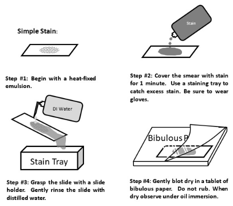
Simple staining
A **simple stain** is when we use a positively-charged dye to stain the negatively-charged bacteria and view them easily under the brightfield lens
90
New cards
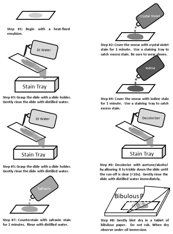
Gram staining
We might use a **gram stain** in a mixed sample of organisms to differentiate cell types. Bacterial cells with gram-positive cell walls are stained purple with the dye crystal violet, while bacterial cells with gram-negative cell walls are counterstained pink with safranin dye.
91
New cards
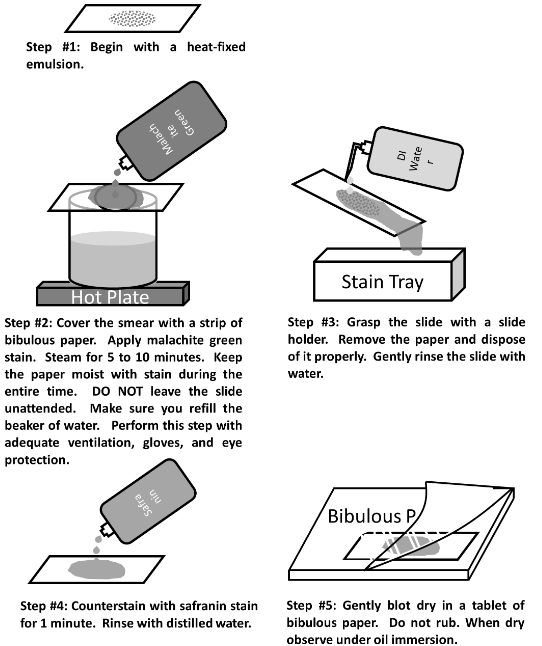
Endospore staining
* An **endospore** is a structure for the preservation of the DNA while conditions are poor
* The heating process makes the spore coat more permeable to the stain
* Endospores are formed when the bacterium is stressed.
* Endospores can be seen in simple and Gram stains, as a clear or empty space in a vegetative cell.
* The heating process makes the spore coat more permeable to the stain
* Endospores are formed when the bacterium is stressed.
* Endospores can be seen in simple and Gram stains, as a clear or empty space in a vegetative cell.
92
New cards
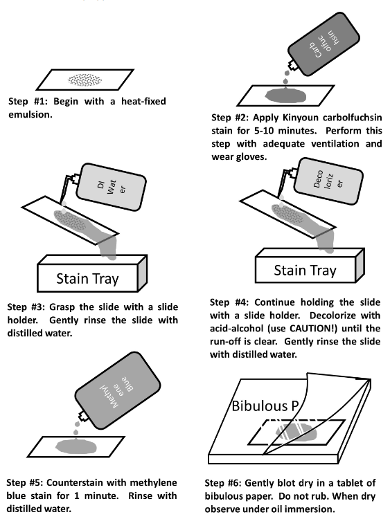
Acid Fast staining
* In the **Acid-Fast stain**, a very concentrated stain solution of kinyoun carbolfuchsin (hot pink) is used in order to penetrate and stain the cell walls
* This stain is a valuable diagnostic tool for finding tuberculosis and nocardial diseases in patient specimens
* *Mycobacterium smegmatis* is acid-fast positive
* This stain is a valuable diagnostic tool for finding tuberculosis and nocardial diseases in patient specimens
* *Mycobacterium smegmatis* is acid-fast positive
93
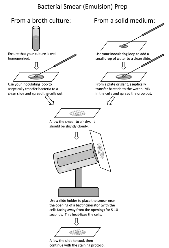
New cards
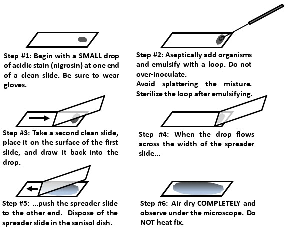
Smear prep from liquid or solid
(photo)
94
New cards
Good smear versus poor smear
A __good smear__ is evenly distributed and not too thick or sparse; almost undetectable to the naked eye → shows it is thin and even
If the smear is too thick the bacteria will not stain evenly. A thick/uneven smear cannot be properly decolorized and might wash off the slide despite fixation; we also won’t be able to see the bacteria clearly (morphology and arrangement) if they’re all piled on top of each other.
If the smear is too thick the bacteria will not stain evenly. A thick/uneven smear cannot be properly decolorized and might wash off the slide despite fixation; we also won’t be able to see the bacteria clearly (morphology and arrangement) if they’re all piled on top of each other.
95
New cards
Heat fixation
We use heat to fix the bacteria onto the slide so it doesn’t wash away with the staining agents and DI water.
96
New cards
Immersion oil
A drop of immersion oil is used ONLY on the 100x objective lens to view microorganisms more clearly since the oil increases the resolving power
Immersion oil has the same refractive index as glass
Immersion oil has the same refractive index as glass
97
New cards
How to observe differential stains
By always using brightfield on a 100X objective lens with oil immersion
98
New cards
What is a differential stain and which differential stains do we use
**Differential stains** use more than one stain, and cells will have a different appearance based on their chemical or structural properties.
* Gram staining
* Endospore staining
* Acid-fast staining
* Gram staining
* Endospore staining
* Acid-fast staining
99
New cards
What is a mordant
A **mordant** is a substance that helps fix a stain to its target
100
New cards
What does it mean if an organism is gram + or gram -
**Gram positive** cells have thick layers of a peptidoglycan (a carbohydrate) in their cell walls; **Gram negative** bacteria have very little.