Unit 4, Chapter 2E, Knowt, by Batoul Mortada, Alena Namoo, and Tasnim Uddin
1/41
Earn XP
Description and Tags
Advanced Human Anatomy PALIA project
Name | Mastery | Learn | Test | Matching | Spaced |
|---|
No study sessions yet.
42 Terms
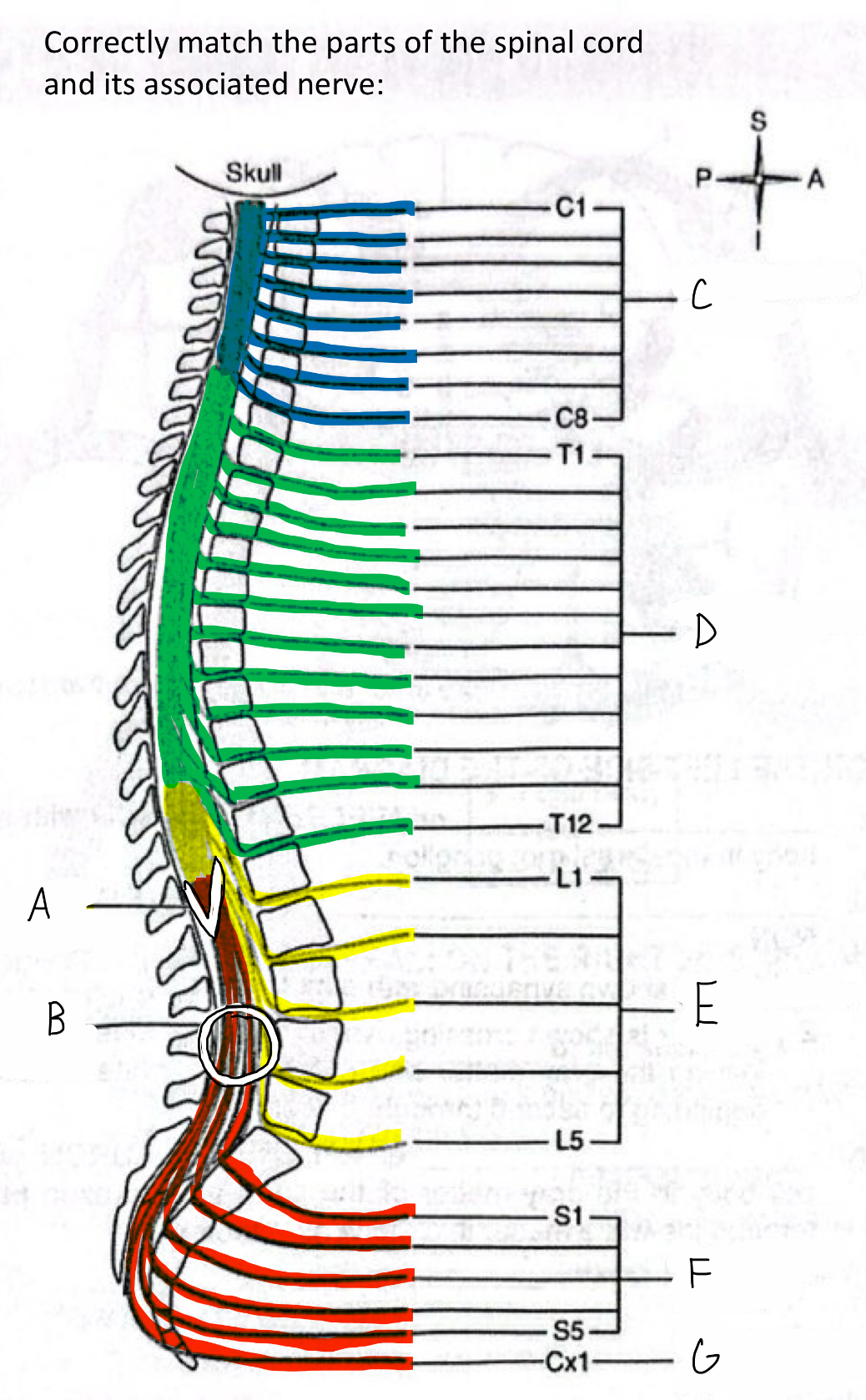
What anatomical structure is labeled B in the diagram?
Cauda Equina
The spinal cord is continuous with which part of the brain?
The medulla oblongata.
What does the pia mater adhere to?
The surface of the brain and spinal cord.
Where are the spinal cord enlargements located?
In the cervical and lumbosacral regions.
What limbs are associated with the spinal cord enlargements?
Cervical: upper limb; Lumbosacral: lower limb.
True or False:
The Dorsal Ramus supplies the entire anterior trunk and appendages.
FALSE.
The Ventral Ramus supplies the entire anterior trunk and appendages.
The Dorsal Ramus supplies all the structures of the back.
What is the conical-shaped lower end of the spinal cord called?
Conus medullaris.
What does the cauda equina consist of?
Very long roots of the lower spinal nerves that descend in a bundle from the conus medullaris.

What anatomical structure is labeled A in the diagram?
Conus Medullaris

What is labeled #2 on the diagram?
Subarachnoid space.
What structure is found below LV2 in the vertebral canal?
The cauda equina.

What anatomical structure is labeled D in the diagram?
Thoracic Nerves (T1-T12)
Where does the arachnoid mater extend?
It surrounds the brain, extends through the foramen magnum, and passes inferiorly through the vertebral canal to the coccyx.

What anatomical structure is labeled F in the diagram?
Sacral Nerves (S1-S5)

Which meninx is colored red in the diagram?
Arachnoid mater.
What are meninges?
Three layers of connective tissue that surround and protect the brain and spinal cord.
What is another name for Interneuron?
Internuncial Neuron.
Name the three layers of the meninges from outermost to innermost.
Dura mater, Arachnoid mater, Pia mater.

What anatomical structure is labeled E in the diagram?
Lumbar Nerves (L1-L5)
What type of tissue composes the dura mater?
Tough fibrous connective tissue.
How much of the vertebral canal does the spinal cord occupy?
The upper 2/3 of the vertebral canal.

What anatomical structure is labeled C in the diagram?
Cervical Nerves (C1-C8)
What is the space external to the dura mater called?
Epidural space.
What is found in the epidural space?
Adipose tissue (in the form of globules of fat) and the vertebral venous plexus.
Which meninx is the intermediate layer?
Arachnoid mater.
Where does the dura mater extend?
It surrounds the brain, extends through the foramen magnum, and courses inferiorly to the coccyx in the vertebral canal.
What space lies between the arachnoid mater and pia mater?
Subarachnoid space.
What is contained in the subarachnoid space?
Cerebrospinal fluid (CSF).
What is the innermost meninx?
Pia mater.
What type of tissue composes the pia mater?
Very delicate connective tissue.

Which meninx is colored green in the diagram?
Pia mater.
Where does the Pia mater extend?
It surrounds the brain, extends through the foramen magnum, and passes inferiorly to the coccyx.
To what level does the spinal cord extend inferiorly?
To the level of LV2.
Which is larger, the Dorsal Ramus or the Ventral Ramus?
Ventral Ramus.

What is labeled #1 on the diagram?
Epidural space.
Fill in the blank:
__________ is an area of cell bodies, dendrites, and unmyelinated axons within the CNS.
Gray matter
What is the texture and structure of the arachnoid mater?
Delicate, loose, netlike membrane.
What is the Dorsal Root Ganglion?
An enlarged portion of the dorsal root that contains cell bodies of sensory neurons.
True or False:
A motor neuron is the same as an efferent neuron.
TRUE.
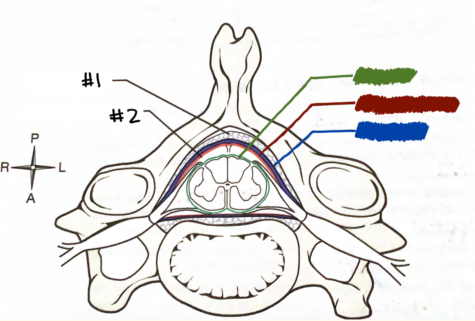
Which meninx is colored blue in the diagram?
Dura mater.
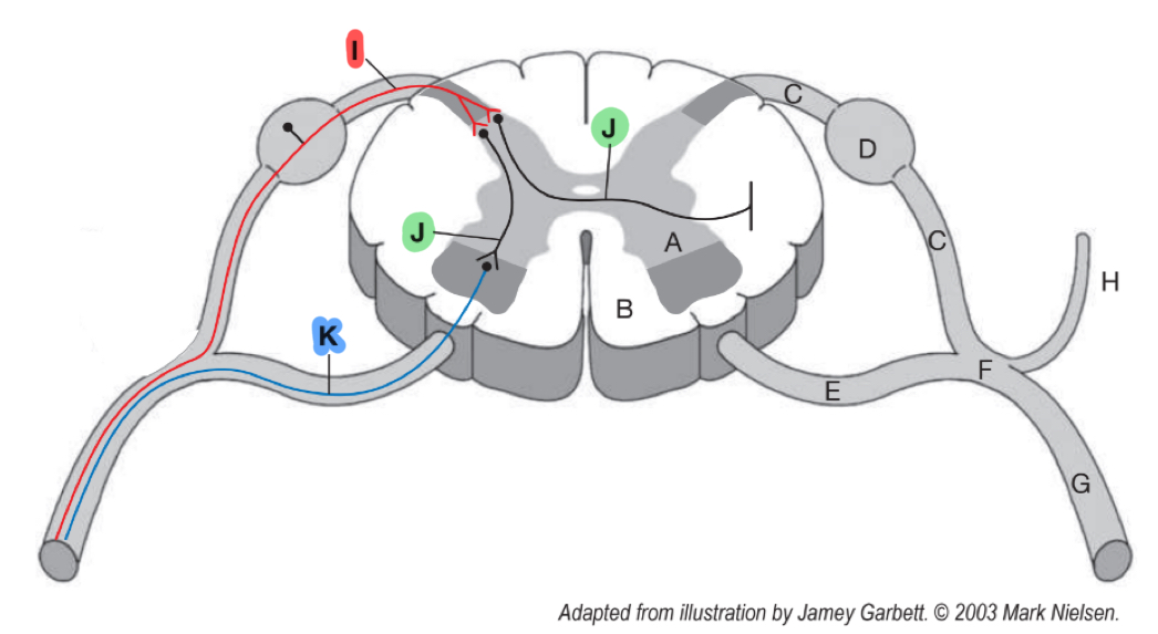
What anatomical structure is labeled I in the diagram?
Sensory (afferent) Neuron.

What anatomical structure is labeled G in the diagram?
Coccygeal Nerves (Cx1)