Retina Anatomy 2
1/64
There's no tags or description
Looks like no tags are added yet.
Name | Mastery | Learn | Test | Matching | Spaced |
|---|
No study sessions yet.
65 Terms
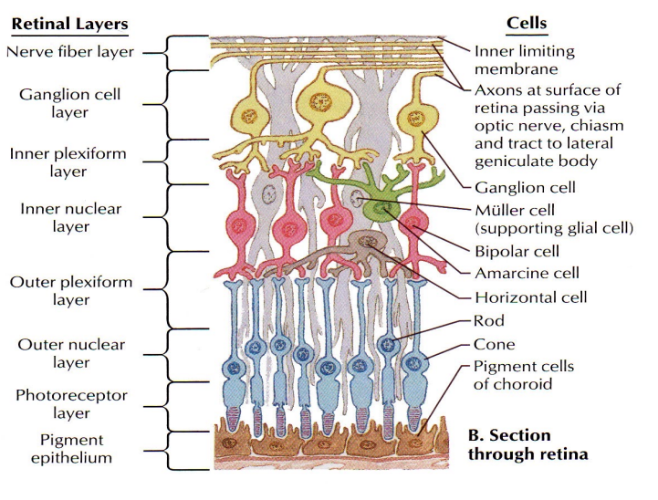
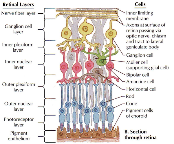
Photoreceptors (Rods And Cones)
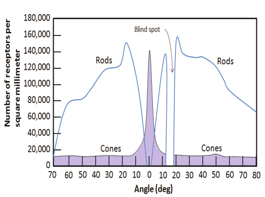
• About 120 million total rods in the retina
• About 6 million total cones in the retina
Number Of Photoreceptors
• Density of rods is greater than cones throughout the retina except at the macula (Cones are concentrated most at the macula especially at the fovea and foveola)
• No rods are found at the foveola (Only cones)
• Rod density is greatest concentrically 4.5 mm or 15 degrees from the foveola
• Total number of both rods and cones decrease toward the ora serrata
• No photoreceptors are found at the optic nerve (Thus physiologic blind spot)
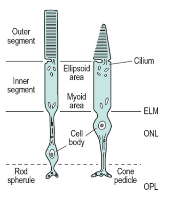
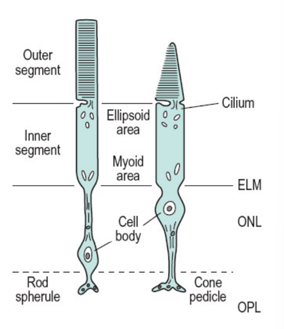
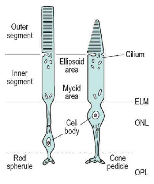
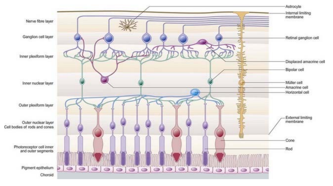
6 Parts To The Photoreceptor (From Closest To The RPE To The OPL)
RPE
IDZ
Outer Segment
Cilium
Inner Segment
Outer Fiber
Cell Body
Inner Fiber
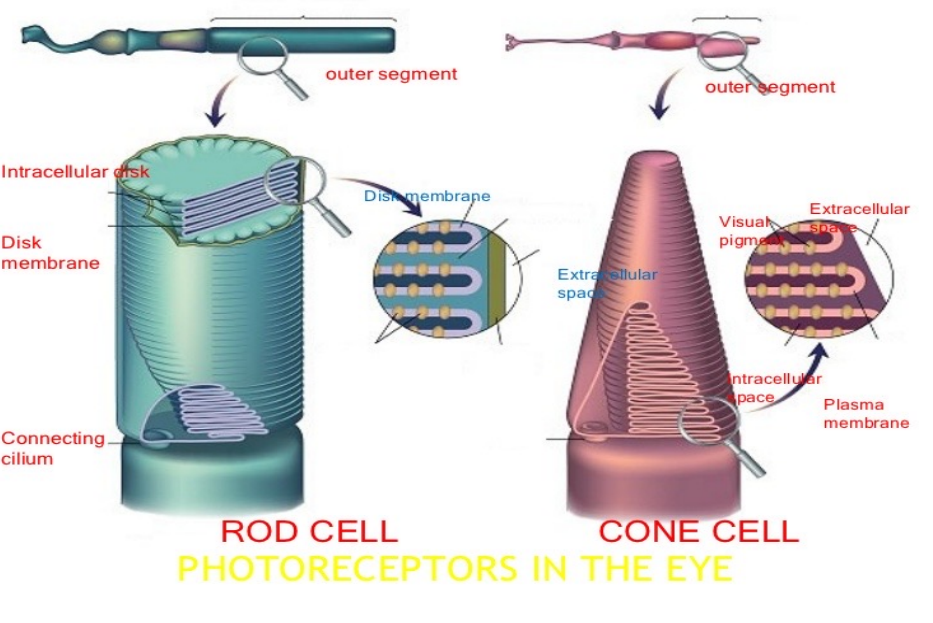
Outer segment
•Made up of a stack of discs
• Each disc is a flattened membrane sac
• Visual pigment molecules are located in these membranous discs
• Shape of outer segment gives rods and cones their name
• The base of the outer segment is in contact with the cilium and the tip of the outer segment is in contact with the interdigitation zone
Cilium
Connects inner segment with outer segment
Inner segment
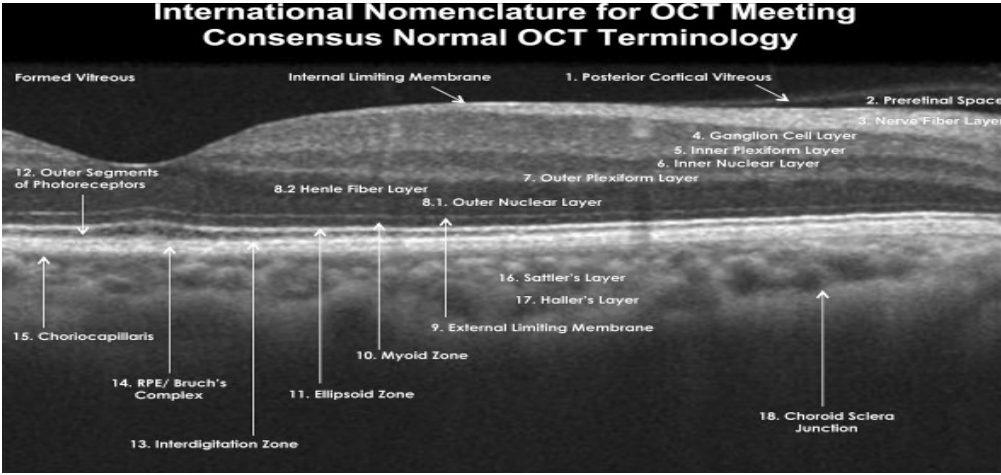
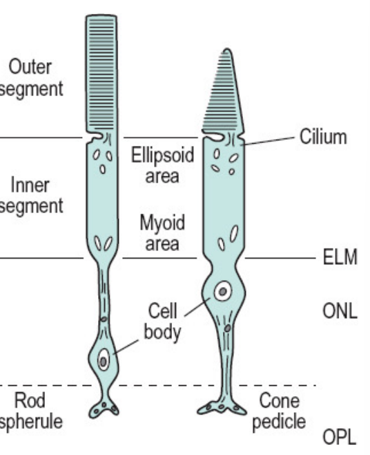
• Made of two parts: Ellipsoid zone and Myoid zone
• Ellipsoid zone is nearest to the outer segment and is directly connected to the cilium. The ellipsoid zone contains mitochondria which provides energy to the photoreceptors
• Myoid zone is nearest to the cell body and directly connected to the outer fiber. The myoid zone contains the other organelles and protein synthesis occurs here
Outer Fiber
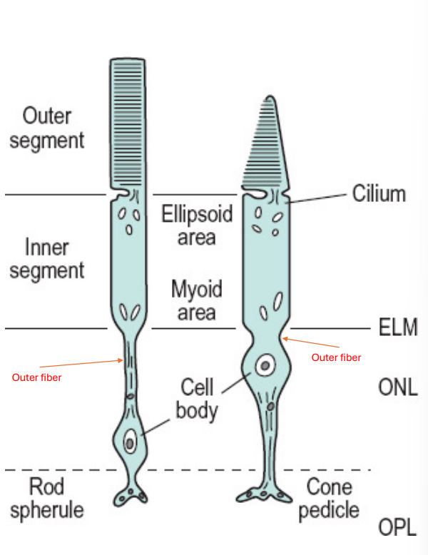
•Connects the inner segment to the cell body
Cell body
•Contains the nucleus
Inner fiber
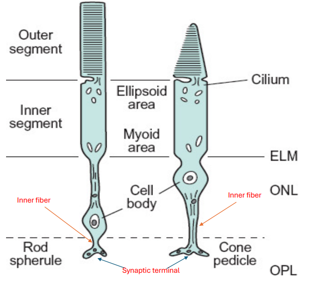
Composed of axons with microtubules that connect the cell body to the synaptic terminals
Rods
40-60 microns long
There is only 1 type of rod (Based on visual pigment)
Rods tend to be overall longer than cones but thinner than cones
More active in dim conditions (Scotopic vision). Most effective after 20-30 minutes of dark adaptation
Responsible for sensing contrast, brightness, and motion
Outer Segment Of Rods
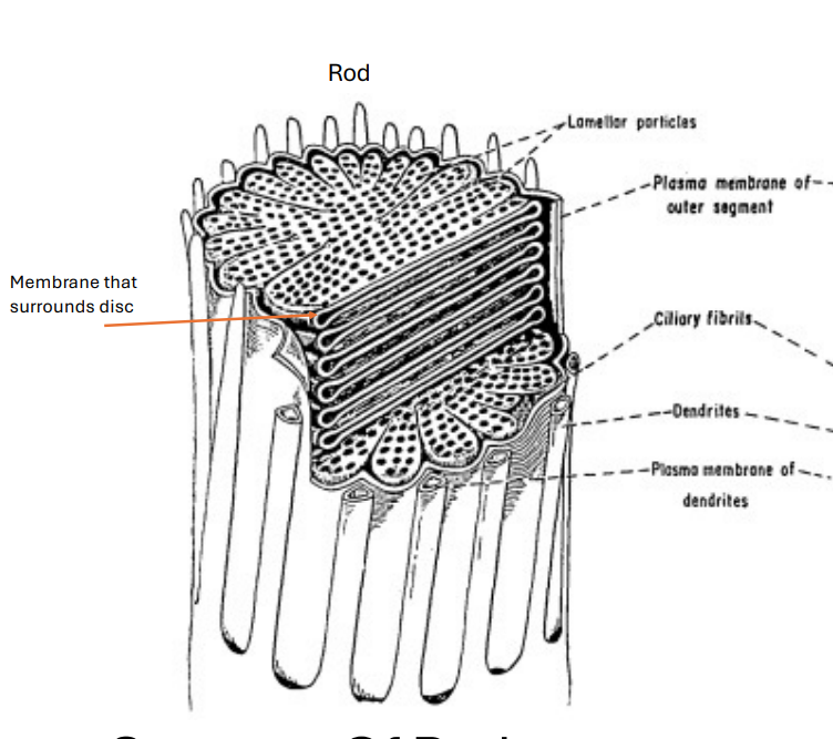
• Discs are found in the outer segment and are for the most part uniform in width (About 600-1000 discs per rod)
• Discs are separate from surrounding, adjacent discs
• Visual pigment is found in each disc
• The outer segment of the rod is enclosed by a plasmalemma (A plasma membrane which surrounds a cell)
• The plasmalemma is separate from the membranes that surround the discs in the outer segment
• Rod outer segments are longer than cone outer segments (*Exception: Cone outer segments are just as long as rod outer segments at the macula, especially the foveal region)
Rod Visual Pigment
• Rhodopsin, found in each disc, is the photosensitive pigment of a rod
• There is only 1 type of rhodopsin
• Since the visual pigment is the same in all rods, there is only 1 type of rod (Monochromatic) which has a maximum wavelength absorption of 498nm
Rod Outer Segment Renewal System
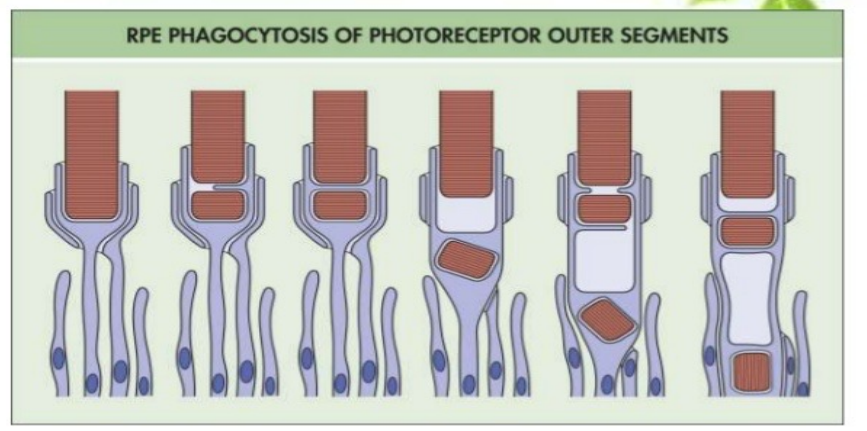
• Components of the disc membrane are made in the inner segment which will move to be a part of the discs in the base of the outer segment
• Old discs are displaced outward by formation of new discs
• Old discs come off the tip of the outer segment and the RPE cells will phagocytize these old discs
• Discs are shed regularly especially in the early morning or in bright environments (Constantly renewed)
Rod (Other Components)
• The rod inner segment is thinner than the cone inner segment (Contributes to the shape of a rod as well)
• The rod inner and outer segment are about the same width, which are connected by the cilium
• A rod’s ellipsoid area of the inner segment contains less mitochondria than a cone’s ellipsoid area of the inner segment
• The rod’s outer fiber is longer than a cone’s outer fiber
• Rod nuclei tends to be found further from the outer retina or external limiting membrane than cone nuclei
• Rod cell body and nuclei are smaller than those of a cone
• The cell body is connected to the synaptic complex (Called a spherule) by the inner fiber which is shorter than in a cone
• A rod’s spherule is smaller than a cone’s pedicle
• Rod spherules connect with bipolar cells, horizontal cells, and other rod spherules and cone pedicles {SAME}
• The neurotransmitter released by the rod spherule is called glutamate (Excitatory neurotransmitter) {SAME}
Cones
40-50 microns long
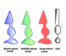
There are 3 types of cones (Based on visual pigment)
Cones tend to be overall shorter than rods but thicker than rods
More active in bright conditions (Photopic vision)
Responsible for sensing fine resolution, spatial resolution, and color
Cone Outer Segments
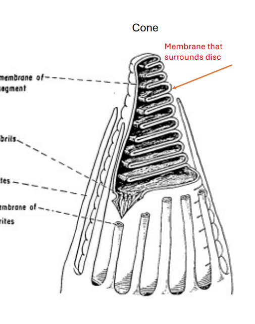
• Discs are found in the outer segment with the discs at the base being wider than at the tip (Main reason for the shape of the outer segment of a cone)
• There are 1000-1200 discs per cone
• Discs are connected to each other (Not separate)
• The outer segment of the cone is enclosed by a plasmalemma (A plasma membrane which surrounds a cell)
• The plasmalemma is continuous with the membranes that surround the discs in the outer segment
• Cone outer segments are shorter than rod outer segments. Thus, cone outer segments may not reach the RPE. Microvilli from the apical surface of the RPE are able to reach and surround the cone outer segments (*Exception: Cone outer segments are just as long as rod outer segments at the macula, especially the foveal region)
Cone Visual Pigments
• Photopsin, found in each disc, is the photosensitive pigment of a cone
• There are 3 types of cones
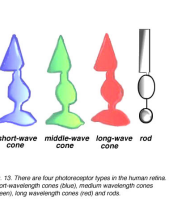
Blue cone (Short wavelength): Maximum wavelength absorption is 420nm
Green cone (Medium wavelength): Maximum wavelength absorption is 531nm
Red Cone (Long wavelength): Maximum wavelength absorption is 588nm
Cone Outer Segment Renewal
• New discs are formed while old discs come off the tip of the outer segment and the RPE cells will phagocytize these old discs
• Discs are shed periodically, typically at the end of the day or in dark environments (Not constantly renewed)
• Not exactly clear how this process happens
Cone (Other Components)
• The cone inner segment is thicker than the rod inner segment (Contributes to shape of a cone as well)
• A cone’s ellipsoid area of the inner segment contains more mitochondria than a rod’s ellipsoid area of the inner segment
• The cone’s outer fiber is shorter than a rod’s outer fiber (Cone outer fiber may even be missing)
• Cone nuclei tends to be found closer to the outer retina or external limiting membrane than rod nuclei
• Cone cell body and nuclei are larger than those of a rod
• The cell body is connected to the synaptic complex (Called a pedicle) by the inner fiber which is longer than in the rod
• A cone’s pedicle is larger than a rod’s spherule
• Cone pedicles connect with bipolar cells, horizontal cells, and other rod spherules and cone pedicles {SAME}
• The neurotransmitter released by cones is called glutamate (Excitatory neurotransmitter) {SAME}
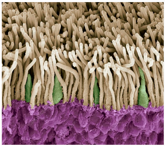
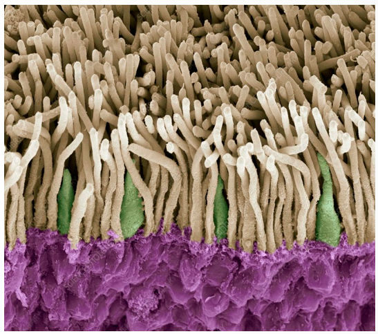
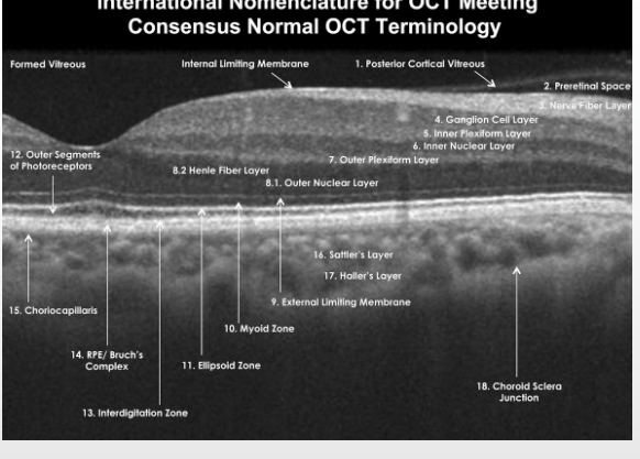
What Is The Most Likely Retinal Location Of The Rods And Cones In The Photo?
Mid-Periphery
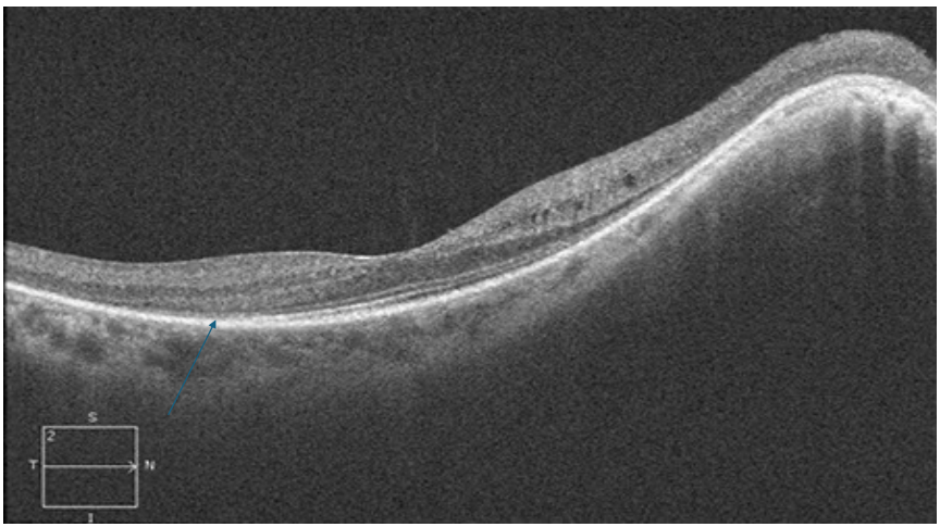
What Is Missing In The OCT Scan?
Ellipsoid zone missing rods
Rod dystrophy
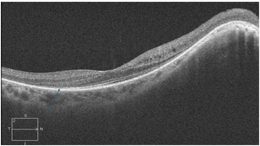

What Is Missing In The OCT Scan?
Ellipsoid zone missing cones in the macula
Cone dystrophy

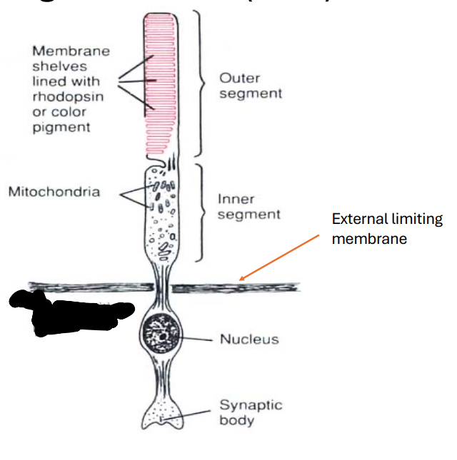
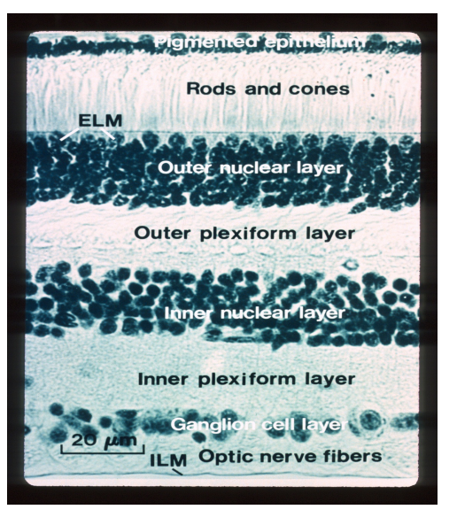
External Limiting Membrane (ELM)
• Not a true membrane
• Composed of zonula adherens junctions (Anchoring junction) between photoreceptor cells as well as photoreceptor and Muller cells at the level of the inner segments
• Function:
• It acts as a metabolic barrier as it prevents passage of large molecules (Small molecules are able to pass)
• It stabilizes the transducing portion of photoreceptors
*Transduction: Process by which light is converted into electrical signals in the rod and cone cells
Outer Nuclear Layer (ONL)
•Contains rod and cone cell bodies (Which contain the nuclei)
•Cone outer fibers are very short or even absent so the nuclei tend to be in a single layer close to the external limiting membrane
•Cell bodies of rods are found in different rows inner to the cone cell bodies because rod outer fibers tend to be longer
• Rod nuclei greatly outnumber cone nuclei throughout the retina except at the fovea and especially at the foveola where there are only cone nuclei
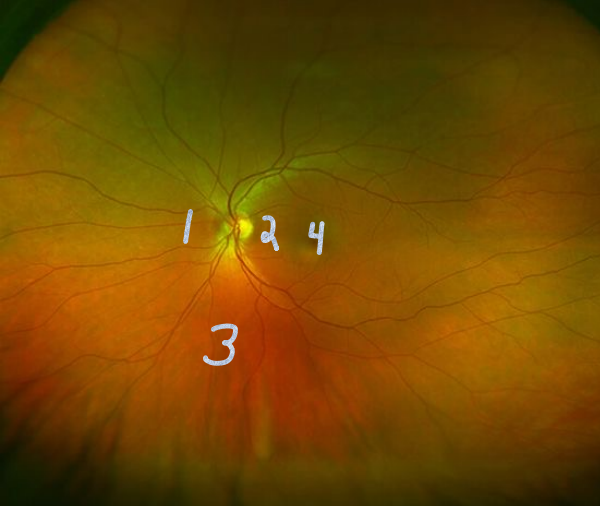
Outer Nuclear Layer (ONL) Thickness Levels
Thickest at the foveola and fovea where 10 rows of cone nuclei can be found (About 50 microns thick)
8-9 rows thick nasal to the optic nerve of both rod and cone nuclei (About 45 microns thick)
1 row of cones and 4 rows of rods thick typically found in the rest of the retina (About 27 microns thick)
Thinnest just temp to the optic nerve where it is 4 rows thick of both rod and cone nuclei (About 22 microns thick)
4.1.3.2 (thinnest)
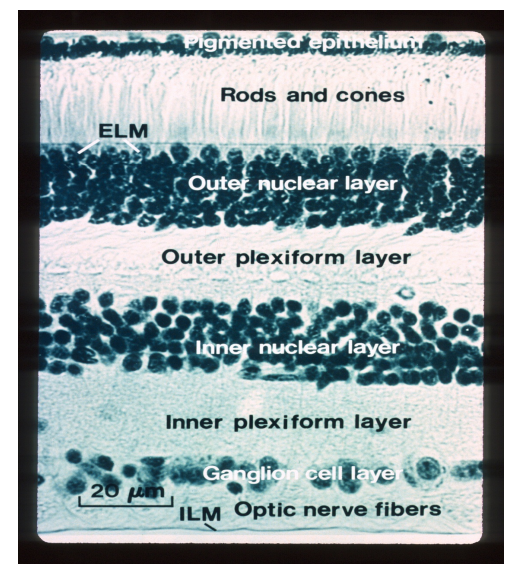
Outer Plexiform Layer (OPL)
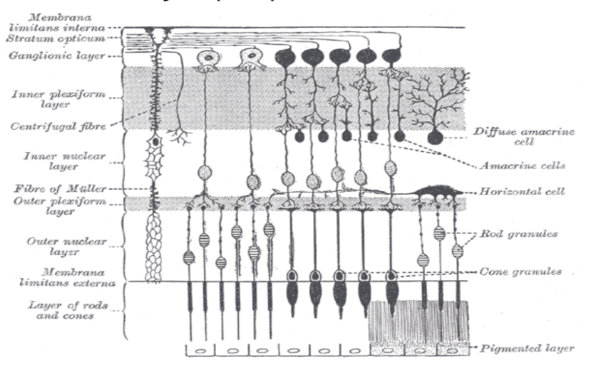
• Also known as the outer synaptic layer (Synaptic junction between photoreceptors and second order neurons which includes bipolar cells and horizontal cells)
• The inner fibers of rods and cones make up the outer aspect of the OPL and the synaptic terminals make up the inner aspect of the OPL
• Thickest in the macula region (About 51 microns)
• Prevents spread of fluid to other layers of the retina
This is the last layer where parts of a photoreceptor can be found
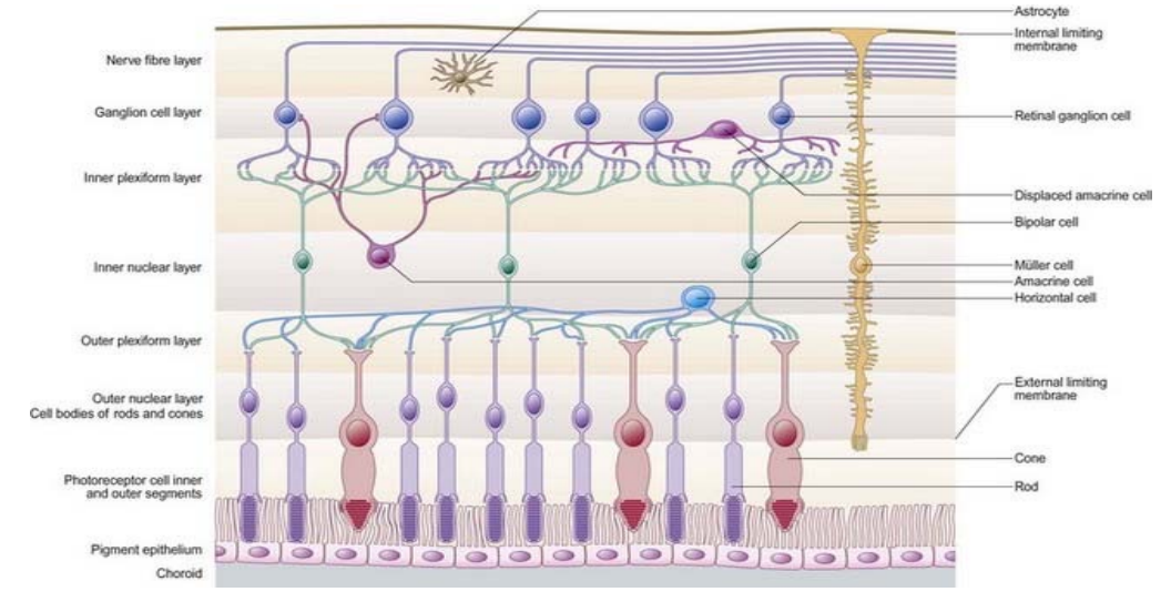
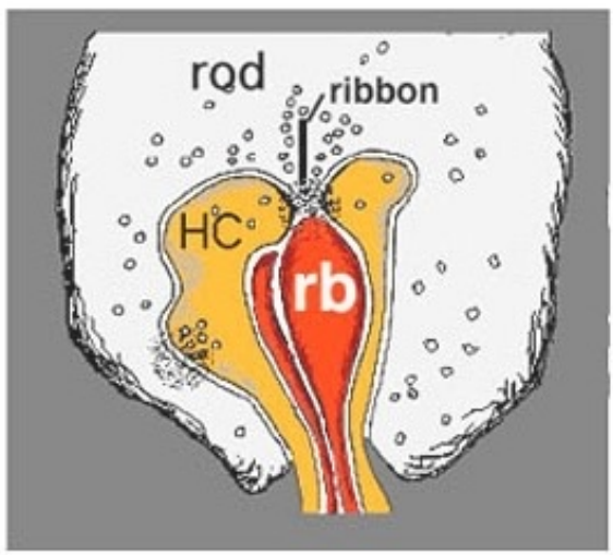
Rods In The Outer Plexiform Layer (OPL)
• Rod spherules synapse with bipolar cell dendrites and horizontal cell axons (Total synaptic complexes per spherule: About 2-7)
• Rod spherules have deep invaginations where the synapses occur
• Each invagination in a rod spherule contains 1 or more rod bipolar dendrites surrounded by two horizontal cell axons
• A membranous plate called a synaptic ribbon is found on the rod spherule adjacent to the invagination
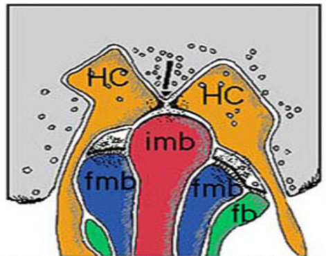
Cones In The Outer Plexiform Layer (OPL)
• Cone pedicles synapse with bipolar cell dendrites and horizontal cell dendrites (Total synaptic complexes per pedicule: about 25)
• Cones pedicles have superficial invaginations where the synapses occur
• Each invagination in a cone pedicle contains an invaginating midget bipolar dendrite surrounded by two horizontal cell dendrites (”Triad”)
• Outside and adjacent to this triad and on the flat surface of the pedicle, there is also at least 1-2 flat midget bipolar dendrites
• Other types of flat bipolar dendrites (Not midget) can also be found outside of the invaginations
• A membranous plate called a synaptic ribbon is found on the rod spherule adjacent to the invagination
Other Synapses In The Outer Plexiform Layer That Doesn’t Involve Invaginations Of Rod Spherules And Cone Pedicles
Horizontal cells synapse with bipolar dendrites through gap junctions
Horizontal cells synapse with other horizontal cells through gap junctions
Rod spherules synapse with cone pedicles and other rod spherules through gap junctions
Cone pedicles synapse with rod spherules and other cone pedicles through gap junctions
(Exception: Blue cones or short wavelength cones do not have gap junctions with other cones and rods)
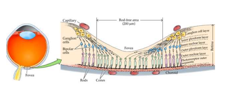
Henle Fiber Layer
Henle Fiber Layer is the name of the outer plexiform layer that is present from the foveola to the perifovea
• Why? Cone pedicles, neurons, and axons are displaced laterally at the fovea and especially at the foveola
When light hits the fovea, want as little obstruction as possible
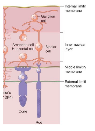
Middle Limiting Membrane (in outer plexiform layer)
• Composed of desmosome-like attachments called synaptic densities that are found within the branching, interwoven bipolar dendrites and horizontal cells
• This membrane prevents leaking from the inner retinal vasculature from getting in the outer retina
• In fact, the inner retinal vasculature usually does not extend beyond the middle limiting membrane
Inner Nuclear Layer (INL)
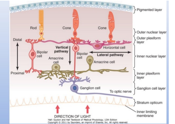
•Contains cell bodies of horizontal cells, bipolar cells, amacrine cells, interplexiform neurons, Muller cells, and displaced ganglion cells
•Composed of 8-12 rows of closely packed nuclei
*This layer disappears at the foveola
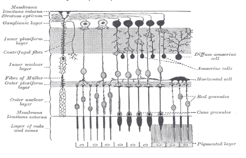
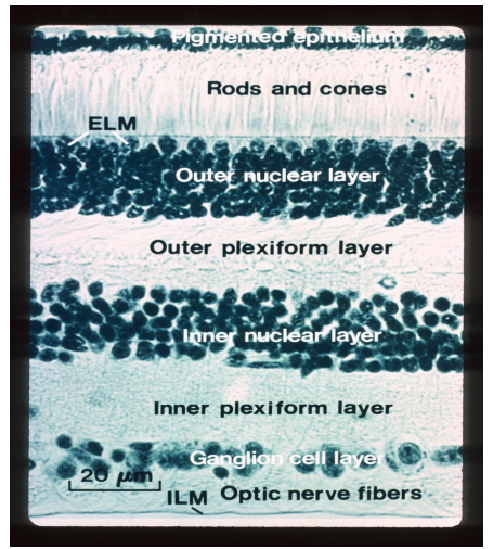
Inner Plexiform Layer (IPL)
• Also known as the inner synaptic layer (Synaptic junction between second-order and third-order neurons which includes the axons of bipolar cells and dendrites of ganglion cells for cones and between second-order, third-order, and fourth order neurons which includes the axons of bipolar cells, amacrine cells, and dendrites of ganglion cells for rods)
• The axon of the invaginating midget bipolar cell ends in the inner half of the inner plexiform layer and the flat midget bipolar cell ends in the outer half of the inner plexiform layer
• There is also synapses between amacrine processes and bipolar axons, amacrine processes and ganglion cell dendrites, amacrine processes and other amacrine processes, and amacrine cells and interplexiform neurons
• Prevents spread of fluid to other layers of the retina
*This layer disappears at the foveola
Plexiform Layers Strength
The inner plexiform layer tends to be stronger than the outer plexiform layer
Ganglion Cell Layer (GCL)
There are 1.12 million-2.22 million ganglion cells
Contains the cell body (Nucleus) of a ganglion cell
Each ganglion cell is separated by Muller cells
*This layer disappears at the foveola
Ganglion Cell Layer (GCL) Thickness
• Single cell thick except at the macula where it is 8 to 10 cells thick and just temporal to the optic nerve where it is 2 cells thick
• The number of ganglion cells decreases significantly further out in the peripheral retina
Convergence Of Rods Into The Ganglion Cell Layer (GCL)
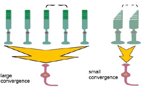
• There is greater convergence of rods than cones into a ganglion cell (Think of a bottleneck) Why? Due to shear number of rods compared to cones
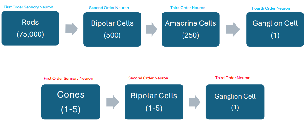
• About 75,000 rods synapse with 500 rod bipolar cells and 250 AII amacrine cells before they converge into a single ganglion cell. This allows increased sensitivity to light and motion
• A rod pathway involves a 4 neuron chain due to involvement of amacrine cells

Convergence Of Cones Into The Ganglion Cell Layer (GCL)
• About 1-5 cones synapse with bipolar cells and a small amount of cone bipolar cells synapse with a single ganglion cell
• In fact, in some areas there is a 1:1 ratio between cones and ganglion cells. For example, a single midget bipolar dendrite may come in contact with one cone pedicle and its axon then synapses with one ganglion cell. This allows the ability to discriminate detail.
• A cone pathway involves a 3 neuron chain

Rods and Cones Convergence Into The Ganglion Cell Layer (GCL)
don’t know exact numbers
rods > bipolar cells > amacrine cells > ganglion cell
cones > bipolar cells > ganglion cells
Nerve Fiber Layer (NFL)
• Consists of the ganglion cell axons that should be unmyelinated
• These axons run parallel to the retina surface and go to the optic disc and through the lamina cribosa
• Thickest at the margins of the optic disc
*This layer disappears at the foveola
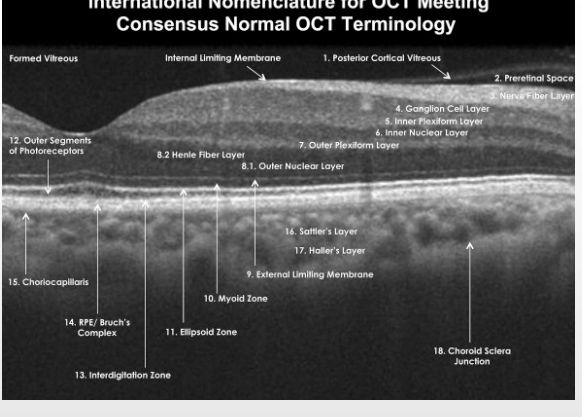
Which Eye Does This OCT Belong To?
Right
Optic nerve on the right (can tell due to the nerve fiber layer being thicker)
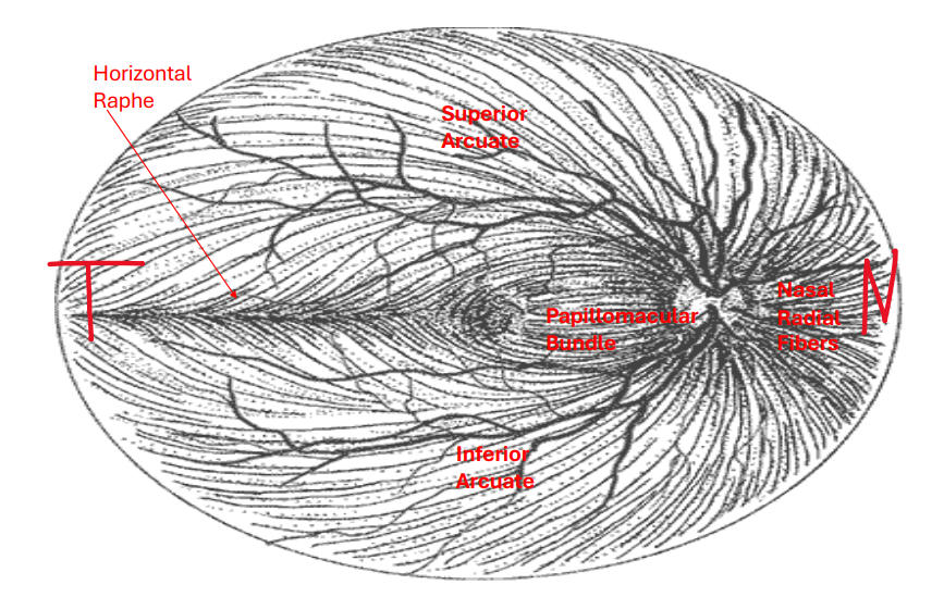
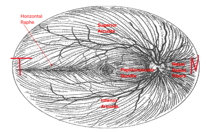
4 Nerve Fiber Layer (NFL) Bundles
*The horizontal raphe divides the superior arcuate fibers from the inferior arcuate fibers

Papillomacular fibers
Radiate to the optic disc from the macula and thus carries information that determines visual acuity.
It is the thinnest nerve fiber layer bundle
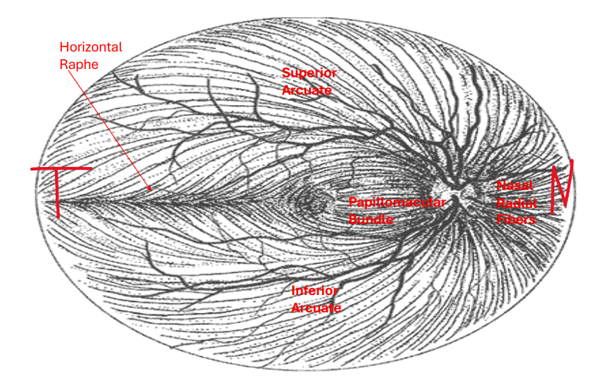
Nasal radial fibers
Brings information from the nasal retina to the optic nerve
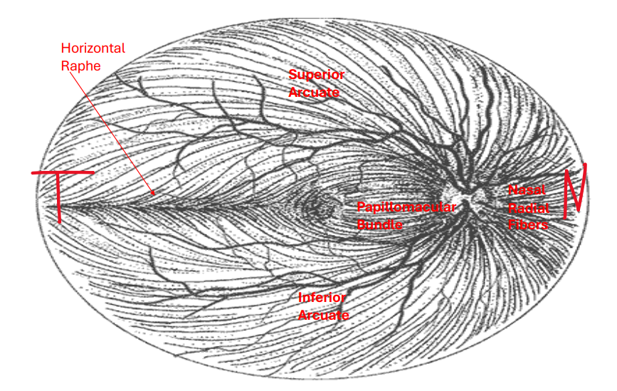
Superior arcuate fibers
Brings information from the temporal retina to the optic nerve
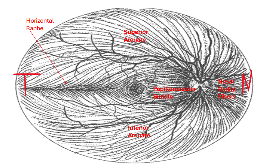
Inferior arcuate fibers
Brings information from the temporal retina to the optic nerve
Appearance Of Flame Hemorrhages
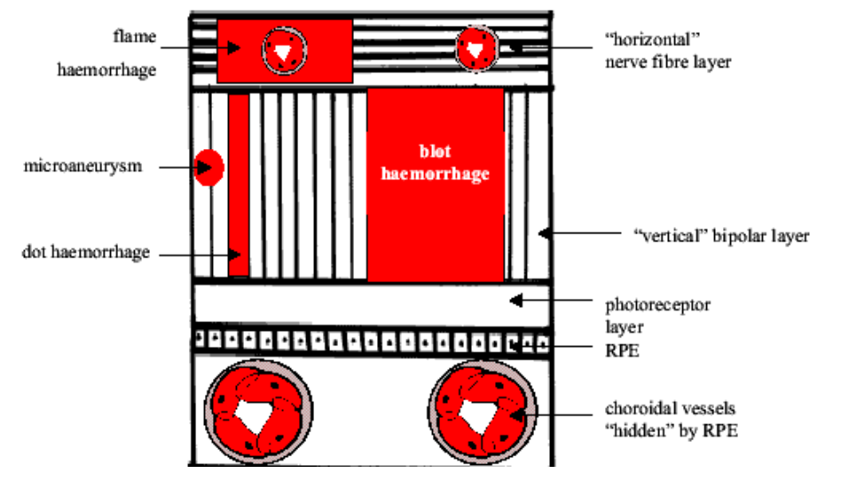
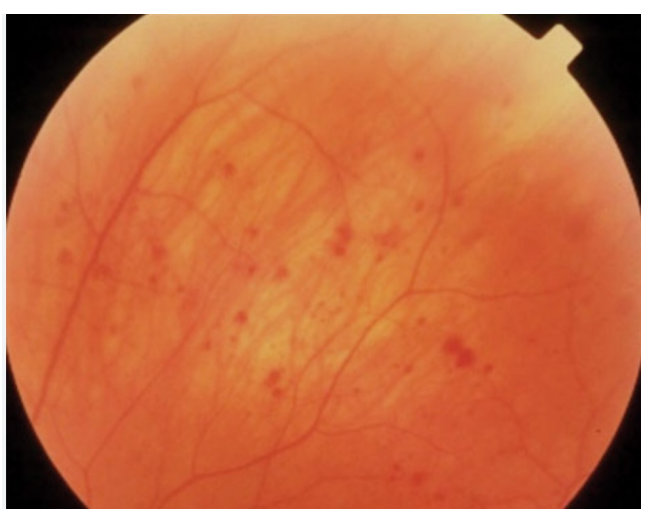
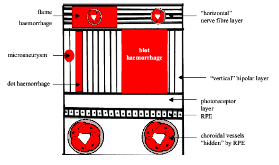
If leaking occurs due to a vascular disease such as diabetes or hypertension, blood will pool in a horizontal and linear formation in the nerve fiber layer due to the horizontal arrangement of ganglion axons in the NFL and appear as a flame hemorrhage
Appearance Of Dot/Blot Hemorrhages
If leaking occurs due to a vascular disease such as diabetes or hypertension, blood will pool in a vertical and linear formation in the inner nuclear layer due to the vertical arrangement of neurons and nuclei in the INL and appear as a dot/blot hemorrhage
Internal Limiting Membrane (ILM)
• Innermost boundary of the retina that adheres to the posterior hyaloid face of the vitreous
• The internal limiting membrane is composed of terminations of Muller cells called footplates as well as collagen fibrils, proteoglycans, other glial cells, and basal lamina
• Eventually becomes continuous with the internal limiting membrane of the ciliary body
*Reflections off the ILM give the retina a bright sheen when looking with ophthalmoscopy especially when the patient is young (Less apparent as a patient gets older). The sheen seen at the macula tends to be circular
Bipolar cells
There are 35.68 million bipolar cells in the retina
The highest concentration of bipolar cells is found in the fovea region
A bipolar cell is composed of a single axon with multiple terminals, a cell body with a large nucleus, and a single dendrite with multiple terminals
The second order neuron in the visual pathway
Relays information from the photoreceptor cells to the horizontal, ganglion, and amacrine cells
Receives synaptic feedback from amacrine cells
Releases the excitatory neurotransmitter glutamate from its’ synaptic terminals
*There are 11 total types of bipolar cells (All are associated with cones except the rod bipolar cell)
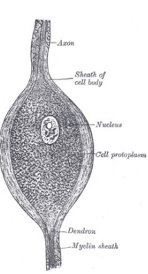
Rod Bipolar Cells
• Only bipolar cell that connects to rods
• Composed of a large cell body
• First appears about 1mm from the fovea and continues into the retina periphery
• The dendrite of a single rod bipolar cell comes in contact with 15-20 rods in the central retina but about 80 rods in the peripheral retina which leads to better sensitivity but less recognition of detail
• The axon of a rod bipolar cell typically synapses with amacrine cells which then synapses with ganglion cells
Midget Bipolar Cells
• Only contacts one cone in the central retina
• Can contact multiple cones in the peripheral retina
• Composed of a small cell body
• Can be either flat or invaginating
Flat Midget Bipolar Cells
• Makes contact with only the flat area of the cone pedicle
• In the central retina, each flat midget bipolar dendrite only contacts one cone (Leads to better recognition of detail)
• In the peripheral retina, each flat midget bipolar dendrite typically comes in contact with 2-3 cones
• The axon synapses with ganglion cells of all types
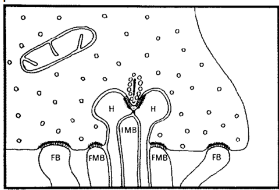
Invaginating Midget Bipolar Cells
• Makes contact with only the invaginating area of the cone pedicle
• Flanked on both sides by a horizontal cell dendrite (Triad)
• Each pedicle can have up to 12 to 25 invaginating areas and thus 12 to 25 triads
• In the central retina, each invaginating midget bipolar dendrite only contacts one cone (Leads to better recognition of detail)
• In the peripheral retina, each invaginating midget bipolar dendrite typically comes in contact with 2-3 cones
• The axon of the invaginating bipolar cell synapses with the dendrite of a single midget ganglion cell
Diffuse Cone Bipolar Cells
• Also called flat bipolar cells or brush bipolar cells
• Makes contact with only the flat area of the cone pedicle
• In the central retina, each diffuse cone bipolar dendrite comes in contact with about 5 other neighboring cones
• In the peripheral retina, each diffuse cone bipolar dendrite comes in contact with about 10-15 neighboring cones
*Only contacts cones that are in close proximity to each other
Blue Cone Bipolar Cells
• Makes contact with only the flat area of the cone pedicle
• Each blue cone bipolar cell can synapse with up to 3 cone pedicles
• Contacts widely spaced rather than neighboring cones
Giant Cone Bipolar Cells
• Makes contact with only the flat area of the cone pedicle
• Has a giant dendrite that branches into 3 trees which clusters into multiple processes (Ends up being size of a cone pedicle)
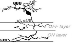
Ganglion Cells
• Third order neuron with cones and Fourth order neuron with rods
• Can be bipolar or multipolar (A single axon with more than one dendrite)
• Can have different cell sizes, branching characteristics, termination of dendrites, and sizes of dendritic tree
• Second cells in the visual pathway to respond with an action potential (Depolarization which will lead to a neuron firing)
*Amacrine cells in the visual pathway are the first to respond with an action potential
Ganglion Cells Pathway
• Each ganglion cell has a single axon which runs parallel to the inner surface of the retina
• Rods have to converge (Share) ganglion cells more so than cones
• The axon releases the excitatory neurotransmitter glutamate at its synaptic cleft
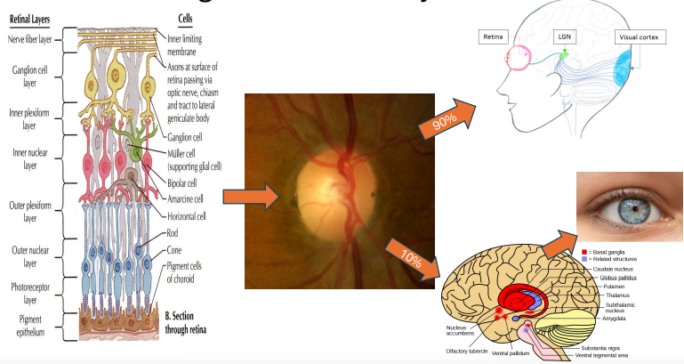
• Axons come together at the optic nerve
• Termination for 90% of these axons end up in the LGN where information will be carried to the visual cortex
• Termination for 10% of these axons end up in subthalamic areas that are involved with pupillary reflexes
Newer Classification Of Ganglion Cells
•Based on the lateral geniculate nucleus layer in which they terminate
•There are P, M, and K cells
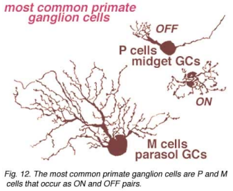
P Cells (Ganglion cells)
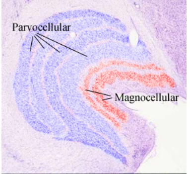
• Called P cells because they terminate in the parvocellular layers of the LGN
• The majority of ganglion cells are P cells (80%)
• The majority of input is from cones (Specifically the medium and long wavelength cones)
• The majority of P cells can be found in the central retina
• Composed of small cell bodies
• Axon conduction is fast
• Specialized for determining color vision, detail, and form
P1 Cells
• Most common P cell
• Also known as a midget ganglion cell
• Small cell with single dendrite
• P1 ganglion cells are exclusively connected to only one midget bipolar cell (Invaginating or flat) which might be connected to only one cone receptor (This allows processing of high contrast detail and color resolution)
• P1 cells would most likely be found in the fovea region
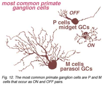
M Cells
• Called M cells because they terminate in the magnocellular layers of the LGN
• Sometimes called a parasol ganglion cell because synapses are made over a wide area
• The majority of input is from rods
• The majority of the M cells are found in the peripheral retina (Only about 5% of the ganglion cells in the central retina are composed of M cells)
• Composed of large cell bodies
• Axon conduction is slow
• Specialized for seeing motion
K Cells
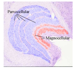
• Called K cells because they terminate in the koniocellular layers of the LGN (Found between the P and M layers)
• The majority of input is from short wavelength cones
• Found throughout the retina
• Thought to be involved with some aspect of color vision but function is not fully understood
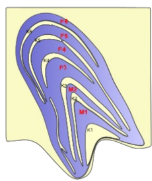
What Visual Field In Clinic Specifically Tests For Function Of M Cells?
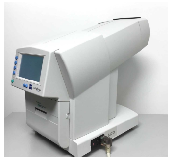
FDT - Frequency Doubling Technology
