Special Echo Applications
1/52
There's no tags or description
Looks like no tags are added yet.
Name | Mastery | Learn | Test | Matching | Spaced | Call with Kai |
|---|
No analytics yet
Send a link to your students to track their progress
53 Terms
3D echo advantages
Elimination of geometrical assumptions when evaluating cardiac chamber volumes and mass
Quantification of complex geometric shape volumes with accurate volumetric evaluation of regurgitant lesions and shunts with color Doppler
Viewing structures from any perspective with a realistic presentation of the heart valves
Assessment of lesions in simultaneous multiplanes and multi-slice mode
Assessment of regional LV wall motion and quantification of systolic dyssynchrony
All 3-D echo images are acquired in what is termed “volumes” derived from thousands of ____
voxels
voxels
geometrically cubed area (cm3) compared to the flat 2-D pixel in convenJonal echo.
operating frequencies of 3D echo
2.5-5 MHz as high as 7 MHz for TEE
Real-time narrow 3D section
beat by beat view with a wider image plane than standard 2-D imaging that can be rotated to view from different perspectives; looks like a thick slice
Real-time 3D zoom volume-rendered images
full volume image of an enlarged area of interest that is rotated to show the structure of interest in a “surgical' view
3D Full volume
multiple beat volumetric imaging stitches together narrow volumes of data over several cardiac cycles; primarily used for accurate software-assisted 3-D quantification of chamber size and volume displacement (EF)
Simultaneous multple plane method
simultaneous display of two 2-D image planes has the ability to adjust the rotation angle, tilt, and elevation of the second image
3D color doppler imaging
uses real-time or full-volume color Doppler data acquisition, but at frame rates lower than for imaging data; new higher voxel rate capabilities using HVR (high volume rate) settings enable the capture of better images during flow cycles like regurgitation, and the possibility to quantify regurgitant jets from 3-D dataset
3-D image displays,
• Volume rendered 3-D images
• Surface-rendered images
• Wireframe images
• Simultaneous display of multiple 2-D images
• Graphic display of 3-D parameters versus time
Focused or limited 3D echo scanning protocol
TTE
Complete 3D evalution scanning protocol
TTE
Clinical Applications of 3-D Echocardiography
• Chamber quantification; LV volumes and EF, RV volume, LA volume
• Cardiac valves evaluation for stenosis and regurgitation
• Guidance of transcatheter procedures
How is LV function described?
measures of EF, regional wall motion changes, and assessment of diastolic filling
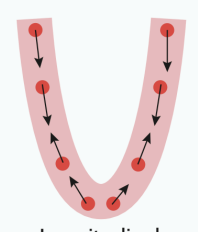
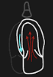
longitudinal direction
the base moves toward the apex
radial direction
walls thicken
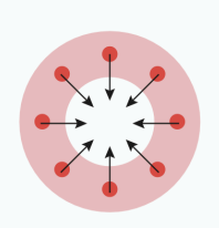
circumferential direction
cavity size decreases perpendicular to long axis of the chamber

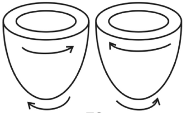
Torsion
the apex and the base rotate in opposite direction during contraction
displacement
the distance a cardiac structure or myocardial element moves between consecutive image frames (cm)
velocity
the speed of movement of a cardiac structure or myocardial element (m/S)
strain
the fractional change in length of a myocardial segment (+/- %)
strain rate
the rate of change in strain (unit per 1 sec)
rotation
the circular motion of the LV myocardium around its long axis (°)
twist
the absolute difference in rotation between LV base and apex (°)
torsion
the gradient in rotation angle from base to apex (°/cm)
Doppler Flow Velocity (PW)
measures low-amplitude, high velocity signals from moving blood cell


Doppler tissue velocity (DTI/TDI)
measures high-amplitude, low velocity signals from myocardium

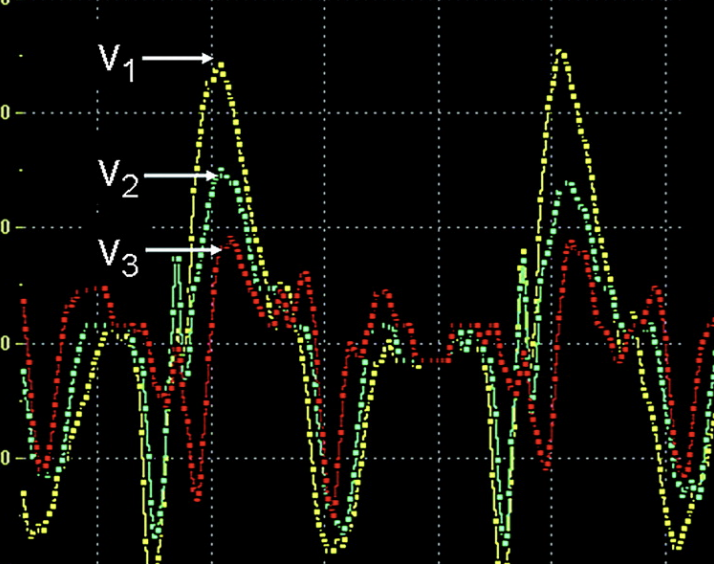
Tissue doppler strain rate
based on difference in TDI velocity between sample volumes divided by distance between them
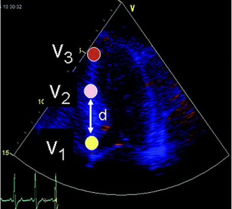
formula for strain rate
SR =(V1- V2)/D (s-1)
Tissue doppler strain
a measure of deformation - the difference between the final length and original length divided by the original length, as a %
formula for strain
Strain =(l - lo) / lo x 100
negative strain
shortening = contraction
positive strain
lengthening = relaxation
During systole, strain ____ rapidly until end-systole is reached
decreases ↓
During diastole, a rapid ____ in strain occurs during early phase of diastolic filling followed by a plateau in diastasis, then another increase with atrial contraction
increase ↑
there zero strain (baseline) at ______
end-diastole = ventricle is fully relaxed, no deformation
maximum negative strain occurs at _____
end-systole
Speckle tracking strain imaging
tracks the motion of “speckles” on myocardium
speckles
markers created by the interference patterns that are generated by the reflected ultrasound; stable, allowing the tracking of myocardial motion.
kernel
the region of speckles being tracked in a single frame
advantages of speckle tracking over DTI
simpler data acquisition
lack of angle dependence
direct measurement of strain
multiple simultaneous measurements
ability to perform analysis after image aquisition
Longitudinal strain (GLS)
AP4, AP2, AP3
circumferential strain (shortening)
PSAX at mid ventricle
Radial strain (thickening)
PSAX
contrast echocardiography
the injection of an agent into the bloodstream that increases the echogenicity of the blood or myocardium, improving visualization of cardiac chambers and myocardial tissue.
main purpose of contrast echo
To enhance ultrasound images by producing opacification of cardiac chambers or increasing the density of the myocardium.
contrast agents are used for right-heart opacification
Agitated saline, followed by a contrast agent mixed with non-agitated saline for improved enhancement.
contrast agents are used for left-heart and myocardial opacification
solubility fluorocarbon gas in stabilized microbubbles encapsulated with denatured albumin or monosaccharides
ultrasound instrument settings during contrast imaging
Lower mechanical index (MI) to about 0.5
Set focal depth to middle/near field
Use a lower transducer frequency
Increase overall gain and dynamic range
*helps minimize microbubble destruction
applications of contrast echo
Detection of intracardiac shunts
Enhancement of Doppler signals
Left-ventricular (LV) opacification
Myocardial perfusion assessment
intracardiac echocardiography (ICE)
invasive, catheter-based imaging modality that allows visualization of cardiac structures and blood flow with doppler
instrumentation of ICE
catheter-like probe inserted through IVC, RA, RV from femoral vein
Applications
guiding device closure of IAS
guiding radio frequency Pv ablation
monitoring transcatheter valve replacement
peri-interventional imaging of AoV/MV