Neuroanatomy
1/26
There's no tags or description
Looks like no tags are added yet.
Name | Mastery | Learn | Test | Matching | Spaced | Call with Kai |
|---|
No analytics yet
Send a link to your students to track their progress
27 Terms
• Practicing a skill (like juggling) can change our brain?
Any skill changes the brain. • Voxel-based morphometry (VBM) associated juggling with increased grey matter density in the visual cortex (Gerber et al., 2014).
• We only use 10% of our brain at any one time?
This is a neuromyth (Scientology used it once). • We use all of our brain, but with varying levels of activation.
• Some neurons in the brain fire only in response to specific people (celebrities such as Bill Clinton or Jennifer Aniston)?
Supported by evidence (“Grandmother cells”). • Single cell measures with epilepsy patients (Quiroga et al., 2005). • However, not accepted by all.
• Brain training games can improve your general cognition?
Insufficient evidence. • Brain training games usually improve skills particular to it. • However, there is no strong evidence of generalization.
Used in dementia research. Makes you better at that specific task.
Socializing is actually much better. Individuals who socialize more are better in later life
• Some people can’t see motion or colour after brain damage?
• Correct. • Motion blindness (akineptosia) associated with damage to primary visual cortex. • Cortical colour blindness (achromatopsia) associated with damage to occipital and temporal lobes (e.g., Bouvier & Engel, 2006).
Ancient neuroscience
Trepanation (drilling of the skull to expose the brain) is the oldest known form of surgery. • 5-10% of skulls of the Neolithic Period (farming - metallurgy). • Survival rate > 50% • Treatment of “epilepsy, animistic possession by evil spirits, or mental illness, expressed by errant or abnormal behavior” (Faria, Surg Neurol Int, 2013). • Later for treatment of hematoma.
ancient egypt neuroscience
• Edwin Smith Papyrus (ca. 1,600 BCE) • 48 clinical case studies • 27 head traumas • 13 damage to the brain (“marrow of the skull”) • Case 20: Language impairment “…if thou ask of him concerning his malady and he speak not to thee while copious tears fall from both his eyes… an ailment not to be treated.”
ancient greece and middle ages neurosciences
Ancient Egypt (ca. 1,600 BCE): The heart is the seat of the soul • Ancient Greece (ca. 400 BCE): The brain controls the body. • European Middle Ages (ca. 1,100 CE): “Accordingly, there are two brains in the head, one which gives understanding, and another which provides sense perception.” • Meanwhile, surgery in the Islamic world associated mental deficits with the brain.
reneissance neuroscience
More refined anatomical descriptions in the renaissance, refinement of mechanistic theories of brain activation (fluids, vibration). • Development of electrotherapy in the 18th century, discovery of excitability of neurons by electricity. • In the 19th century, brain lesions associated with behavior changes, e.g. in aphasia.
The Brain input output
The brain is an input-output device: • Input: sensory, chemical, biological • Output: Behaviour (actions) • It does this by: • Association • Memorization • Pattern recognition • Categorization • Anticipation • Simulation • Much of this work takes place between and within networks of activation.
Basic function of neurons
Neurons excite or inhibit each other
They have a mylin sheath which helps insulte the energy and be more efficient
The astonishing hypothesis
The ‘Astonishing Hypothesis’: we are nothing but the coordinated firing of billions of neurons (Francis Crick, 1994). • The mind is the computational functioning of the brain (common modern assumption). • Neurons are the basic units of information processing in the brain (see de-Wit et al. 2016).
Methods: lesion studies
Paul Broca’s (1824-1880) patient “Tan”: Seemed to understand language, but could only say few words.
Methods: single cell recordings
Use of electrodes on the brain to pick up electrical signals. • Hubel and Wiesel – V1 ‘edge responses’
(cat able to see movement of a black dot going in and out of his vision on the powerpoint slid
• Place Cells (O'Keefe, 1976; O'Keefe and Conway, 1978) • Nobel prize 2014 (rats remember the locations of the decision they took inside the maze
What are limitations of lesion and single cell recording studies?
Neuroimaging: Positron emission tomography (PET)
Tracing of radioisotopes injected into the subject (e.g. Oxygen-15) • Can detect changes in metabolism, e.g. as the result of activation of neurons. • Spatial resolution: 4-5 mm • Temporal resolution: 5-10 seconds • E.g., Zeki et al. (1991) • Contrasts
Blood Oxygen Level Dependent (BOLD)
Functional Magnetic resonance imaging (fMRI)
Detects hydrogen in the brain. • High spatial resolution: 1 mm3 • Temporal resolution (limited by hemodynamic response): 3s • Can be lowered to 100ms depending on experimental design
The Fusiform Face Area (Kanwisher et al., 1997) “The evidence from neurological patients is powerful but limited in anatomical specificity; however, functional brain imaging allows us to study cortical specialization in the normal human brain with relatively high spatial resolution and large sampling area”
Localized area in 12 out of 15 participants • Contrasts: faces vs. objects • Uses a ‘block’ design • Most evident on the right • This ‘ROI’ (region of interest) is then used in subsequent analysis.
Types of MRI/fMRI analysis
fMRI – Average BOLD – ROI vs whole brain – MVPA (Multi-Voxel Pattern Analysis) – fMRIa (adaption) – Connectivity (Dynamic Causal Modelling) • MRI – Structural – DTI (Diffusion Tensor Imaging / Tractography)
TMS - Transcranial Magnetic Stimulation
Magnetic fields excite neurons • Can be used to inhibit or excite neurons • Pitcher et al. (2009) identified areas for body, face and object perception.
Risk of wrong interpretation of TMS
TMS is increasingly used in Cognitive Neuroscience to study functional contributions of a stimulated brain region to cognitive and perceptual processing. TMS-related behavioural effects are often interpreted as reflecting selective disruption of processing primarily within the stimulated region itself. This approach is now being extended by studies that combine TMS with concurrent neuroimaging measures, such as fMRI. We discuss some recent combined TMS-fMRI studies and their implications for TMS investigations of cognition and perception. An emerging theme is that TMS does not affect only the stimulated region, but can also influence remote brain areas interconnected with the stimulation site. Such ‘network’ effects of TMS can be anatomically specific, but also context-dependent, changing with the current functional state of the targeted network rather than simply reflecting just fixed, context-invariant anatomical connectivity. Perceptual and behavioural effects of TMS may correspondingly involve TMS influences on remote interconnected brain regions, not solely on the stimulated region itself. Thus, TMS can now be used to study the consequences of functional interactions between the stimulated region and other parts of the network. This may lead beyond strictly modular views of brain function, that emphasize functional properties of single brain areas, towards new perspectives on how functional interactions between remote but interconnected brain regions may support perception and cognition.
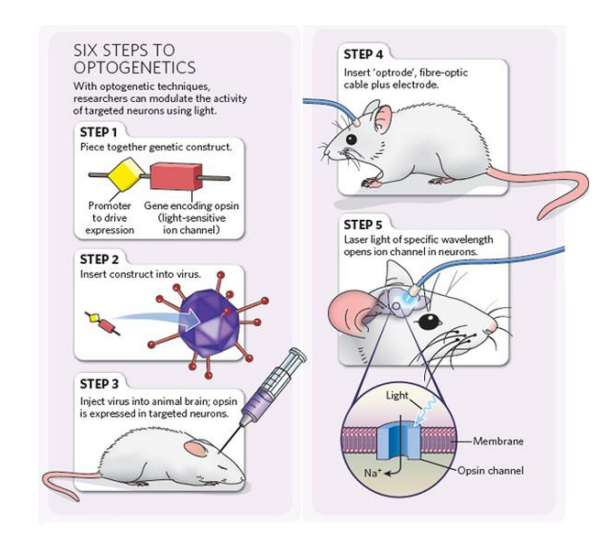
Optogenetics
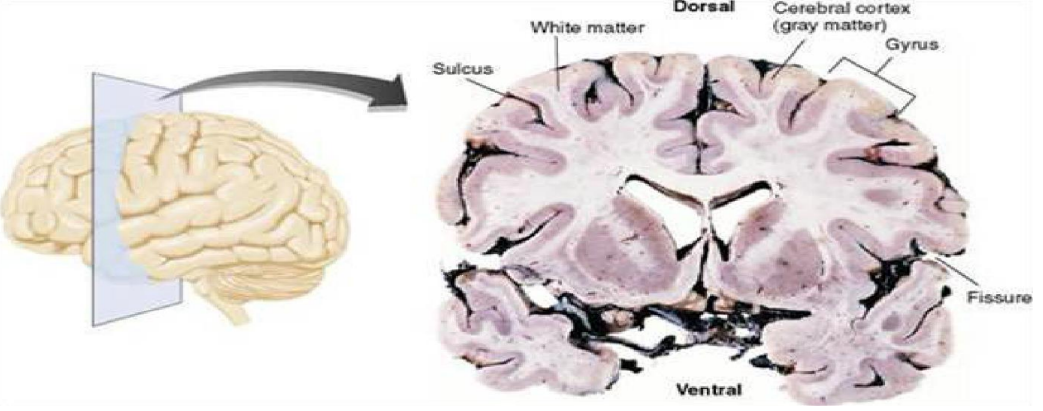
Sulcus/gyrus & White/Grey matter
The sulcus is the space between thegyrus
The connections if the brain is the white atter, so lesiosn there case much more damage.
with bigger brains there are more folds to fit more volume
Goals… DLPFC
Area of the brain especially associated with ‘goal directed behavior’ – D = Dorsal – L = Lateral – P = Pre – F = Frontal – C = Cortex
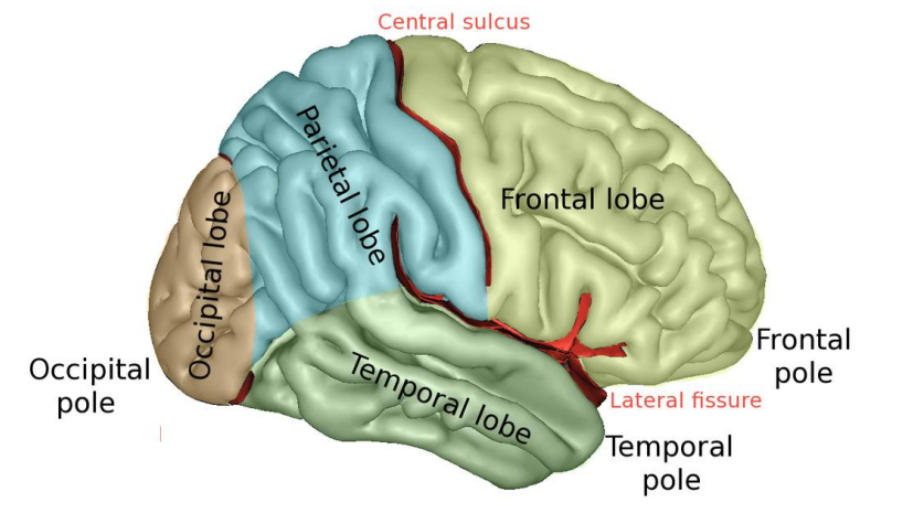
anatomy of brain
The central sulcus is the dominant boundary between frontal and pariatal lobe
Lateral fissure aka perisylvian sulcus.
Frontal lobe is for decision making, complex motor skills and central executive. The occipital lobe has no clear outside sulcus The brain evolved back to front , inside out. Temporal lobe = auditory system, semantics knowledge Pariatel = reading and sensory skills
Basic Neuroanatomy
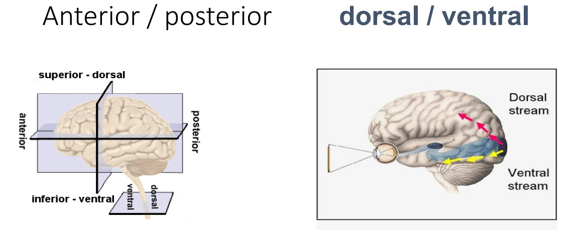
lateral vs medial
soral stream is the WHERE strea. ventral is the WHAT stream
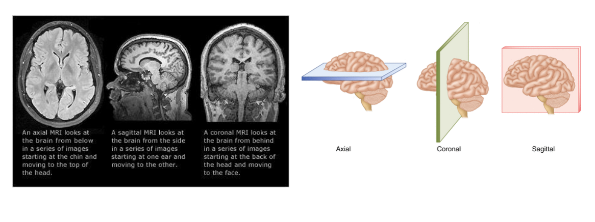
Axial, sagittal and coronal images