ANTC47: Leg
1/32
There's no tags or description
Looks like no tags are added yet.
Name | Mastery | Learn | Test | Matching | Spaced | Call with Kai |
|---|
No analytics yet
Send a link to your students to track their progress
33 Terms
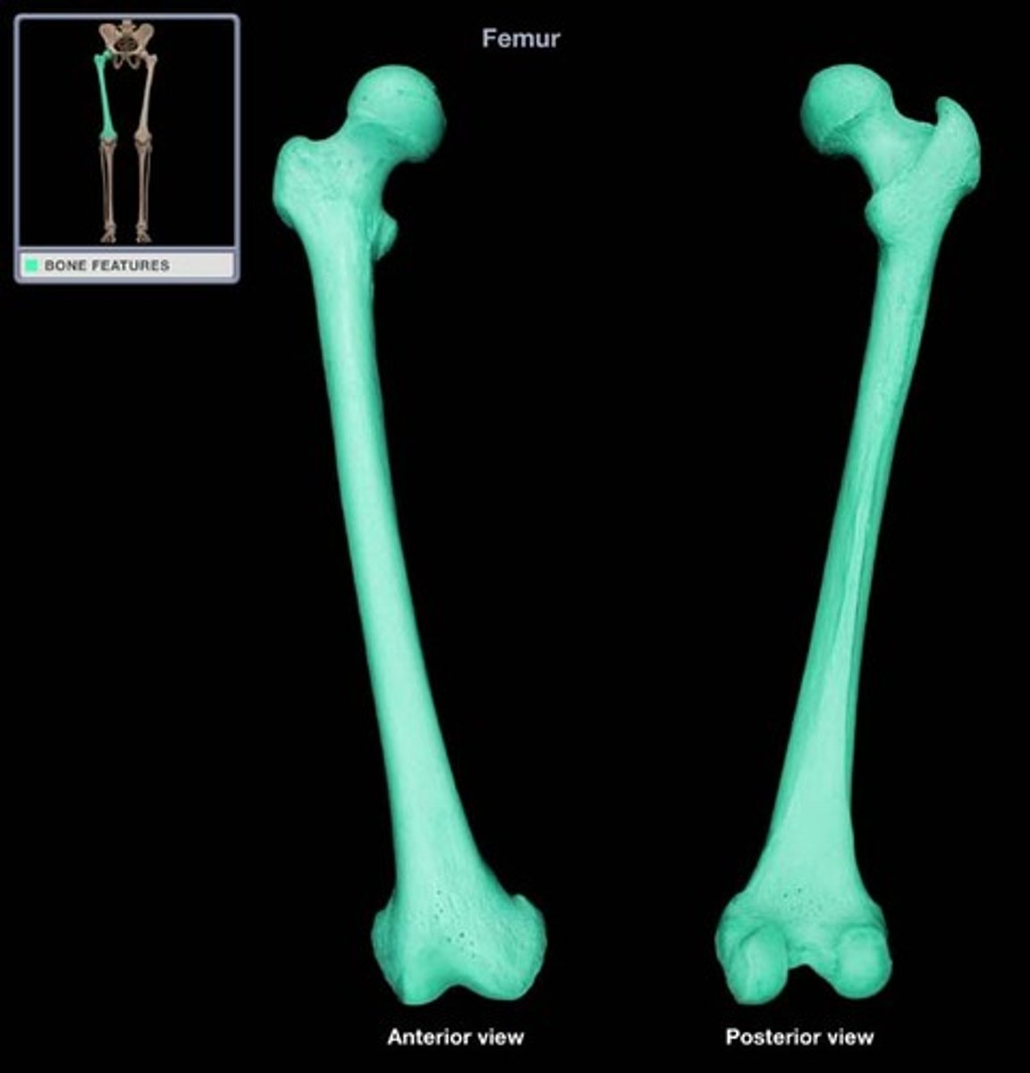
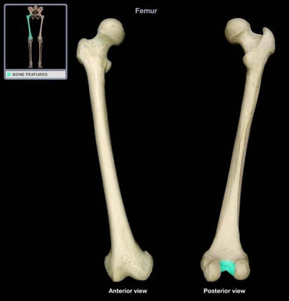
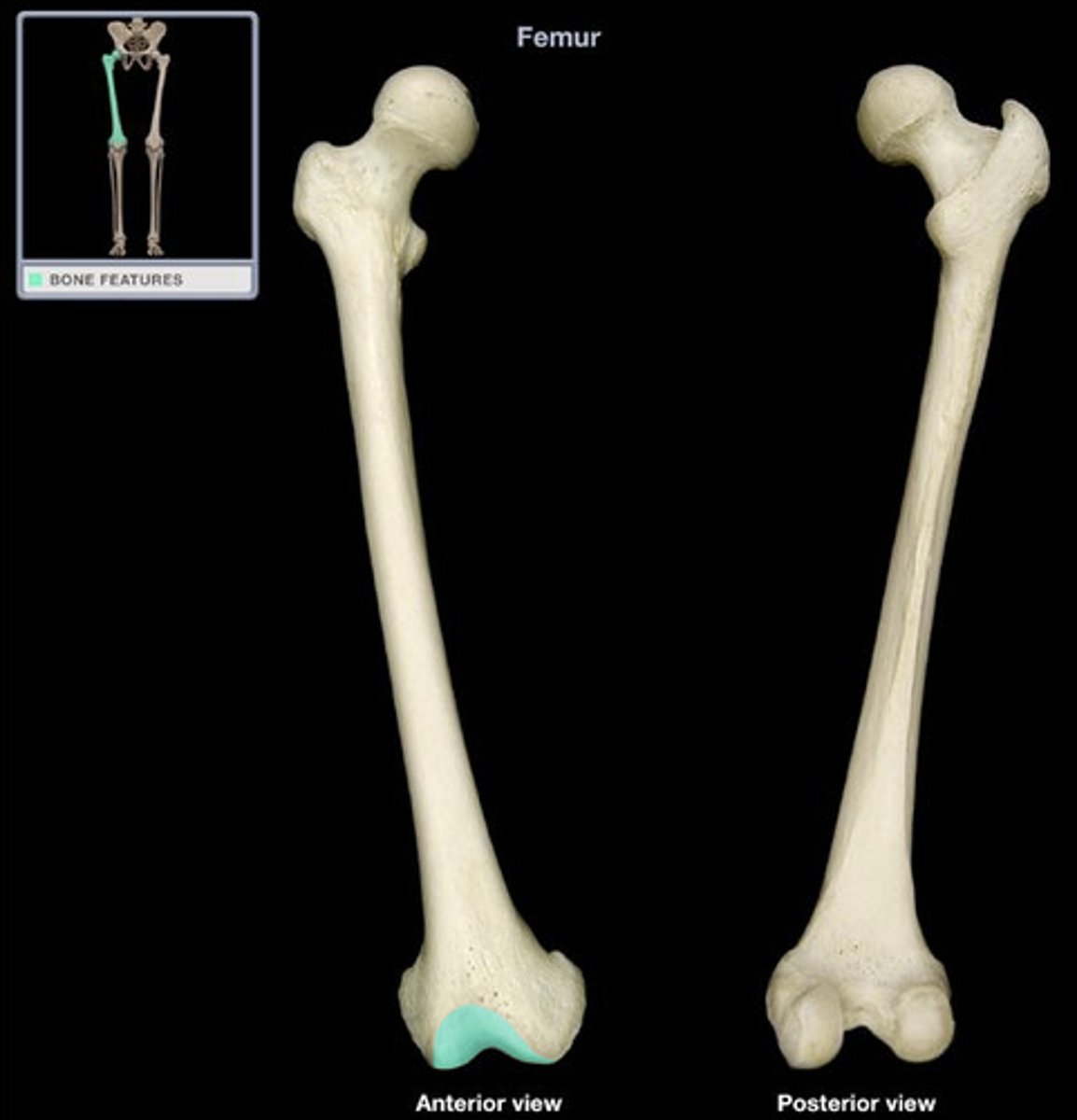
Femur
Proximal leg bone; the longest and strongest bone in the body
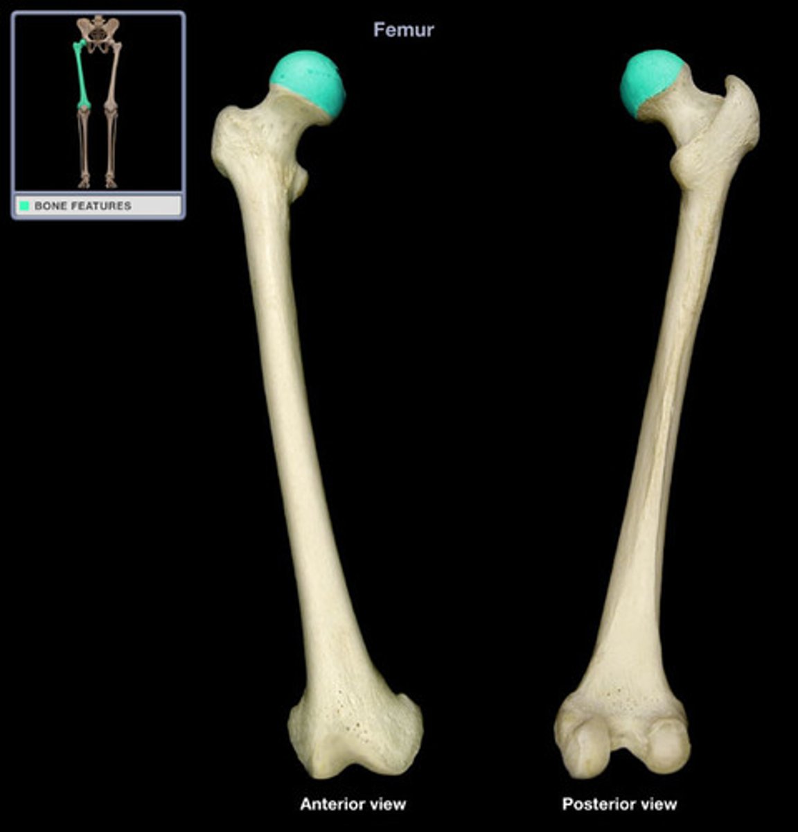
Femoral Head
The proximal end of the femur that articulates with the acetabulum to form the hip joint.
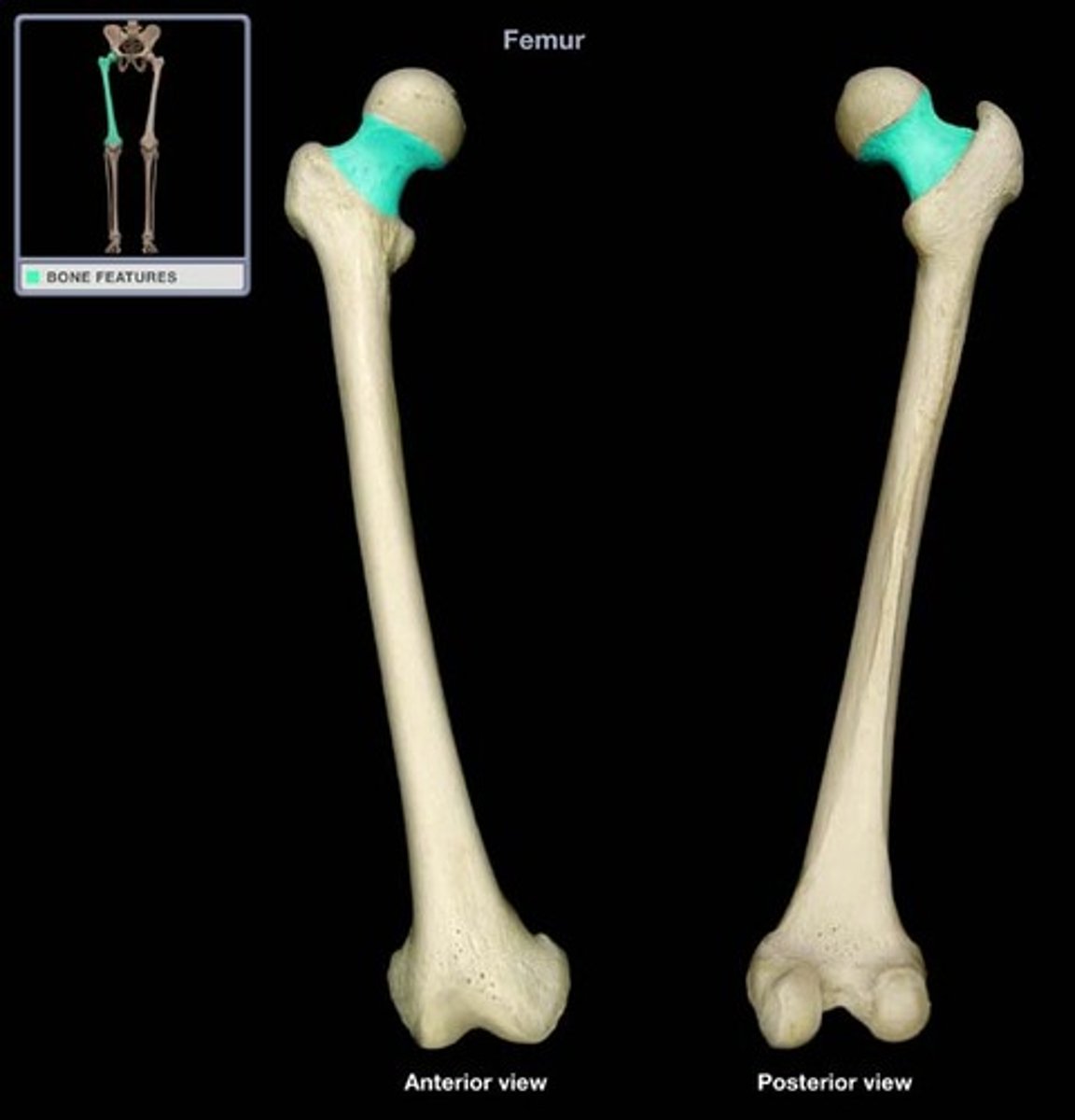
Femoral Neck
Section of bone that joins head of femur to the shaft of the femur
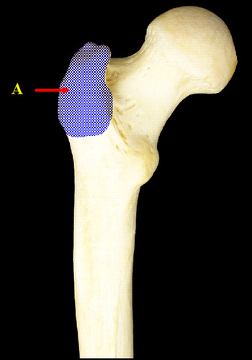
Greater Trochanter
Large, blunt non articular prominence on the lateral, proximal part of the femur
Intertrochanteric Crest
Raised area formed posteriorly between the Greater and Lesser Trochanters
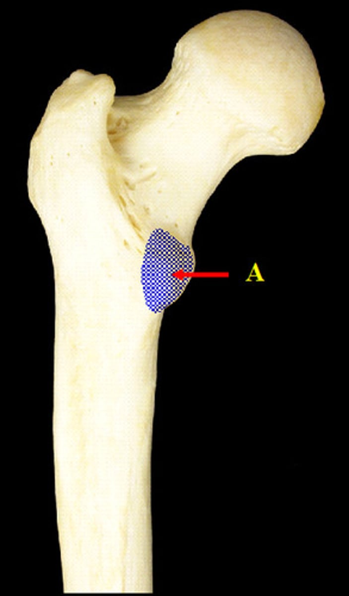
Lesser Trochanter
The smaller blunt, prominent, protrusion of the posterior femoral surface, just under the point where the neck of the Femur joins the shaft
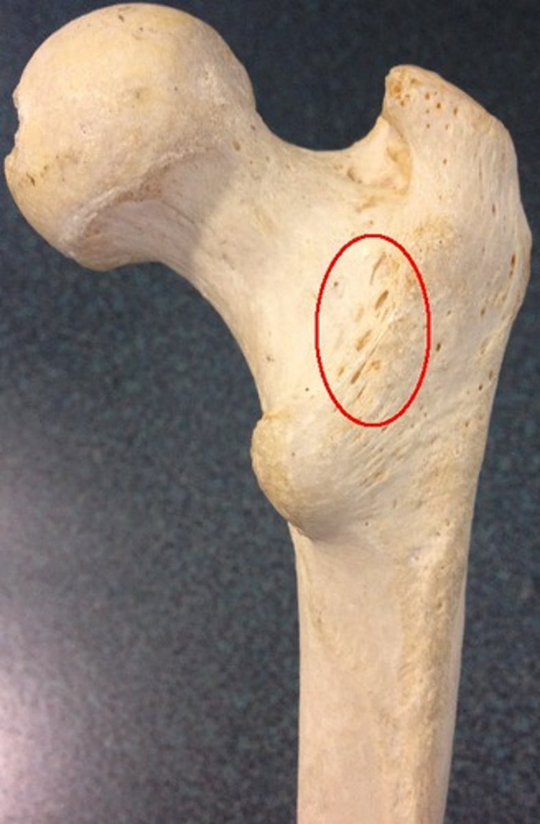
Linea Aspera
Long, wide, roughened and elevated ridge that runs along the posterior end of the shaft of the femur
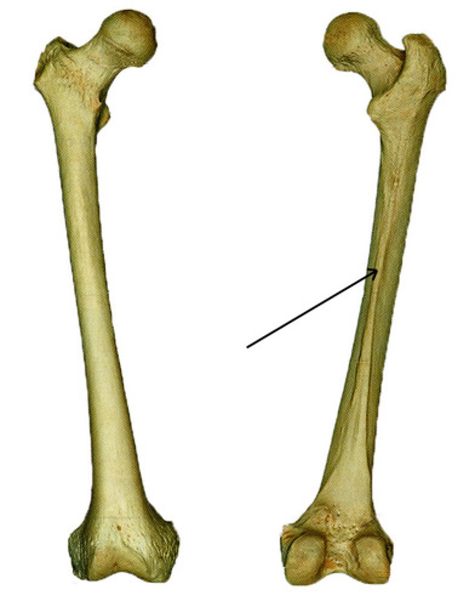
Gluteal Line
The slightly raised ridge on the posterior of the Femur, above the Linea Aspera

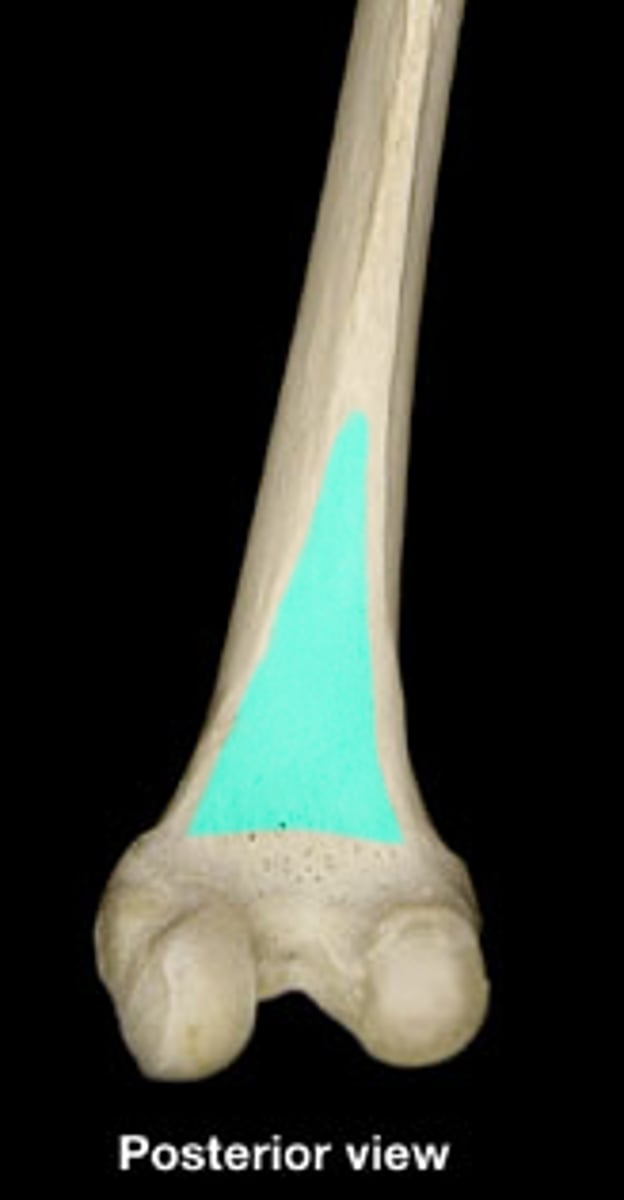
Popliteal Surface
Smooth, triangular area on the posterior distal femur where the lips of linea aspera diverge
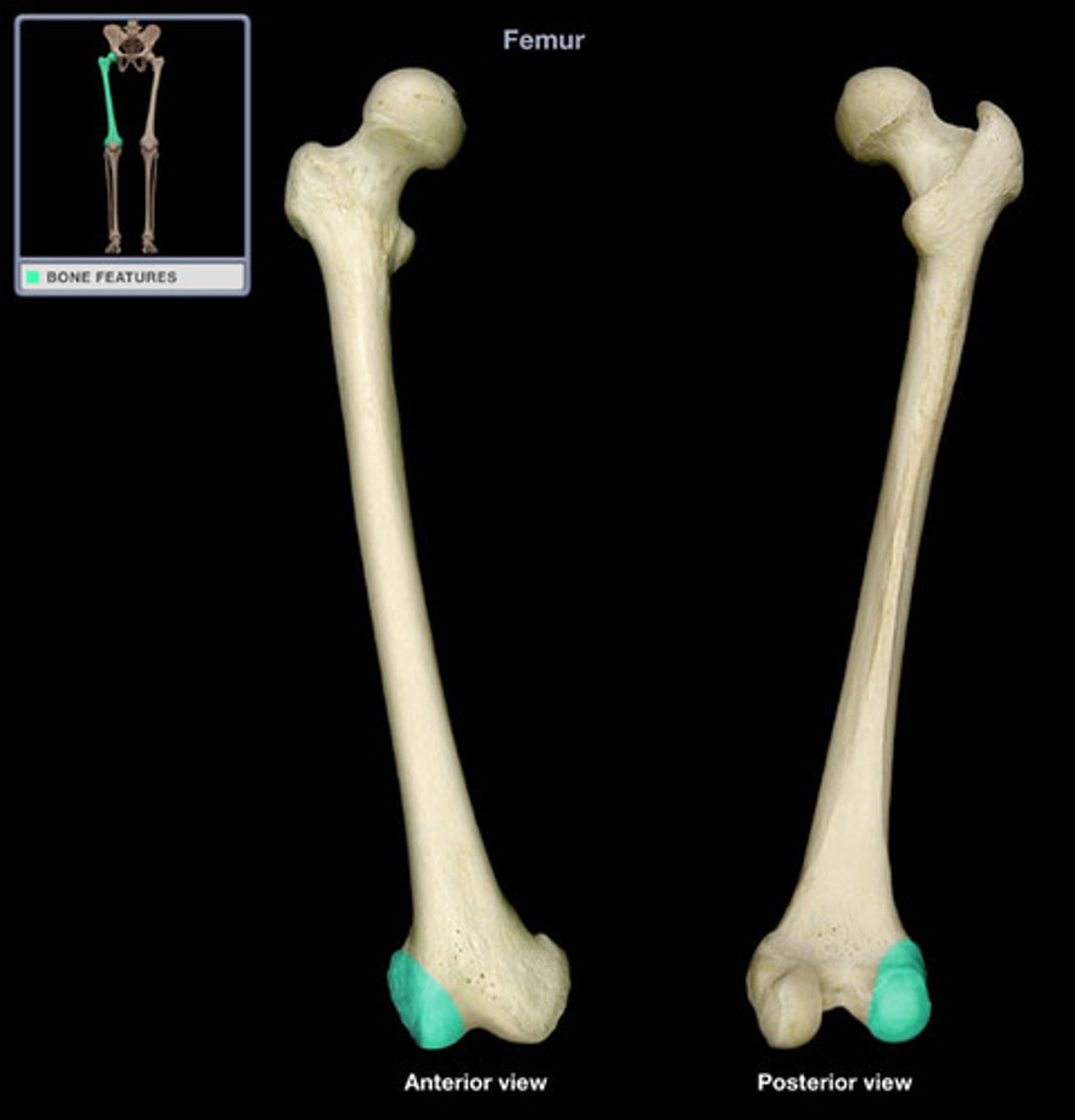
Lateral condyle of Femur
Lateral protrusion of bone on the distal end of the Femur that articulates with the Tibia.
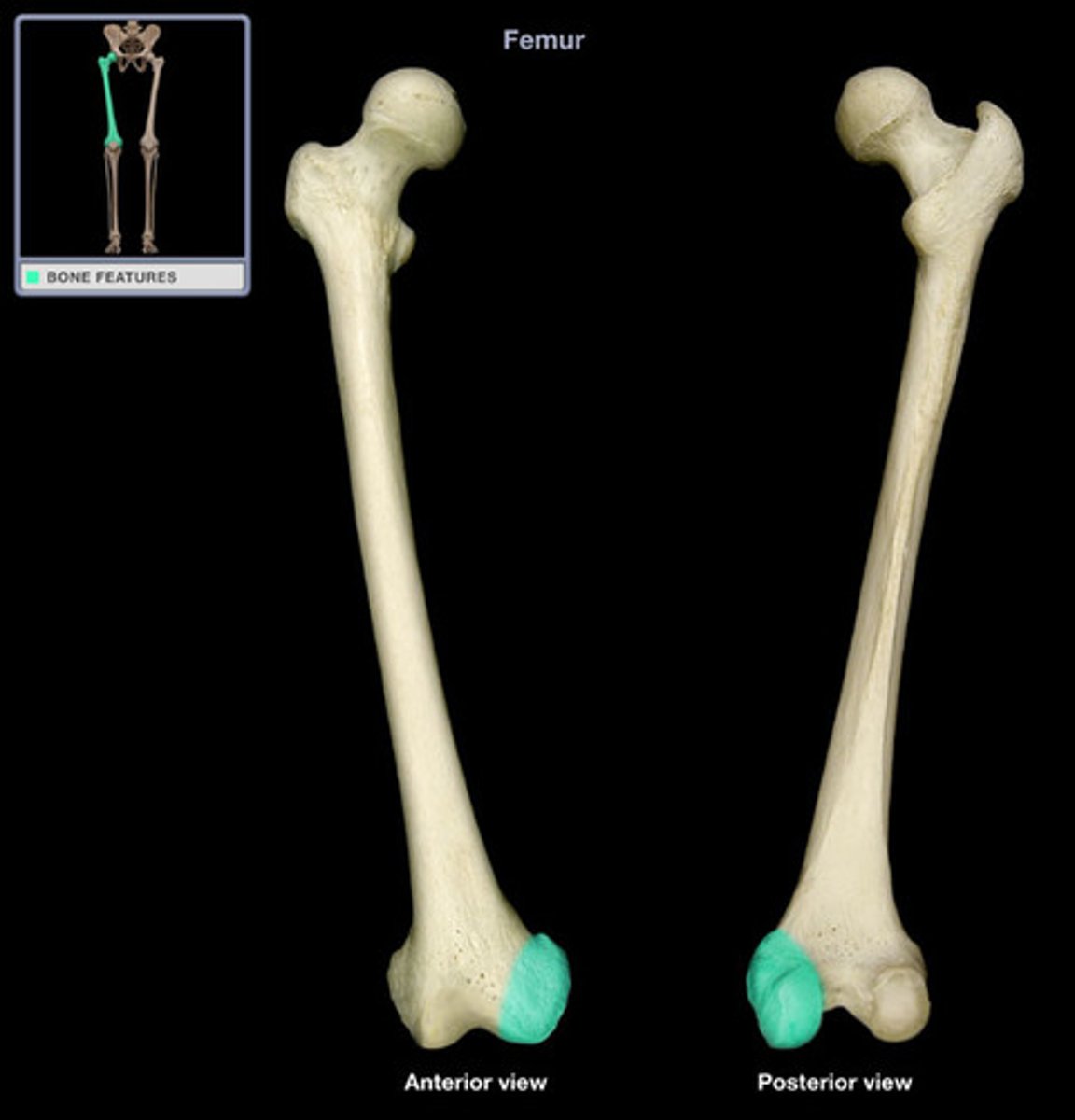
Medial Condyle of Femur
Medial protrusion of bone on the distal end of the Femur that articulates with the Tibia.
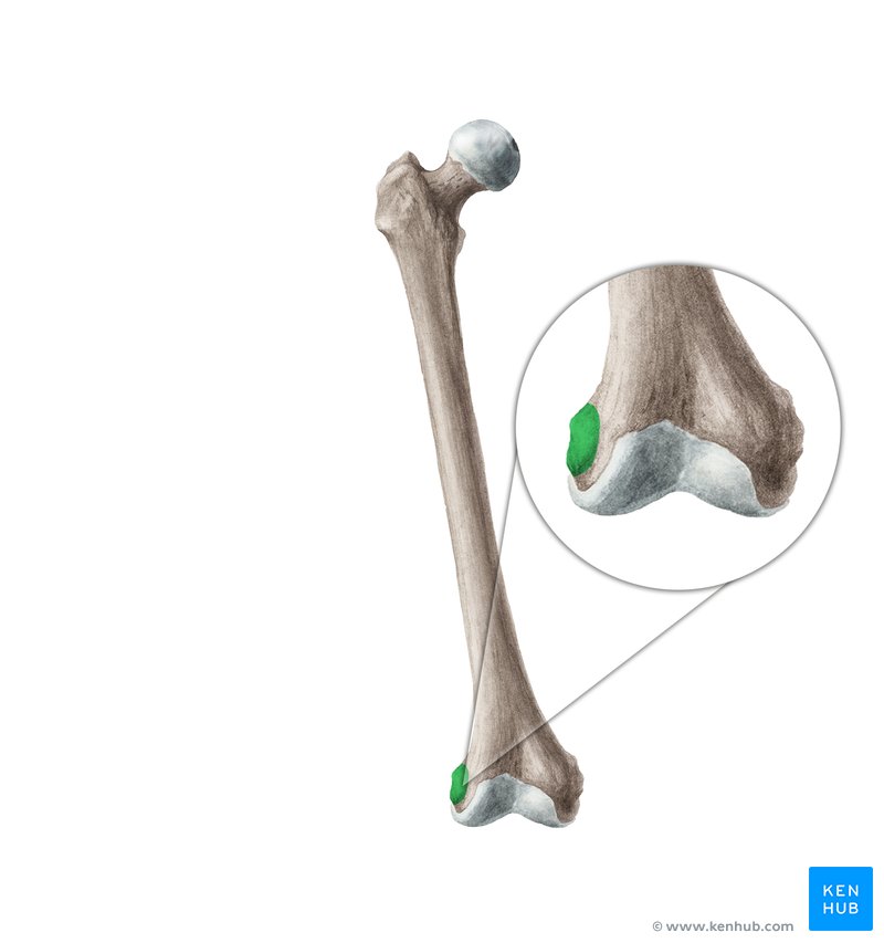
Lateral Epicondyle
Raised, roughened area of the Femur located on the lateral side of the Lateral Condyle
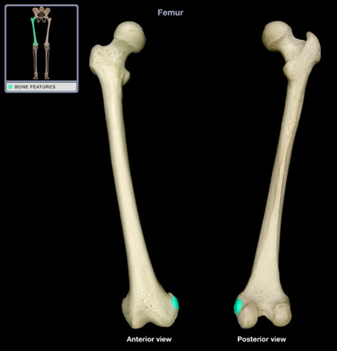
Medial Epicondyle
Raised, roughened area of the Femur located on the medial side of the Medial Condyle
Intercondylar Fossa
Deep depression between the condyles on the Femur
Patellar Surface
Notched articular area on the anterior side of the distal end of the Femur
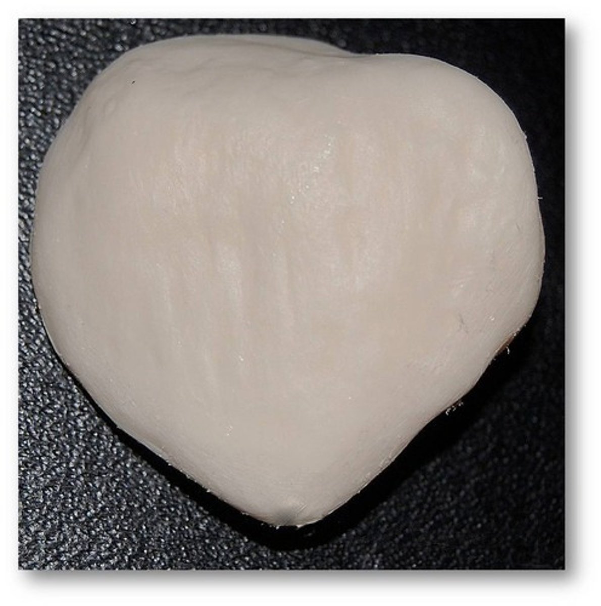
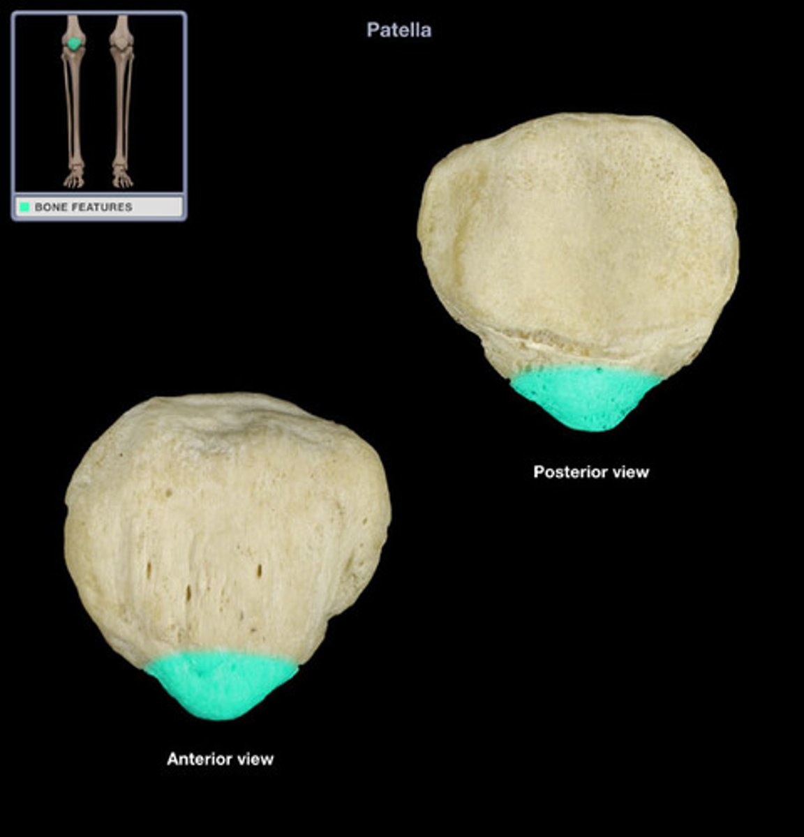
Patella
Knee cap bone
Apex
Point at the distal end of the Patella
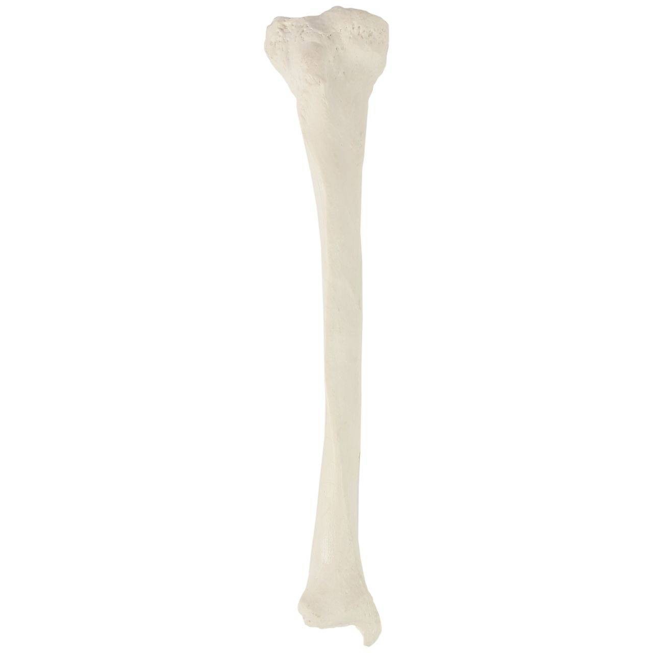
Tibia
Larger distal leg bone
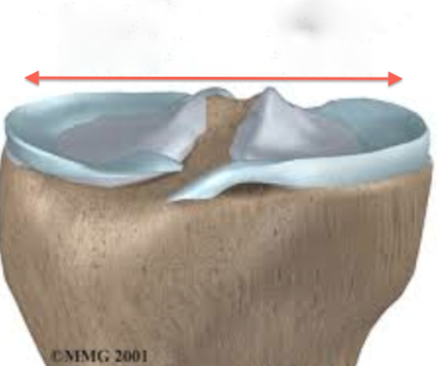
Tibial Plateau
The proximal flat portion of the tibia.
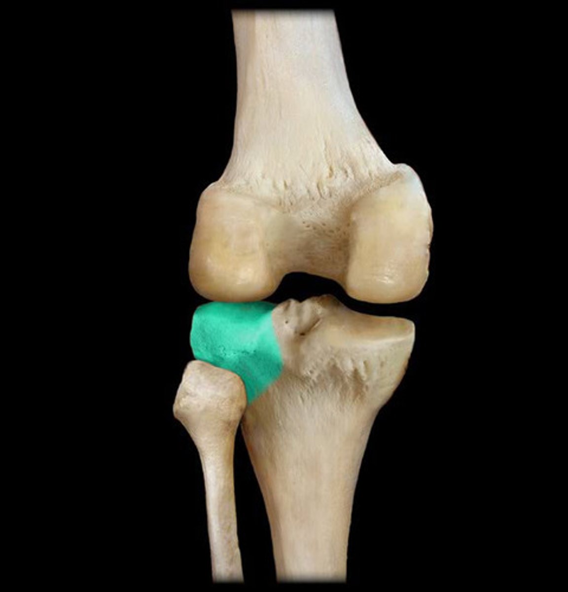
Medial Condyle of Tibia
Indented medial articular surface on the proximal end of the Tibia that articulates with Femur
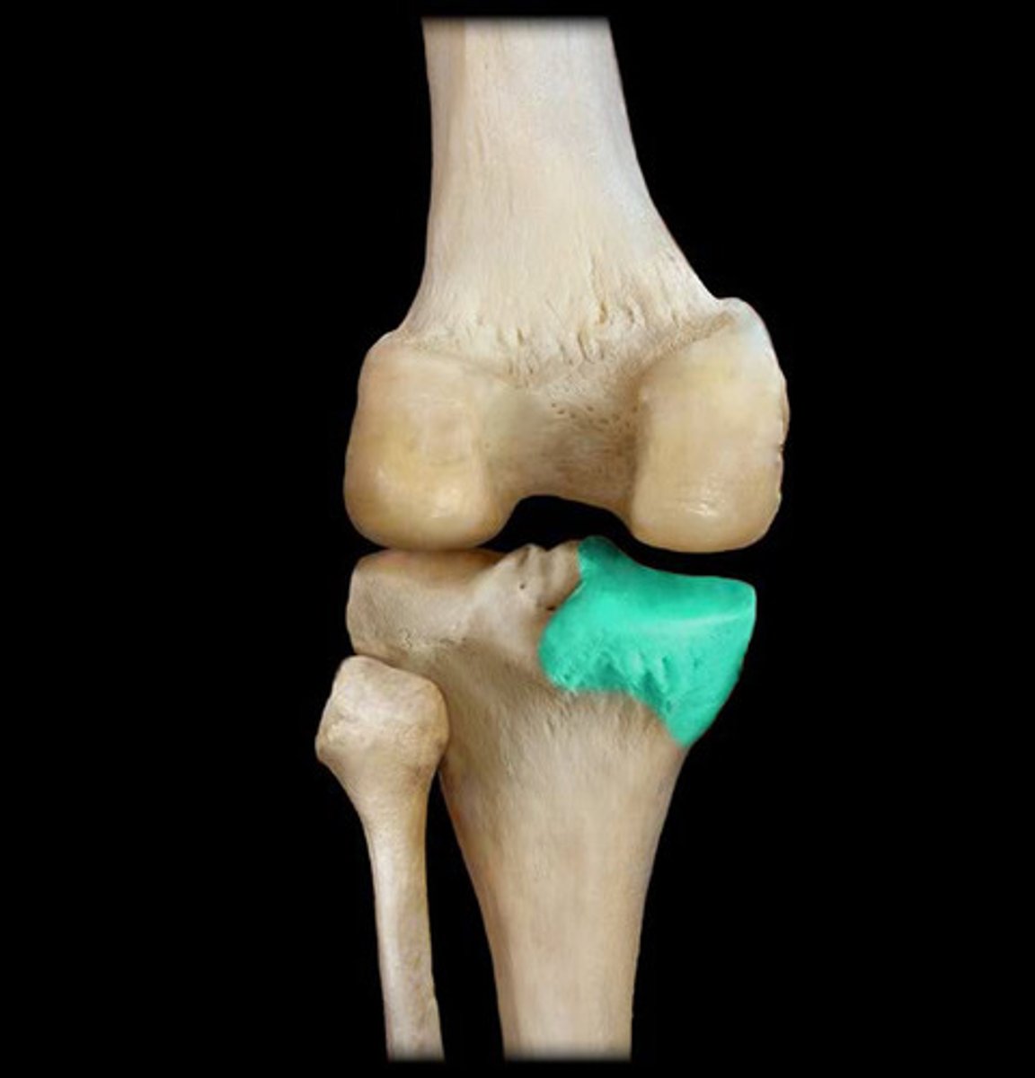
Lateral Condyle of Tibia
Indented lateral articular surface on the proximal end of the Tibia that articulates with Femur
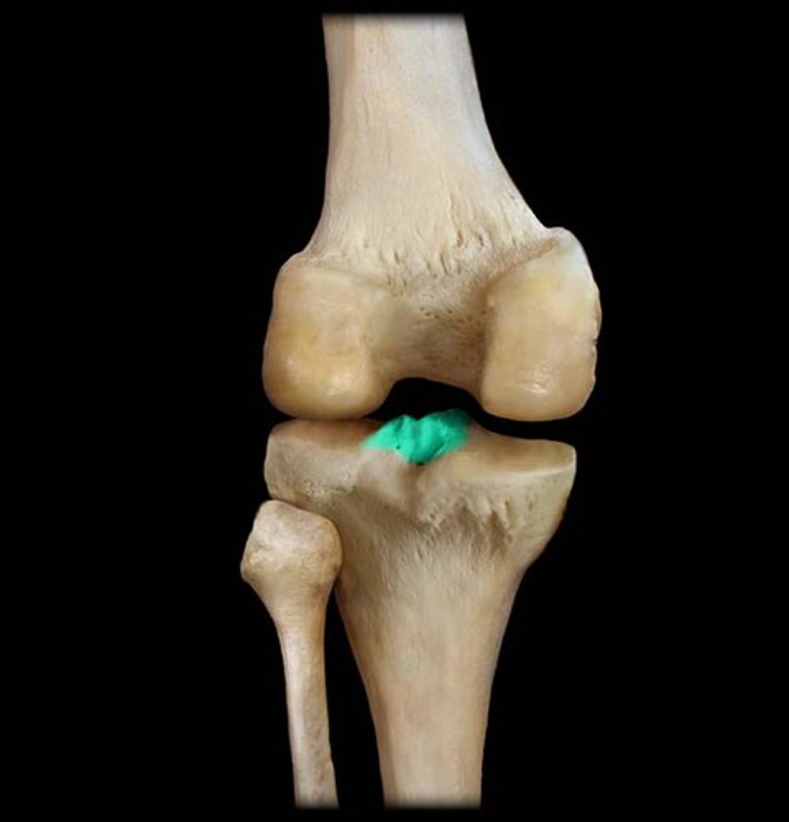
Intercondylar Eminence
Projection of bone located between the two condyles on the Tibia
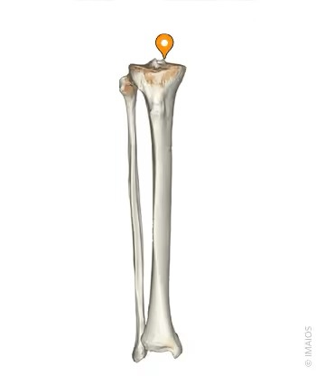
Medial Intercondylar Tubercle
Bump on the proximal surface of the Tibia that makes up the medial side of the Intercondylar Eminence.
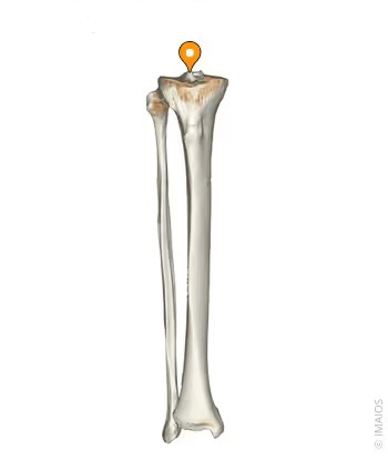
Lateral Intercondylar Tubercle
Bump on the proximal surface of the Tibia that makes up the lateral side of the Intercondylar Eminence.
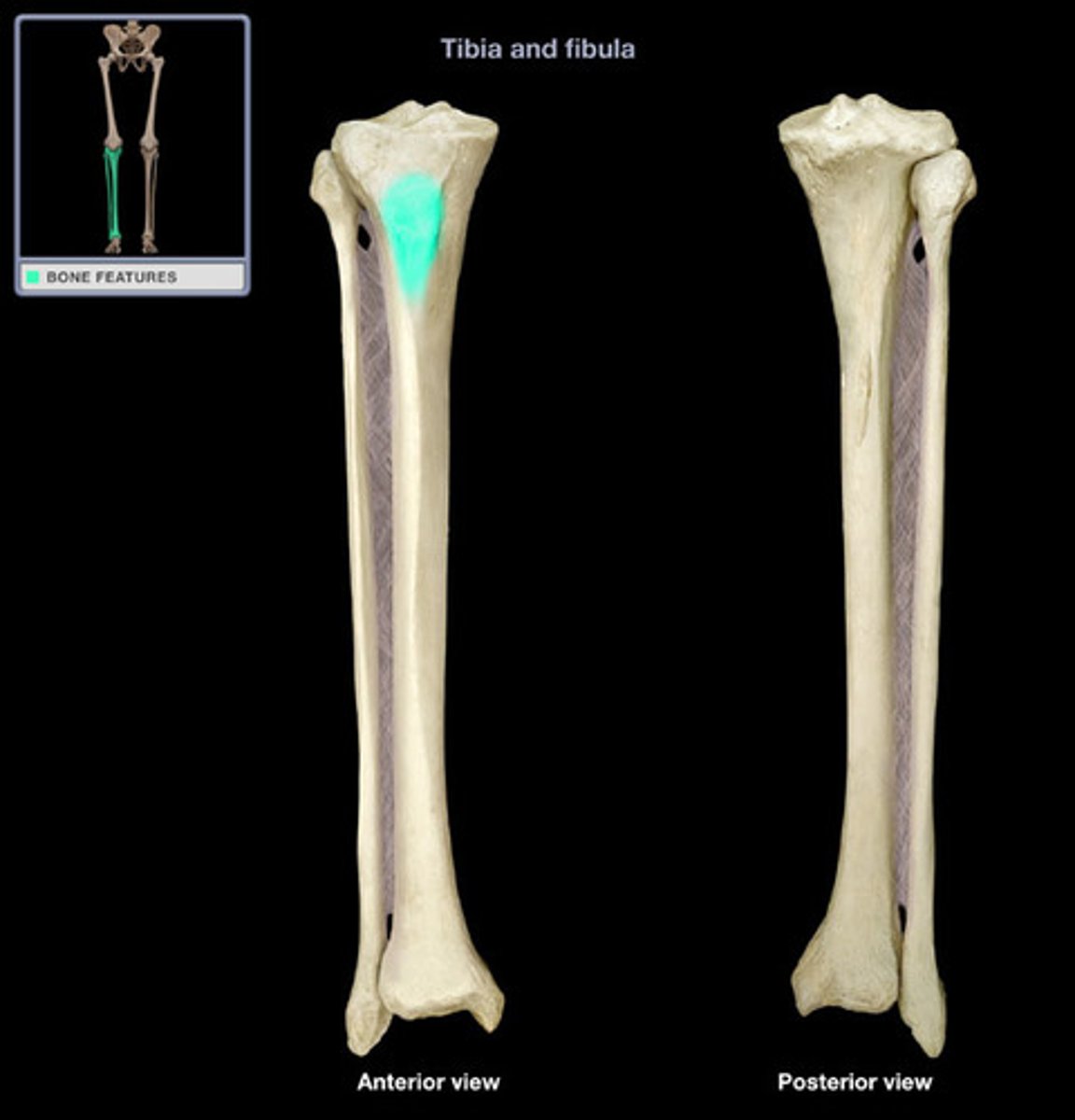
Tibial Tuberosity
Slight protrusion on the anterior side of Tibia, below the Plateau. Acts as an insertion of for ligaments and muscles
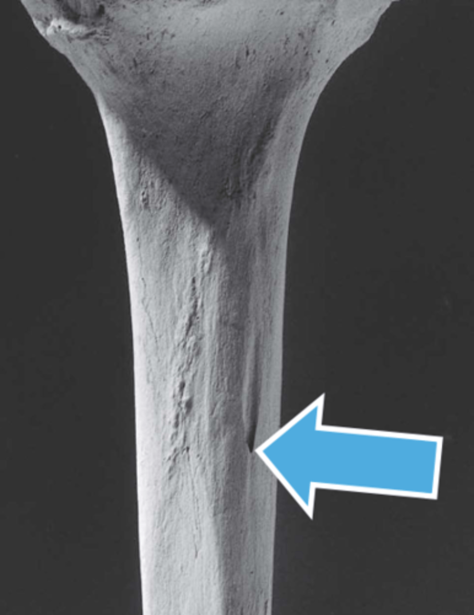
Nutrient Foramen
Small hole on the posterior side of the Tibia, about 1/3 down the shaft
Anterior Crest of Tibia
Ridge of bone along the shaft of the Tibia

Medial Malleolus
Distalmost extension of the Tibia that forms one of the bumps of the ankle.

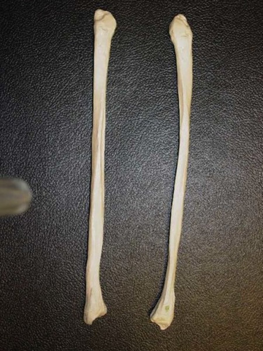
Fibula
Smaller distal leg bone
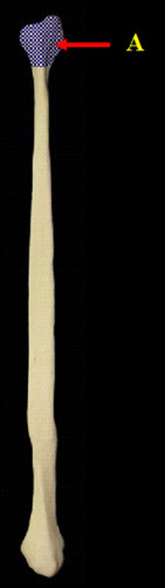
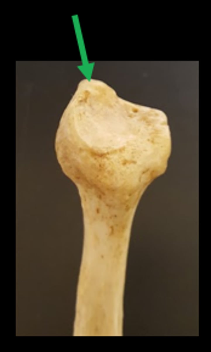
Head of Fibula
Bulbous proximal end of the Fibula
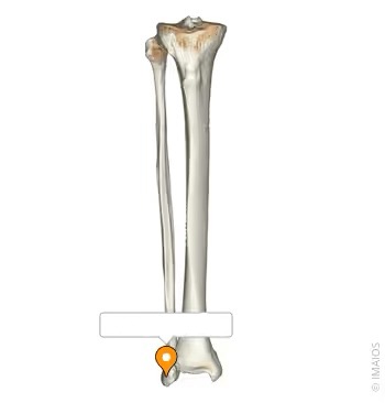
Lateral Malleolus
Distalmost point of the Fibula that forms one of the bumps of the ankle
Styloid Process
Pointed apex of the head of the Fibula
Peroneal Groove
Very slight depression on the distal end of the Fibia that houses tendons
