Cardiovascular System BIO 202 Lab 2
 Call Kai
 Learn
Learn Practice Test
 Spaced Repetition
 Match
Match Flashcards
 Knowt Play
1/64

There's no tags or description
Looks like no tags are added yet.
Name | Mastery | Learn | Test | Matching | Spaced | Call with Kai | Chat |
|---|
No analytics yet
Send a link to your students to track their progress
65 Terms
hemolytic disease of newborn
mother’s antibodies developed through previous exposure to Rh+ blood are transferred through the placenta to the fetus
treatment = phototherapy (exposure to UV light) to degrade bilirubin
exchange transfusion to completely replace infant’s blood
gene
unit of genetic information (segence of DNA nucleotides)
codes for a specific trait → protein
alleles
alternative forms of the same gene
one allele is inherited from mom and other allele from dad
gene pair = 2 alleles
genotype
the genes (alleles) responsible for a trait
the genetic code
phenotype
the physical manifestation of the alleles
Which alleles are codominant
A & B
Which allele is recessive?
O
One mate is genotype AO and the other is BO. What are the possible genotypes of their offspring? What are the phenotypes?
genotypes = AO, AB, BO, OO
phenoypes = A, AB, B, O
Where is the heart located?
between the 3rd rib and diaphragm
posterior to the sternum
thoracic cavity (mediastinum)
What side does the base of the heart point?
towards the right
What side does the apex of the heart point?
towards the left
What is the heart’s covering that separates it from surroundings?
pericardium (parietal and visceral)
Name the layers of the heart (superficial to deep)
Superficial
pericardium
Fibrous pericardium
parietal layer of serous pericardium
pericardial cavity
epicardium (heart wall)
visceral pericardium
myocardium
endocardium
Deep
myocardium
composed of cardiac muscle (cardiomyocytes)
thickness proportional to workload (left ventricle thicker than right)
myocardium vortex = muscle spirals around the heart
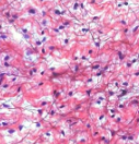
loose areolar connective tissue
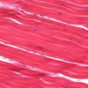
dense regular connective tissue
dense irregular connective tissue

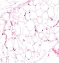
adipose tissue
myocardium’s fibrous skeleton
framework of collagenous and elastic fibers
concentrated on the left ventricle
3 functions
structural support
anchors cardiomyocytes
electrical insulator
cardiomyocytes
a muscle cell, especially a cell of cardiac or smooth muscle
uninucleate (1 nucleus)
blunt, end to end connection
intercalated discs = join individual cells allowing electrical and mechanical connections
complex of fascia adherens, desmosomes and gap junctions (specialized for direct ionic, electrochemical signal transfers)
endocardium
endothelial cells ( simple squamous epithelial cells) line up this layer of the heart
identical to endothelial cells of tunical interna of blood vessels
In what position are both auricles visible?
anteriorly
In what position is the coronary sinus and origins of the pulmonary veins visible?
posteriorly
4 chambers of the heart
right atrium
right ventricle
left atrium
left ventricle
What structure separates the right and left atriums?
interatrial septum
What structure separates the right and left ventricles?
interventricular septum
Where can pectinate muscles be found?
in right and left atria and auricles
Where can trabeculae carnae be found?
in ventricles
papillary muscles are a subtype of these
How do atrioventrical valves work when open?
chordae tendineae are slack
papillary muscle is relaxed
How do atrioventrical valves work when closed?
chordae tendineae are taut
papillary muscle is contracted
During diastole, atrioventricular valves are
passively open
Atrioventricular valves _ with ventricular contraction
close
this is due to pressure
What holds valves closed with blood?
sinuses
artery
vessel that carries blood away from the heart
vein
vessel that carries blood towards the heart
True or false: Arteries always carry oxygenated blood
False
What structure carries deoxygenated blood away from the heart and to the lungs?
pulmonary arteries
What structure carries oxygenated blood back to the heart?
pulmonary veins
pulmonary circuit
carries blood to and from the lungs
systemic circuit
carries blood to and from all tissues in the body
What is the first step of blood flow through the heart?
blood enters right atrium from superior and inferior venae cavae
What is the second step of blood flow through the heart?
blood in right atrium flows through right AV valve into right ventricle
What is the third step of blood flow through the heart?
contraction of right ventrical forces pulmonary valve open
What is the fourth step of blood flow through the heart?
blood flows through pulmonary valve into pulmonary trunk
What is the fifth step of blood flow through the heart?
blood is distributed by right and left pulmonary arteries to lungs, where is unloads CO2 and loads O2
What is the sixth step of blood flow through the heart?
blood returns from lungs via pulmonary veins to left atrium
What is the seventh step of blood flow through the heart?
blood in left atrium flows through left AV valve into left ventricle
What is the eighth step of blood flow through the heart?
contraction of left ventricle (simultaneous with contraction of right ventricle and opening of pulmonary valve) forces aortic valve open
What is the ninth step of blood flow through the heart?
blood flows through aortic valve into ascending aorta
What is the tenth step of blood flow through the heart?
blood in aorta is distributed to every organ in the body, where it unloads O2 and loads CO2
What is the final step of blood flow through the heart?
blood returns to heart via venae cavae
coronary vessels
arise from the ascending aorta
branching patterns varies
all originate from left and right coronary arteries
conduction system
SA nodes fires
Excitation spreads through the atrial myocardium
AV node fires
Excitation spreads down AV bundle
Purkinje fibers distribute excitation through ventricular myocardium
electrocardiogram (EKG/ECG)
detects abnormal heart rhythms due to damage to conduction system or electrolyte imbalances
p wave
atrial depolarization, electrical signals moves from SA node to AV node
right atrium → left atrium
QRS complex
depolarization of R/L ventricles
happens quickly
T wave
ventricular repolarization
absolute refractory period
beginning of QRS → maximum of T wave
cardiac cycle
all events concerning blood flow that occur from the beginning of one heartbeat to the beginning of the next
continues w/o input from CNS (intrinsic)
input only increases or decreases the frequency of the cycle
5 major stages of cardiac cycle
early diastole (dub sound)
atrial systole
isovolumentric ventricular contraction (lub sound)
ventricular ejection
isovolumetric ventricular relaxation
diastole
semilunar valves close, all of heart relaxed in early stages
dub sound
atrial contraction, but much of ventricular filling is passive
AV valves open
semilunar valves closed when AV valves open
systole
Isovolumentric contraction
atria relax
ventricles contract forcing the AV valves shut
lub sound
isovolumetric = ventricles contract, but do not eject blood
ventricular ejection
occurs when there is enough pressure to force the semilunar valves open
isovolumetric relaxation
no blood enters ventricules, pressure
AV valves are closed during all of systole
Aortic arch branches
ABCS
Ascending aorta
Brachiocephalic trunk - supplied blood to arm, neck, and head
L. Common Carotid
L. Subclavian
brachiocephalic vein (left and right)
returns blood from the arm, neck, and head to a great vein
branches off from superior vena cava
right and left subclavian artery
have intenral thoracic artery to feed the front of the ribs via the anterior intercostal aa