Soft Tissue Neck Pathology
 Call Kai
 Learn
Learn Practice Test
 Spaced Repetition
 Match
Match Flashcards
 Knowt Play
1/16

There's no tags or description
Looks like no tags are added yet.
Name | Mastery | Learn | Test | Matching | Spaced | Call with Kai | Chat |
|---|
No analytics yet
Send a link to your students to track their progress
17 Terms
Lymphoma
Lymphoma is a cancer that affects the lymph nodes, spleen, thymus and bone marrow. There are two types: Non-Hodgkin lymphoma and Hodgkin Lymphoma. The soft tissue neck has many lymph nodes and therefore can be affected by lymphoma.
Appearance on MRI:
•
T1 images: a mass associated with Lymphoma will have low signal intensity.
•
T2 images: lymphoma will have a higher signal intensity.
•
STIR images: lymphoma affected tissue will be bright.
•
Lymphoma nodes will typically enhance with contrast.
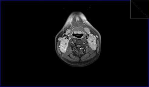
T1 FS axial post contrast demonstartes lymphoma.
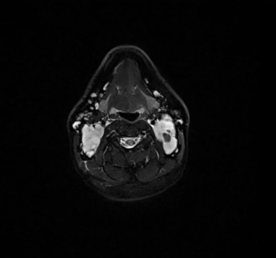
STIR axial image showing lymphoma
Parotid Gland Tumor
Parotid gland tumors may be benign or malignant. Benign tumors are usually palpable, discrete, and mobile. Malignant tumors commonly present as a palpable lump or mass.
Appearance on MRI:
•
Lesions are best identified on T1-weighted images amid the bright signal of parotid fat.
•
Benign tumors are very bright on T2-weighted images.
•
Malignant tumors may have mixed signal intensities on T2-weighted images
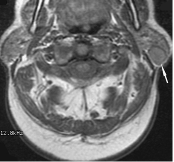
Axial T1 shows a parotid tumor.
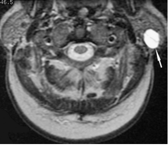
Axial T2 shows the same parotid tumor.
Warthin’s Tumor
Warthin’s tumor, also known as Warthin’s adenoma, is a benign salivary gland tumor that primarily affects the parotid gland.
Appearance on MRI:
•
T1 images will appear as a well-defined mass with low to intermediate signal intensity
•
Post-contrast images will show strong enhancement; if the mass is cystic, there will be rim enhancement.
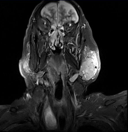
STIR coronal showing a Warthin’s tumor.
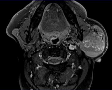
Axial T1 FS post contrast shows enhancement within a Warthin’s Tumor.
Epidermoid Cyst
An epidermoid cyst is a skin lesion arising from the outermost layer of skin and commonly occurs on the face and neck, but they can occur anywhere on the skin.
Appearance on MRI:
•
T1 images: Cysts will appear as well-defined lesions with low signal intensity
•
T2 images: Show cysts with high signal intensity.
•
STIR images: Help determine the extent of surrounding tissue involvement.
•
Post Contrast images: Will not show enhancement.
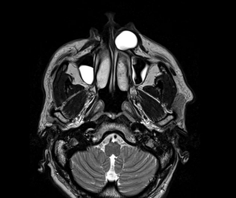
Axial T2 shows Epidermoid cyst as well as sinus congestion in the maxillary sinuses.
Glomus Tumor
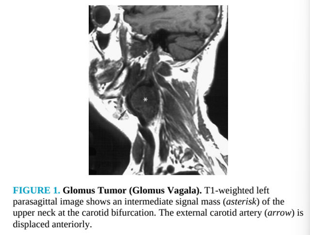
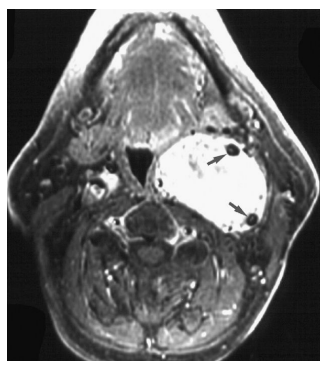
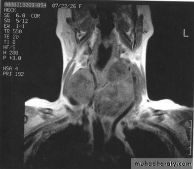
A glomus tumor is a benign, slow-growing, hypervascular lesion arising from paraganglion cells in the extracranial head and neck.
Appearance on MRI:
•
T1 images show mixed signal mass with signal voids.
•
T2 images will show high signal.
•
Post contrast T1 images with show hyperintense signal with voids.
Thyroid Goiter
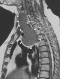
A thyroid goiter is an enlargement of the thyroid gland that causes swelling in the anterior neck. This type of goiter is also called a nontoxic goiter, as it is not related to overproduction of thyroid hormone or malignancy.
Appearance on MRI:
•
Normal thyroid gland appears with intermediate signal on T1-weighted images.
•
Normal thyroid gland appears with higher signal on T2-weighted images.
•
T2 weighted imaging is also good for evaluating compression or deviation of the trachea and intrathoracic extension of the goiter.
Peritonsillar Abscess
A peritonsillar abscess (PTA) is an accumulation of pus in the tonsillar bed with medial displacement of the tonsil.
Appearance on MRI:
•
T1-weighted images appear hypointense to isointense to surrounding muscles.
•
Hyperintense on T2-weighted images.
•
Gadolinium-based T1-weighted images show an enhancing rim of the abscess.
Submandibular Salivary Gland Abscess
A submandibular salivary gland abscess is a mucus-filled retention cyst derived from obstructed or traumatized salivary ducts.
Appearance on MRI:
•
Hypointense on T1-weighted images
•
Hyperintense on T2-weighted images
•
Post-Contrast T1 Enhancement