Lesson 1 Cells of the Nervous System 1
1/125
There's no tags or description
Looks like no tags are added yet.
Name | Mastery | Learn | Test | Matching | Spaced |
|---|
No study sessions yet.
126 Terms
The human brain contains two basic types of cells
Neurons and glia
What is the main functional unit of the brain? (The signaling units of the nervous system!)
Neurons
What is outnumbered by glia and are the only cells capable of generating the electrical signals known as nerve impulses?
Neurons
What is the support cell of the nervous system that also means “glue”
Glia
Although this is not the main cell type of the nervous system, these various cell types, serve several import roles that allow neurons to function, in part, as they do…?
Glia
In the past neurons were difficult to study because of their small size what size were they?
0.01mm to .05mm
what made it difficult to view nervous tissue under a microscope?
In the past, it was difficult to obtain very thin slices of brain tissue for viewing under the microscope. In addition once cut, the thin slices were not resolvable until stains were developed that could selectively color different parts of the cells in neural tissues.
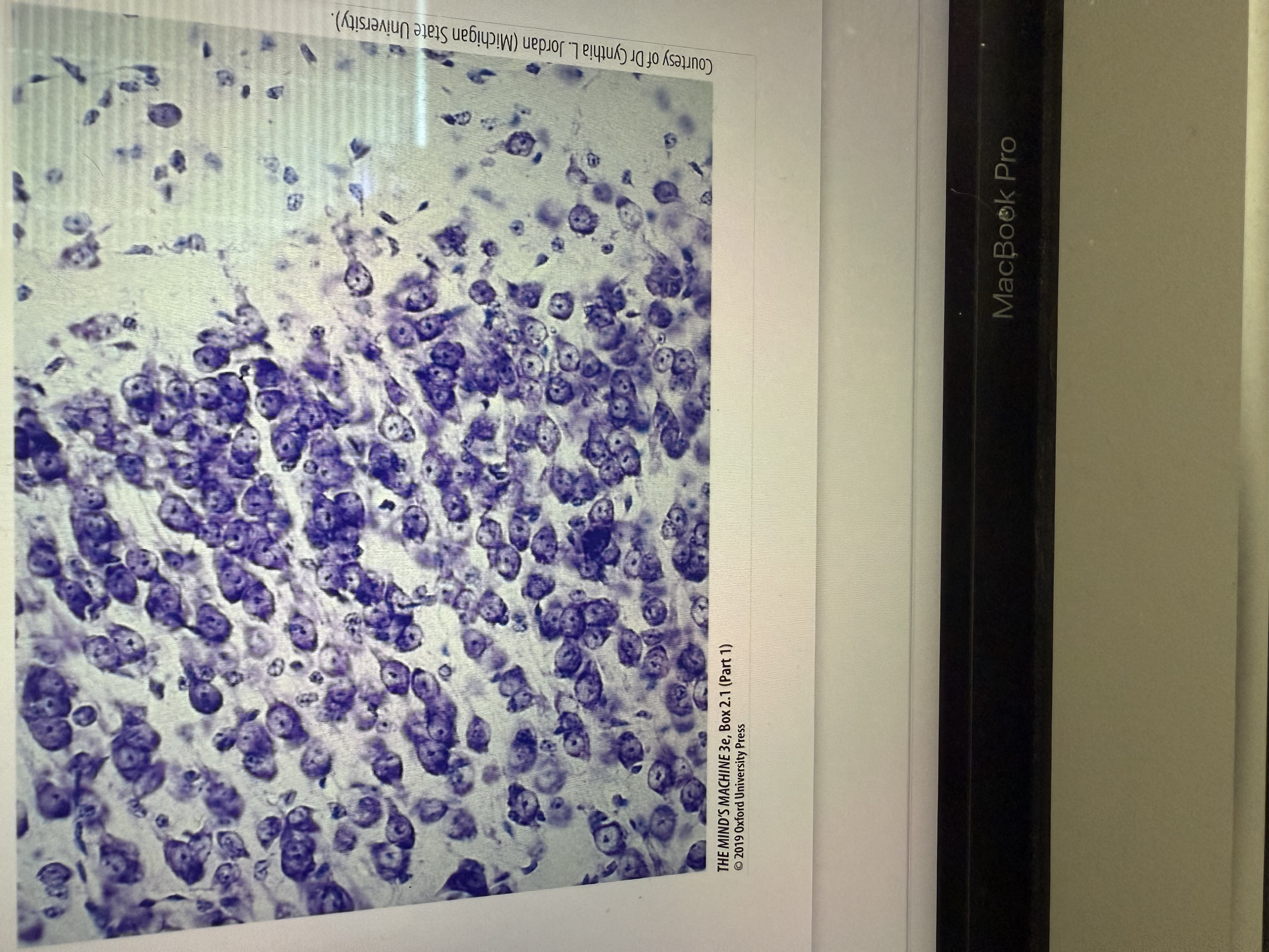
Which picture is a Nissl stain?
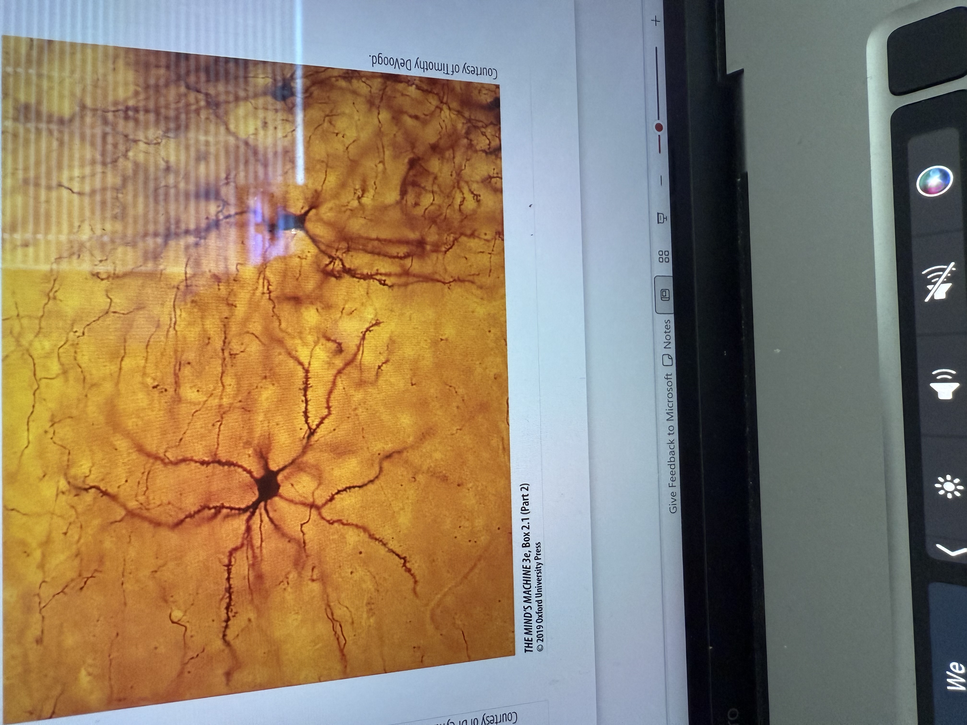
Which picture is a golgi stain?
Neurons are morphological and functionally
Asymmetrical
What serves as the main part of the neuron, housing the nucleus and much of the cellular organelles?
The Soma or cell body
What are extensions of the Soma that receive incoming information?
Dendrites
What is a long extension that carries information away from the Soma towards the axon terminals?
The axon
What in the axon terminal housing transmitters is used to signal other cells?
The terminal boutons (axon terminals)
How do electrical currents within a neuron travel?
From the dendrites to the axon hillock or initial segment (where the axon attaches to the soma)
What does the electrical currents within a neuron generate?
An action potential or “nerve impulse”
Where does the electrical current within a neuron generate an action potential?
at the initial segment
From the axon hillock, where does the action potential travel, and what happens when it reaches the axon terminals (boutons)?
Travels down the axon to the axon terminals, or boutons, Where it results in the release of a chemical neurotransmitter
All neurons basically function the same way, however they can have something different. What is it?
Morphologies (they don’t all look exactly like)
What type of neurons have a single process?
Unipolar neurons (found in the autonomic nervous system)
What kind of neurons have two distinct processes, a dendritic structure and an axon?
Bipolar neurons (Such as sensory neurons)
What kind of neurons typically have a single axon and branching denture structures
Multipolar neurons (In vertebrates, they are the most common)
What else can neurons also be classified as functionally?
Sensory, motor, or interneuron
What do the various compartments within the nervous system include?
The intracellular environment of both neurons and glia cells, the extracellular environment (or interstitial compartment), the cerebrospinal compartment, and the vascular system
What is the watery extracellular [interstitial] environment in the brain called?
the brain extracellular fluid (BECF)
From what compartments is the brain extracellular fluid (BECF) separated, and by which barriers or membranes?
it is separated from the intracellular compartments of neurons and glia by their cell membranes, from the intravascular system by the blood-brain barrier (BBB), and from the cerebrospinal compartment by the blood-cerebrospinal fluid barrier.
What regulates the brain extracellular fluid (BECF) compartment, which plays an important role in neural activity?
The blood-brain barrier (BBB), the cerebrospinal fluid (CSF) barrier, and glial cells.
What is the microenvironment of neurons?
Everything that surrounds the individual neurons
What does the microenvironment include?
The brain extracellular fluid (BECF), capillaries, and glial cells
Where does communication between neurons in the CNS largely occur?
in the neuropil
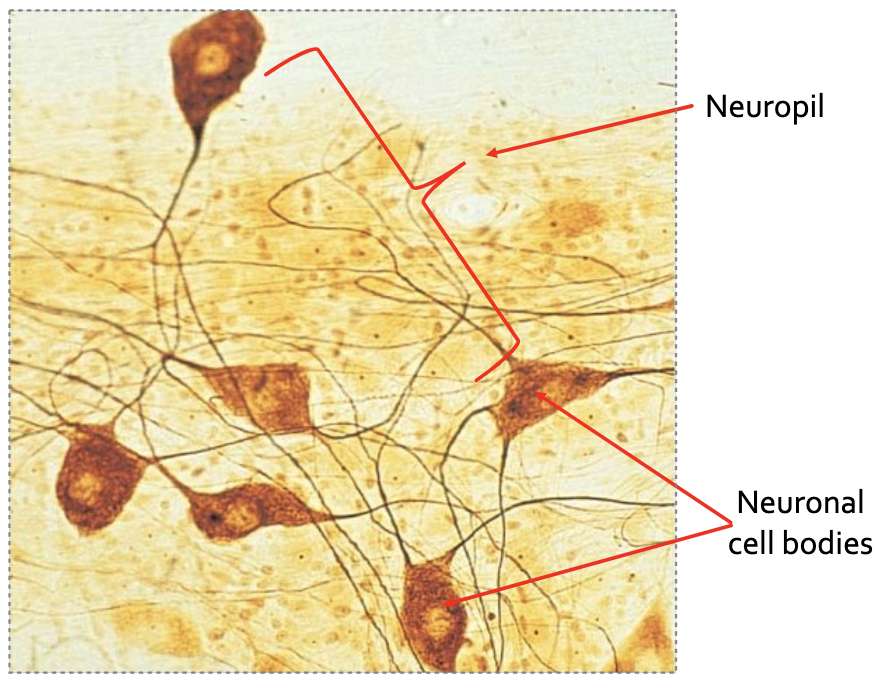
Where is the neuropil located and what is it composed of?
It is located in the area outside of neuronal cell bodies; it is composed of axonal and dendritic branches, and the synapses between them.
What is brain parenchyma?
functional tissue of the brain (i.e., neurons and glia), which is distinguished from the structural or supporting elements of the brain tissue
In what way are neurons similar to other mammalian cells?
they contain two compartments and various organelles.
What are the key features and functions of the nucleus in a neuron?
The nucleus has a double membrane with nuclear pores, contains DNA (and some RNA), is usually centrally located, and serves as the site of transcription.
What are the main features and functions of the cytoplasm in a neuron?
The cytoplasm is the location of most organelles, contains the cytosol (the aqueous part of the cytoplasm), includes some freely soluble proteins, and is the site of translation.
Why do neurons have different organelles?
Different organelles perform distinct functions that enable the neuron to carry out essential life processes.
What are the two types of endoplasmic reticulum (ER) in neurons, and how do they differ?
The endoplasmic reticulum is often attached to the nuclear membrane. Rough ER (rER) is embedded with ribosomes, while smooth ER (sER) is not.
Ribosomes are the site of
protein synthesis
What are the main roles of the Golgi apparatus in neurons?
It is the site of post translational modifications, protein sorting (determining protein destinations), and protein packaging.
What are the main functions of mitochondria in neurons?
They are the site of cellular respiration and ATP production, serving as the main source of energy for the cell.
What does the neuronal plasmalemma do?
separates the cytosol from the interstitial fluid.
What bounds neurons and what is unique about the structure of this boundary?
their plasmalemma, or cell membrane, which is asymmetric in structure like all biological membranes.
From what fluids is the interstitial (extracellular) fluid physically separated?
The intracellular fluid (inside the cell) and from the fluid inside the vascular system.
What does the lipid membrane of a neuron provide, and what is it impermeable to?
a hydrophobic barrier that is impermeable to most water-soluble substances.
What enables the plasmalemma to separate the intracellular and extracellular environments?
its structure
The neuronal membrane is largely composed of
phospholipids
What structural feature do phospholipids have?
both a polar end (charged) and a nonpolar end (uncharged)
In the phospholipid bilayer, how are the polar (charged) ends of phospholipids oriented?
face outward toward the polar watery environments on both the inside and outside of the cell.
What forms the inner part of the phospholipid bilayer, and what property does it give the membrane?
The inner bilayer is composed of nonpolar (uncharged) tails, creating a hydrophobic inner membrane that prevents charged substances from passing through.
What types of interactions occur between lipids and between lipids and proteins in the membrane?
Many interactions are noncovalent and include electrostatic forces and hydrogen bonding.
What components are found within the cytoplasm of a neuron?
both free-floating proteins and molecules, as well as membrane-bound organelles.
What types of proteins are found freely in the cytoplasm, and how are they organized?
Various proteins, including some enzymes and intracellular signaling molecules (such as second messengers), are found freely in the aqueous part of the cytoplasm. Most are organized into functional complexes, and many are integrated into the cell membrane.
What membranous organelles and systems are found in the cytoplasm of a neuron?
mitochondria and peroxisomes, as well as a complex system of tubules including the endoplasmic reticulum (smooth and rough ER), Golgi apparatus, lysosomes, endosomes, and secretory vesicles. This tubule system is sometimes called the vacuolar apparatus.
How are the major sub compartments of the tubular system in the cytoplasm connected?
They are not anatomically continuous, but they are functionally connected because membranous and luminal materials are transported from one compartment to another by transport vesicles.
Where are most of a neuron’s organelles located?
within the cytoplasm of the cell body.
What is the relationship between the endoplasmic reticulum (ER) and ribosomes, and what role does the ER play in protein processing?
The ER is typically connected to the nuclear membrane and is often embedded with ribosomes, which are the sites of protein synthesis. The ER is involved in processing proteins after they are assembled on ribosomes, including post-translational modifications.
What happens to proteins in the Golgi apparatus, and what are their possible destinations?
proteins undergo further posttranslational modifications, are sorted, and packaged. Their possible destinations are: staying inside the cell, being inserted into the cell membrane, or being exported out of the cell.
What cellular processes occur in the mitochondrion, and what is its main function?
The mitochondrion is the site of the Krebs cycle and the electron transport chain, which are parts of cellular respiration responsible for ATP production. (Glycolysis, however, occurs in the cytoplasm, not in the mitochondria.)
Where does most of what is typically referred to as “cellular respiration” occur, and what important exception should be noted?
inside the mitochondria. However, glycolysis takes place in the cytoplasm outside the mitochondria and does not require oxygen.
What is the end product of glycolysis
pyruvate
What happens to pyruvate, the end product of glycolysis, once it enters the mitochondria?
it is converted into acetyl CoA, which then enters the citric acid cycle (also called the Krebs cycle or TCA cycle).
Where are the electrons gathered from the citric acid cycle and glycolysis transferred, and where is this located?
The electrons are transferred to the electron transport chain, which is located in the inner mitochondrial membrane.
How does the electron transport chain contribute to ATP synthesis, and what role does oxygen play?
The electrons create a hydrogen gradient that drives ATP synthesis. Oxygen serves as the final electron acceptor, combining with hydrogen to form water.
Why do neurons require a constant supply of oxygen and glucose?
Because neurons continuously make ATP due to their high metabolic activity
What role do integral membrane proteins play for the cell?
they provide a way for the cell to communicate with its environment.
What roles can integral membrane proteins serve in cell communication?
Ligand-binding receptors for transmembrane signaling
Adhesion molecules that form physical contacts with the extracellular matrix (e.g., integrins) or other cells
Transport proteins (channels, carriers, pumps) that allow movement of water-soluble substances through the hydrophobic lipid bilayer
Enzymes
Intracellular signaling proteins (e.g., GTP-binding proteins, kinases)
What roles do peripheral membrane proteins play in neurons?
They can participate in intracellular signaling and help form a submembranous cytoskeleton.
How are integral proteins often associated with or connected to the membrane?
by proteins on both sides of the membrane.
What roles do transmembrane proteins that form complexes with intra- and extracellular proteins play?
They help stabilize dynamic contacts and mediate intracellular signaling.
What are cell adhesion molecules (CAMs), and which types are included?
CAMs are proteins involved in cell-to-cell and cell-to-matrix interactions; examples include integrins and cadherins.
What roles do CAMs play in the nervous system?
Nervous system maintenance and development including myelination, synaptic stabilization, neuronal tube formation, neuronal and glial migration, and plasticity.
What are the main protein components of the cytoskeleton?
microtubules, neurofilaments, and microfilaments.
What are microtubules made of, what is their size, and what functions do there structures serve?
(20–25 nm) are composed of α- and β-tubulin protein subunits, have a negative and a positive end, and these structures serve to form the neuron’s shape and assist in transport, especially along axons.
What are neurofilaments (intermediate filaments) , and what is their role and size?
~10 nm, are the “bones” of the cytoskeleton, the most abundant fibrillar component in axons. Unlike microtubules, they are stable, being mostly polymerized in the cell.
What are microfilaments, what are they made of, what size are they and what do they do?
(thin filaments, 3–8 nm) are composed of actin, are concentrated near the cytoplasmic plasmalemma, and like microtubules, undergo cycles of polymerization and depolymerization. They are probably among the most abundant animal proteins in nature.
Into what two main categories are glial cells divided?
Glial cells are divided into microglia and macroglia.
What are microglia, and what is their origin and function?
They are mesodermal in origin, are the immune cells of the nervous system. They respond to various circumstances including antigen presentation and phagocytic cleanup.
What percentage of brain cells are macroglia, and what types are included?
They comprise about 80% of all cells in the brain and include astrocytes, oligodendrocytes, and Schwann cells.
What are the roles of astrocytes?
They help regulate the neuroenvironment and support neuron functioning in several ways.
What is the role of oligodendrocytes?
They are associated with myelination within the central nervous system (CNS).
What is the role of Schwann cells?
They are responsible for myelination within the peripheral nervous system (PNS).
What are the “immune” surveillance cells of the nervous system?
Microglia
What is the role of oligodendrocytes in the CNS?
They are responsible for myelinating axons in the CNS early during neurodevelopment.
How many axonal segments can a single oligodendrocyte myelinate?
It can envelop several axonal segments (internodes).
What is the role of Schwann cells in the PNS?
They are responsible for myelinating axons in the PNS early during neurodevelopment.
How many axonal segments does a single Schwann cell myelinate?
It envelopes a single segment between two nodes of Ranvier.
What are the two main types of astrocytes, and where are they found?
Protoplasmic astrocytes are found in gray matter, where they envelope nerve cell bodies and synapses.
Fibrous astrocytes are found in white matter, where they envelope or contact axons at the nodes of Ranvier.
What common function do both types of astrocytes perform related to blood vessels?
They help form the blood-brain barrier by wrapping around blood vessels entering the brain.
What are the main functions of astrocytes?
Insulate neuronal groups and synaptic connections from each other
Regulate the concentration of K⁺
Take up neurotransmitters, helping regulate postsynaptic activity
Nourish neurons by releasing growth factors
Who first recorded an action potential, when, and from what organism?
Hodgkin and Huxley first recorded an action potential in 1939 from a giant squid axon.
How does an action potential propagate down an axon?
at a set speed and amplitude (Y-value), being regenerated along the axon.
What role do action potentials play in the nervous system?
They are the signals by which the brain receives, analyzes, and conveys all kinds of information.
What determines the information conveyed by action potentials?
The frequency of action potentials, their pattern, and the pathway (circuit) they travel.
What is the knee-jerk reflex an example of?
It is an example of a circuit associated with a simple behavior.
How does the knee-jerk reflex work?
Stretch receptors (muscle spindles) in the quadriceps extensor muscle of the leg detect stretch and send sensory information to motor neurons in the spinal cord, which then send signals to the extensor muscles to contract.
What protective function does the knee-jerk reflex serve?
It helps protect muscles, stretch receptors, and tendons from injury.
Why is the knee-jerk reflex clinically useful?
It shows that neural circuits are intact and functioning properly.
How do divergence and convergence of synapses contribute to neural communication?
ensure proper communication in both sensory and motor pathways.
What is divergence, and where is it most common in the nervous system?
It is when one neuron sends output to numerous cells. It is most common within the input stages of the nervous system.
What is convergence, and where is it most common in the nervous system?
it is when one neuron (e.g., a motor neuron) receives input from numerous cells. It is most common within the output stages of the nervous system.