Lecture 24. GI Infections 5
 Call Kai
 Learn
Learn Practice Test
 Spaced Repetition
 Match
Match Flashcards
 Knowt Play
1/40

There's no tags or description
Looks like no tags are added yet.
Name | Mastery | Learn | Test | Matching | Spaced | Call with Kai | Chat |
|---|
No analytics yet
Send a link to your students to track their progress
41 Terms
Mycobacterium spp. etiology
Aerobic, non-spore forming, gram-positive rods.
what makes Mycobacterium spp. ZN positive
High lipid and mycolic acid content in the cell wall.
due to the ZN positive component of Mycobacterium spp...
they are highly resistant but still susceptible to pasteurization
Common habitats of Mycobacterium spp.
Soil, vegetation, and water.
Media for growing Mycobacterium spp.
Egg-enriched media.
Types of pathogens associated with Mycobacterium spp.
Obligate pathogens, opportunists, and saprophytes.
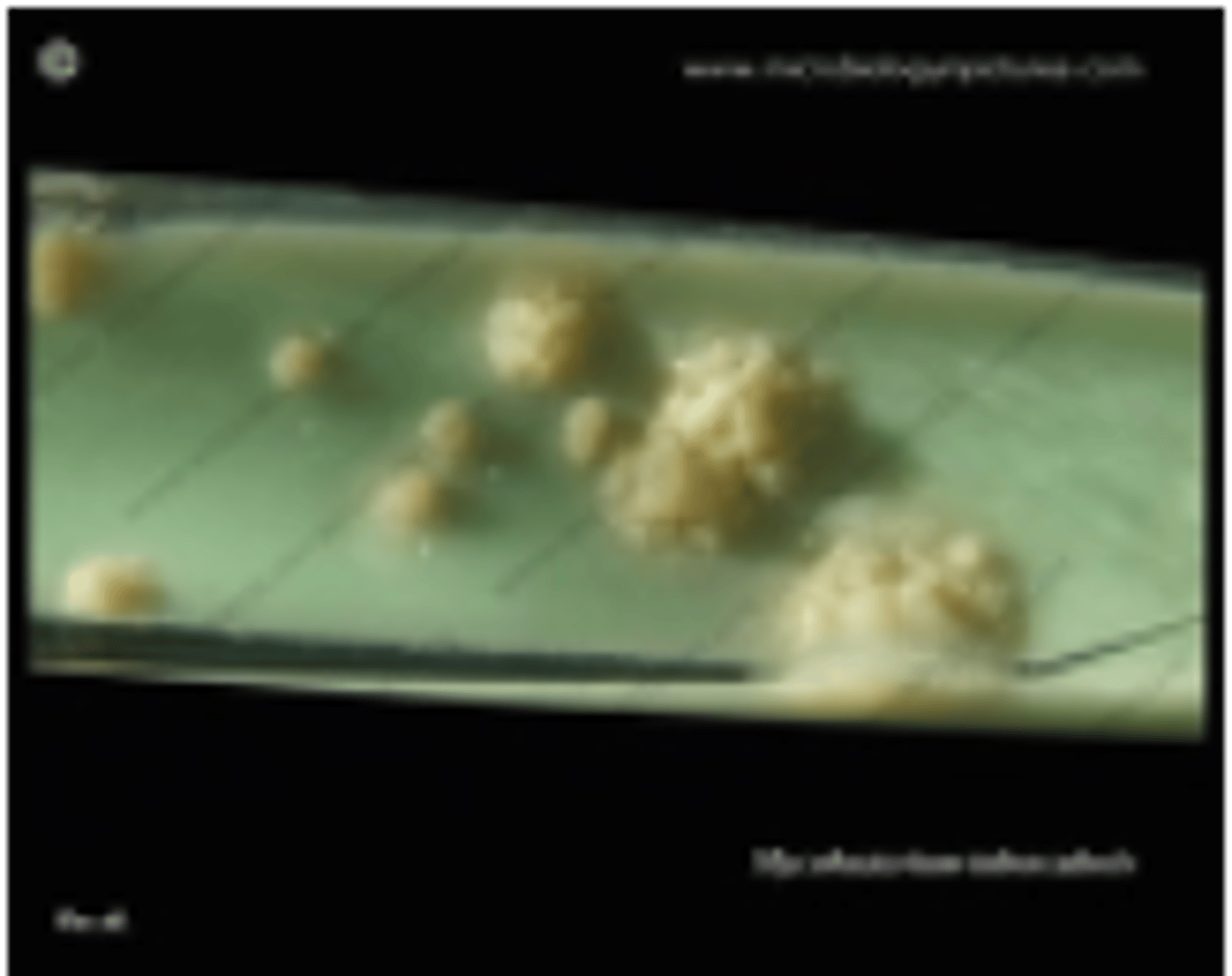
Colony characteristics of M. tuberculosis
Rough colonies, hard to break; grows in 3-8 weeks at 37°C in aerobic conditions; enhanced by glycerol.
M. tuberculosis
what Mycobacterium spp. is indicated in this image that affects humans
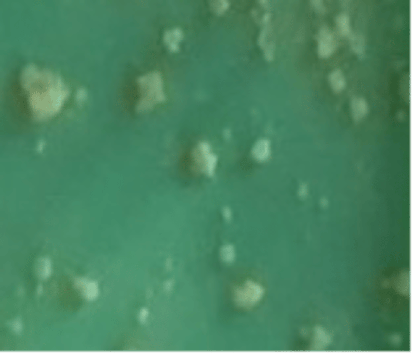
Colony characteristics of M. bovis
Cream colonies, break easily; grows in 3-8 weeks at 37°C in aerobic conditions; inhibited by glycerol, enhanced by pyruvate.
M. bovis
what Mycobacterium spp. is indicated in this image that affects cattle
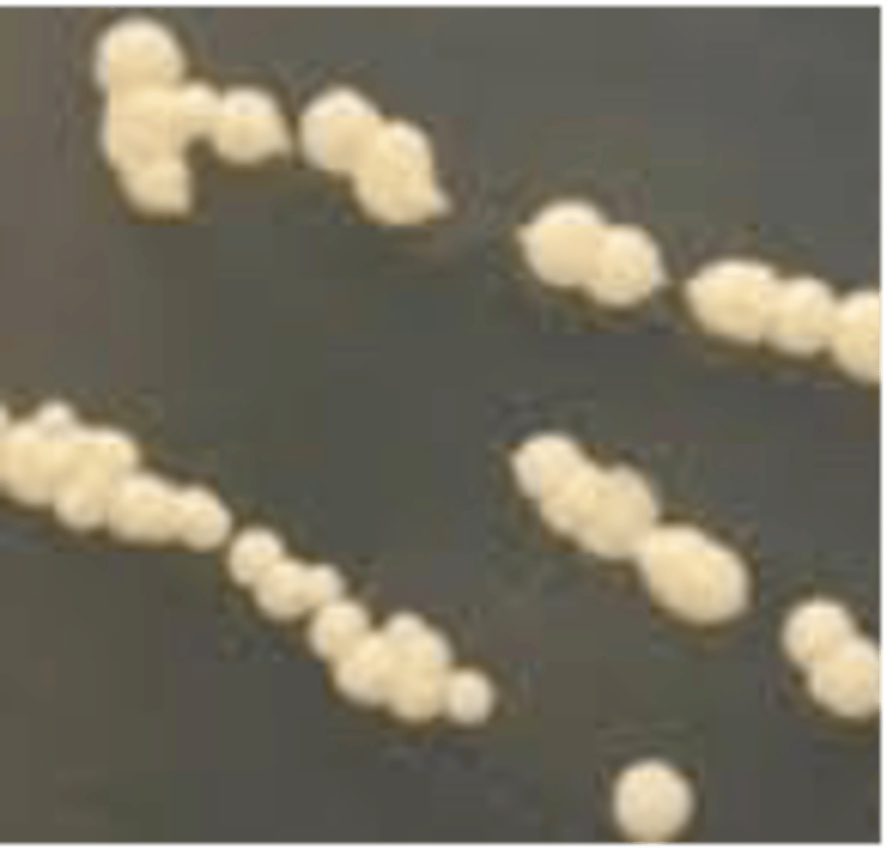
Colony characteristics of M. avium
Sticky, off-white colonies that break easily; grows in 2-6 weeks at 37-43°C; enhanced by glycerol.
M. avium
what Mycobacterium spp. is indicated in this image that affects domestic poultry
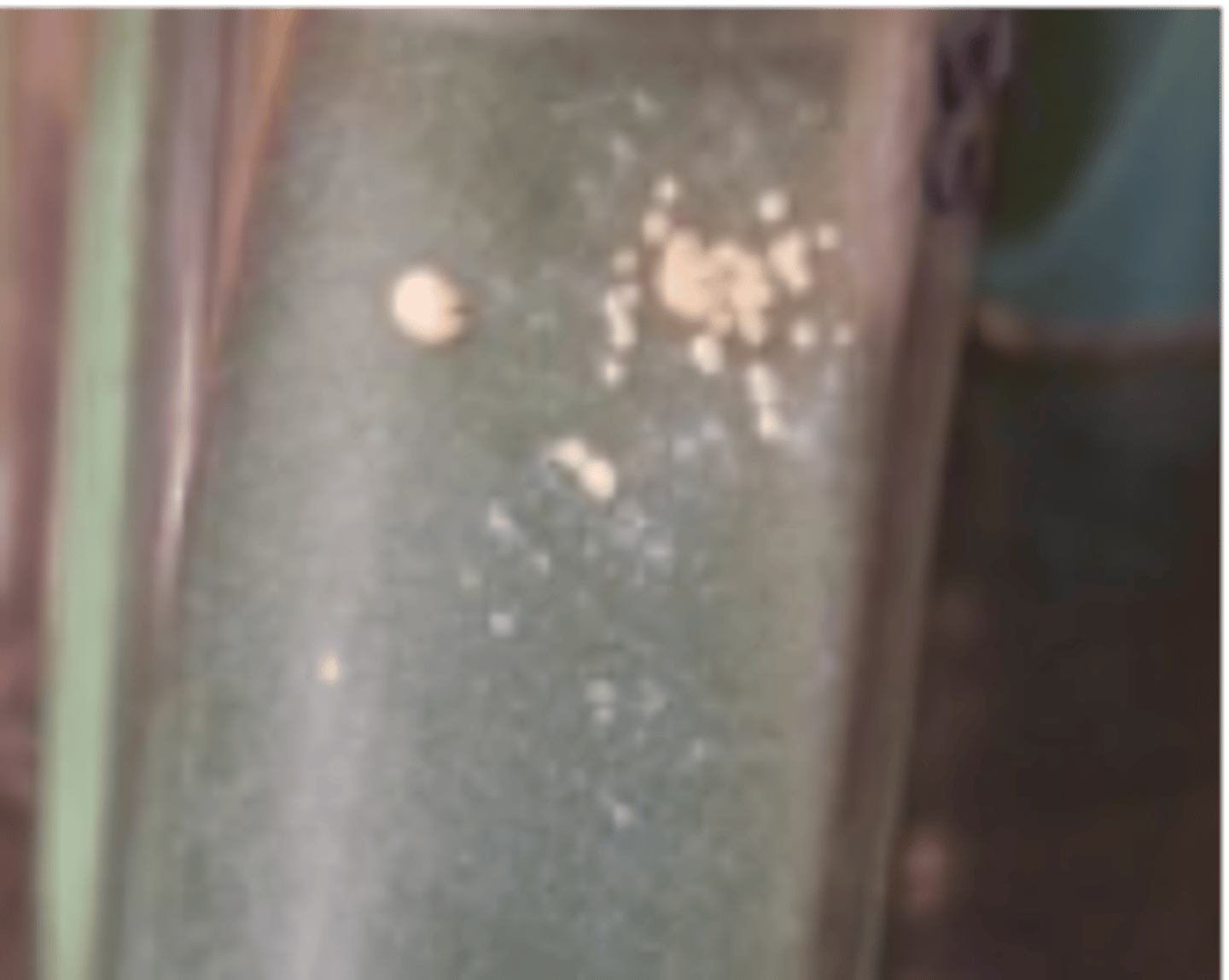
Colony characteristics of M. avium subsp. paratuberculosis
Small, hemispherical colonies with pigment; grows in 16 weeks at 37°C; requires mycobactin.
M. avium subsp. paratuberculosis
what Mycobacterium spp. is indicated in this image that affects ruminants
it is important to remember that tho the M. avium complex is associated with most avian species, it is not associated with....
psittacines
Pathogenesis of Mycobacterium spp.
They multiply intracellularly, causing chronic granulomatous infections.
Cause of mammalian tuberculosis
Mycobacterium tuberculosis complex.
Cause of avian tuberculosis
Mycobacterium avium subsp. avium (serotypes 1-3).
Cause of Johne's Disease
Mycobacterium avium subsp. paratuberculosis (MAP).
Cause of leprosy in humans and cats
Humans: M. leprae; Cats: M. lepraemurium.
Stain used for diagnosing Mycobacterium infections
Acid-fast staining (Ziehl-Neelsen).
Common media for culture of Mycobacterium spp.
Lowenstein-Jensen medium.
Molecular tools for diagnosing Mycobacterium infections
PCR and rRNA sequencing.
Differentiation of M. tuberculosis and M. bovis in the past was done via
Guinea pig and rabbit inoculation.
zoonotic implications of Mycobacterium spp.
M. tuberculosis, M. bovis, M. africanum, M. canettii, M. pinnipedii, M. marinum, M. ulcerans and M. leprae are all zoonotic to humans
Transmission of Johne's Disease
Fecal-oral route, mainly affecting calves.
MAP is brought into the herd by...
subclinical carriers brought into the herd
____% of cows in a herd can be subclinically infected w/ Johne's Disease
50%
Onset of signs of Johne's Disease
Acquired early in life but signs develop at ~2 years old.
Pathogenesis of Johne's Disease
Chronic granulomatous enteritis; bacteria infect Peyer's patches and macrophages in GIT & LNs.
what is the result of infection with Johne's disease that leads to chronic granulomatous enteritis
interference with nutrient uptake and processing
infection w/ Johne's disease
what lead to the physical condition of this cow
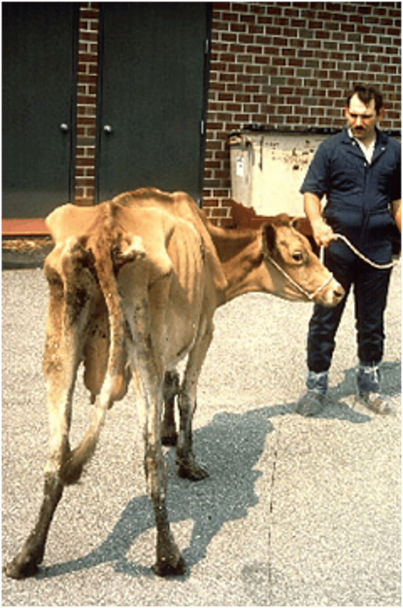
Clinical signs of Johne's Disease in cattle
Persistent diarrhea, weight loss/emaciation, ventral/intermandibular edema, death.
Additional signs in dairy cattle or goats with Johne's Disease
Drop in milk yield.
tubercular lesion
how would you describe the appearance of this intestine
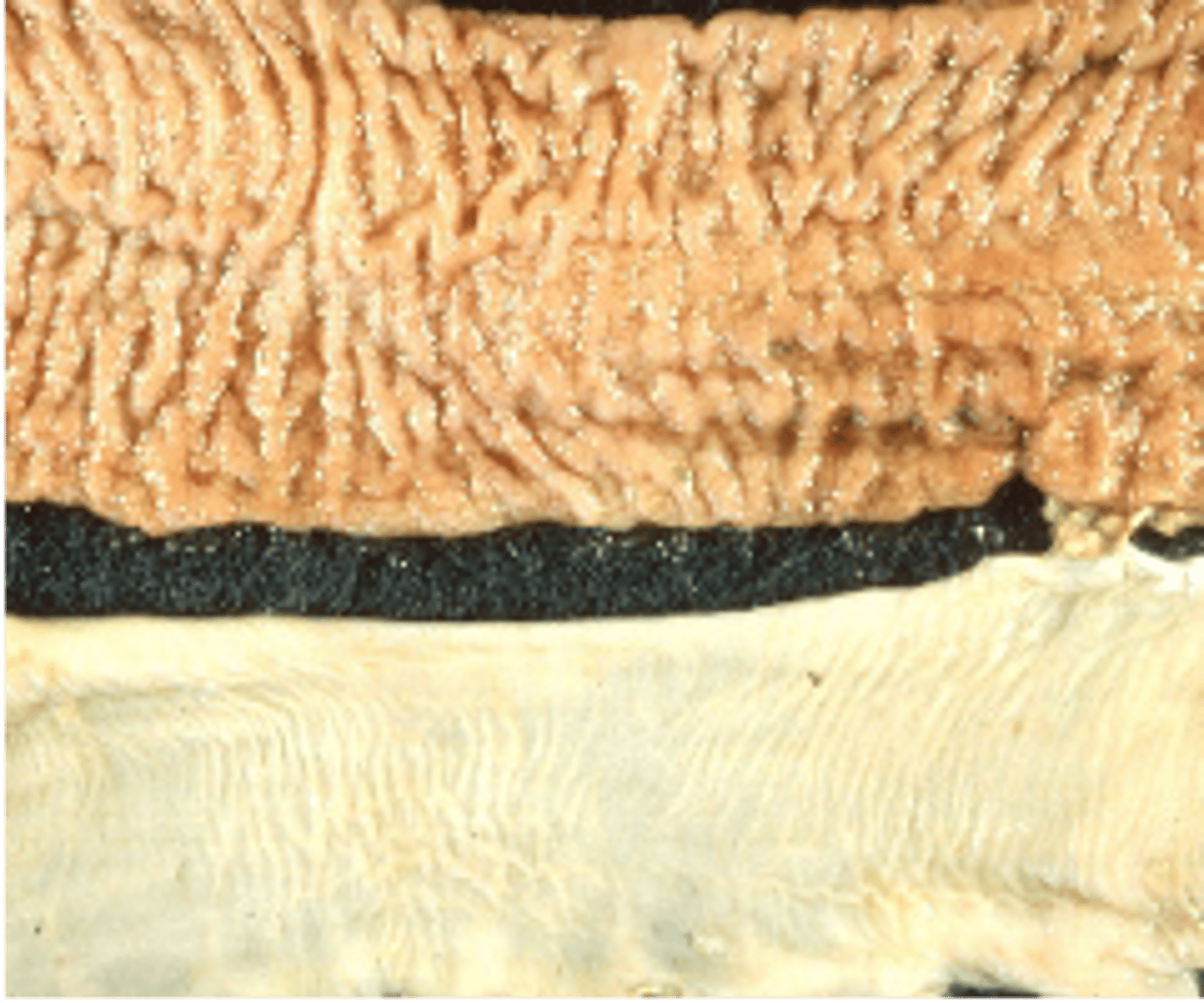
How would you describe the Intestinal (tubercular) lesions seen with Johne's Disease
Thickened, corrugated distal small intestine in addition to edematous neighboring lymph nodes.
Diagnosis of Johne's Disease
1. Histopath: granulomatous enteritis w/ acid-fast macrophages
2. PCR, ELISA, or AGID.
Treatment for Johne's Disease
No real treatment; sometimes self-limiting via cell-mediated immunity.
Control of Johne's Disease
1. Separate young
2. feed MAP-free colostrum
3. remove infection sources
4. disinfect.
Disease caused by Mycobacterium ulcerans
Buruli ulcer in humans, koalas, and possums.
Disease caused by Mycobacterium marinum
Tuberculosis in fish, aquatic mammals, and amphibians.