Anatomy Module 1: Introduction to Human Anatomy
 Call Kai
 Learn
Learn Practice Test
 Spaced Repetition
 Match
Match Flashcards
 Knowt Play
1/64

There's no tags or description
Looks like no tags are added yet.
Name | Mastery | Learn | Test | Matching | Spaced | Call with Kai | Chat |
|---|
No analytics yet
Send a link to your students to track their progress
65 Terms
Muscular system
All muscles; how they move joints
Cardiovascular (arterial/venous) system
Heart + vasculature; pumps blood throughout body
Respiratory (pulmonary) system
Nasal/oral cavity → trachea → lungs (lobes, covered by pleura); site of gas exchange
Digestive (GI) system
Mouth → esophagus → stomach → small intestine (nutrient absorption) → large intestine (waste/water/electrolyte disposal) → rectum/anus
Lymphatic system
Lymph = interstitial fluid (byproduct of blood); mobilizes lipids/proteins from interstitial spaces between cells back into bloodstream
Endocrine system
Hormone production and regulation
Integumentary system
Largest organ system; skin, hair, nails
Nervous system
Master regulatory system — controls heart rate, breathing depth, sweating, hormone release, etc.; coordinates all other systems toward the goal of keeping the body alive and thriving
arteries
carry oxygenated blood away from the heart (~99% of the time — exception discussed later, e.g., pulmonary artery)
veins
carry blood toward the heart
Levels of biological organization
Cell (e.g., osteoblasts, osteoclasts)
Tissue (e.g., bone tissue)
Organ (e.g., a single bone)
Organ system (e.g., skeletal system)
Organism (multiple organ systems → whole human body)
Systemic approach
Studies the body by organ system, looking at each system in isolation across the whole body
Regional approach
Studies all systems together within a specific body region (bones, muscles, tendons, ligaments, vessels, nerves, lymphatics, skin — all studied together for that region).
Clinical approach
Frames anatomy in terms of clinical relevance and consequences of injury/dysfunction rather than pure structure
Imaging approach
Uses medical imaging to understand structural relationships:
X-ray — bone position/connections
MRI — soft tissue relationships
CT scan — differentiates fluid-filled vs. non-fluid-filled structures
Ultrasound — real-time visualization; used in this professor's PA program (students scan each other to locate heart, lungs, etc.)
Advanced/novel method: CT scans of cadavers converted into 3D holograms, viewed via VR goggles, used by orthopedic residents to preview tissue layers before dissection/surgery.
Anatomical position
The standardized baseline reference posture used for all anatomical descriptions, measurements, and documentation.- eliminates ambiguity, serves as reference point, and gives relativity for directional terms
Standing upright
Palms facing forward
Shoulders level
Head facing forward
Feet shoulder-width apart, facing forward
superficial
closer to the outside/external surface
deep
farther inside, beneath another structure
intermediate
between superficial and deep
superficial/intermediate/deep
Describes depth/layering relative to another structure.
Skin (most superficial)
Biceps (intermediate — relative to skin & brachialis)
Brachialis (deep to biceps)
Ligaments (deep to brachialis)
Bone (deepest)
medial/lateral
Describes position relative to the midline of the body (an imaginary line dividing the body into right and left halves).
Clavicle (collarbone): has a medial end (near sternum/midline) and a lateral end (toward shoulder)
Femur (knee): medial condyle (inner bony prominence) vs. lateral condyle (outer bony prominence)
medial
closer to the midline
lateral
farther from the midline
anterior
toward the front
posterior
toward the back
ventral/dorsal
Functionally equivalent to anterior/posterior, but used in specific contexts — especially the spine, hand, and foot
Important: This relationship is defined relative to anatomical position. If you rotate the palm, the ventral surface doesn't "become" dorsal — the terminology stays fixed to the anatomical position reference, not to the momentary orientation of the limb
ventral
anterior (front)
dorsal
posterior (back)
proximal/distal
Describes position relative to the trunk/midpoint of the body (point of attachment/origin), rather than the midline.
Important nuance: Proximal/distal can overlap in meaning with either superior/inferior or medial/lateral, depending on the structure:
Comparable to... | Example |
Superior/Inferior | Shoulder (proximal/superior) vs. elbow (distal/inferior) |
Medial/Lateral | Medial end of clavicle = proximal end; lateral end = distal end |
Think of proximal/distal in terms of a central reference circle at the body's core/trunk — proximal = closer to that center, distal = farther away — rather than strictly "above/below" or "toward/away from midline."
proximal
closer to the trunk/center of the body
distal
farther from the trunk/center of the body
cephalad/caudal
Used specifically for the axial skeleton (spine/vertebral column) — less commonly used for peripheral structures.
Examples:
The head end of the spinal cord (toward the brainstem) = cephalad
The cauda equina (bottom of spinal cord) = caudal end
Functionally interchangeable with superior/inferior, but conventionally reserved for spinal/axial skeleton descriptions. In general conversation, people are more likely to say "superior/inferior" than "cephalad/caudal."
cephalad
toward the head (Latin: "ceph/cephal" = head — same root as biceps/triceps, referring to number of muscle "heads")
caudal
toward the tail/sacrum (Latin: "caudal" = tail)
plane
an imaginary flat surface (think: a sheet of paper) placed through/over a joint that shows the direction in which motion is occurring
Also used in radiology/imaging to describe the view or section of a patient being examined.
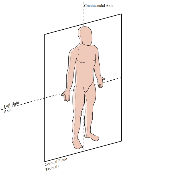
frontal/coronal plane
orientation: viewed from the front
typical motions: abduction/adduction
axis it rotates around: Anterior–Posterior axis (also called frontal/coronal axis)
sagittal plane
orientation: viewed from the side
typical motions: flexion/extension
axis it rotates around: Medial–Lateral axis

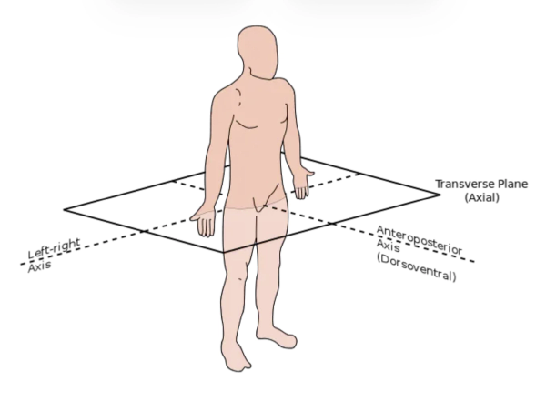
horizontal/transverse plane
orientation: viewed from above, across the body
typical motions: internal/external rotation
axis it rotates around: longitudinal (vertical) axis
Uniplanar/uniaxial joints
move in only 1 plane
Example: Elbow, knee → sagittal plane only (flexion/extension); no meaningful ab/adduction or rotation
biplanar joints
move in 2 planes
Example: Knuckles (MCP joints of fingers) → frontal plane (abduction/adduction = spreading fingers) and sagittal plane (flexion/extension)
multiplanar/multiaxial joints
move in all 3 planes
Example: Shoulder and hip (ball-and-socket joints) — the most mobile joint type, with the greatest number of planes of motion
head and neck motion
Because the head/neck is a central/midline structure (not paired left/right like a limb), its motions get unique names instead of "abduction/adduction":
Moving head to either side (frontal plane) = side flexion
Rotating head to look side to side (horizontal plane) = rotation (simply "right rotation" / "left rotation" — not internal/external, since it moves toward both sides)
side flexion (head)
Moving head to either side (frontal plane)
rotation (head)
Rotating head to look side to side (horizontal plane)
abduction
moving a body part away from the midline
frontal plane
Occurs around the anterior-posterior axis
adduction
moving a body part back toward the midline
frontal plane
Occurs around the anterior-posterior axis
flexion
brings a segment anteriorly
sagittal plane
Applies to: shoulder, elbow, wrist, hip, knee, head/neck
extension
brings a segment posteriorly
sagittal plane
Applies to: shoulder, elbow, wrist, hip, knee, head/neck
flexion/extension exceptions and nuances
Hip: flexion moves the thigh anteriorly (as expected), but the "logic" of anterior/posterior can feel reversed depending on viewing angle — regardless of direction, it is still sagittal plane motion.
Knee: flexion brings the lower leg posteriorly (bending backward), still classified as sagittal plane motion.
Foot/ankle: flexion/extension are renamed:
Dorsiflexion = bringing the top (dorsum) of the foot upward
Plantarflexion = pointing the foot downward (standing on tiptoes), named for the plantar (sole) surface
dorsiflexion
bringing the top (dorsum) of the foot upward
plantarflexion
pointing the foot downward (standing on tiptoes), named for the plantar (sole) surface
flexion/extension
sagittal plane
Applies to: shoulder, elbow, wrist, hip, knee, head/neck
abduction/adduction
frontal plane
Occurs around the anterior-posterior axis
Examples: shoulder abduction/adduction, hip abduction/adduction, finger abduction/adduction
internal (medial)/external (lateral) rotation
horizontal plane
Occurs mainly in multiaxial joints (shoulder, hip)
Important tip: Always assess rotation by watching the movement of the proximal long bone (humerus or femur), NOT the distal segment (forearm/lower leg) — the distal part can be misleading since it "goes along for the ride."
Example: turning the foot outward while the knee is bent actually reflects internal rotation of the femur.
internal/medial rotation
rotating the limb inward
external/lateral rotation
rotating the limb outward
pronation/supination
describe how your feet roll when you walk or run, or how your hands and forearms rotate
Key point: This motion technically occurs in the forearm (rotation between the radius and ulna), not the wrist — the hand/wrist simply moves along with the forearm.
Foot pronation/supination is more complex — it results from multiple joints acting together (not a single joint like the forearm). This will be covered later when discussing foot arches/biomechanics.
supination
palm-up position (think: "holding a bowl of soup")
pronation
palm-down position (think: "dropping the bowl")
protraction
moving a structure forward
retraction
moving a structure backward
protraction/retraction
Occurs at:
Scapula/shoulder blades — protraction (shoulders forward) vs. retraction (shoulders back, like rowing)
Head/neck — forward head motion ("pigeon" motion) vs. pulling head back
Jaw — protracting/retracting the lower jaw
elevation
moving a structure upward (e.g., shrugging shoulders up)
depression
moving a structure downward (e.g., shoulders back down)
functional plane of the shoulder
In addition to the 3 anatomical planes, the shoulder has a functional plane — the natural resting angle of the scapula on the rib cage, angled roughly 30° anterior to the frontal plane.
This is not one of the 3 anatomical planes (not frontal, sagittal, or horizontal) — it's a real-world/biomechanical positioning concept.
Clinical relevance: When asked for the anatomical planes of motion of the shoulder, the standard answers still apply (abduction = frontal, flexion/extension = sagittal, rotation = horizontal).
However, in rehab/orthopedic practice, exercises (e.g., strengthening the rotator cuff/abductors) are performed in the functional plane — because that's the plane in which the shoulder actually moves during real-life/functional activities — rather than strictly on the anatomical frontal plane.