PHRM82400 Exam 4 Learning Objectives
 Call Kai
 Learn
Learn Practice Test
 Spaced Repetition
 Match
Match Flashcards
 Knowt Play
1/54

There's no tags or description
Looks like no tags are added yet.
Name | Mastery | Learn | Test | Matching | Spaced | Call with Kai | Chat |
|---|
No analytics yet
Send a link to your students to track their progress
55 Terms
Understand how cells respond to injury (entire process)
- homeostasis and adaption: hypertrophy, hyperplasia, atrophy, metaplasia, and dysplasia
- cell injury: occurs when stress exceeds adaptive capacity
- reversible injury: cell swelling, fatty change, cell recover if stress is removed
- irreversible injury: mitochondrial dysfunction, membrane damage, leads to apoptosis
- mechanisms of injury: ATP depletion, mitochondrial damage, calcium influx, oxidative stress, membrane permeability defects
- cell death pathways: necrosis, apoptosis
- repair: regeneration, fibrosis
Understand five major cellular adaptions in response to injury or stimuli. Pay close attention to definition, possible causes, outcomes, and the differences in these adaptations.
- hypertrophy
- hyperplasia
- atrophy
- metaplasia
- dysplasia
Hypertrophy
- definition: increase in cell size, leading to enlargement of the organ
- cause: increased world, pressure overload
- mechanism: increased protein synthesis triggered by mechanical stress and hormonal signals
- outcome: can maintain function initially, but prolonged hypertrophy may lead to heart failure
Hyperplasia
- definition: increase in number of cells in a tissue or organ
- causes: hormonal, compensatory, excessive hormonal or growth factor stimulation
- outcome: adaptive, but persistent pathological hyperplasia can predispose to cancer
Atrophy
- definition: decrease in cell size, leading to reduced tissue/organ size
- causes: decreased workload, loss of innervation, reduced blood supply, malnutrition, aging
- mechanism: increased protein degradation and reduced synthesis
- outcome: functional decline, but reversible if cause is corrected
Metaplasia
- definition: reversible change where one adult cell type is replaced by another better suited to withstand stress
- causes: chronic irritation or inflammation
- outcome: protective initially, but may impair function and increase cancer risk
Dysplasia
- definition: disordered growth with variation in cell size, shape, and organization.
- causes: chronic irritation, inflammation, often in metaplastic epithelium.
- outcome: potentially reversible if stimulus removed, but considered precancerous
Understand 8 causes of the cell injury and examples for each of them
- oxygen deprivation: hypoxia and ischemia
- chemical agents: drugs, poisons, CO, abestos
- infectious agents: viruses, bacteria, fungi, parasites
- immunological reactions: autoimmune diseases, sepsis
- genetic defects: sickle cell anemia, familial hypercholesterolemia
- physical agents: trauma, heat, cold, electric shock
- nutritional imbalances: decencies, diabetes, excess nutrition
- radiation: ionizing, non-ionizing, ultraviolet radiation
- aging: accumulation of damage by ROS, loss of telomerase function
The characteristics of reversible vs irreversible cell injury
- reversible: cellular swelling and fatty change
- irreversible: inability to reverse mitochondrial dysfunction and membrane dysfunction
Understand the mechanisms of cell injury (ATP depletion, mitochondria damage, membrane damage, calcium influx, increased ROS)
1. ATP Depletion
Loss of ATP impairs ion pumps, increases lactic acidosis, and halts protein synthesis, leading to cell dysfunction and death.
2. Mitochondrial Damage
Damage opens permeability pores, reduces ATP production, and releases cytochrome c to trigger apoptosis.
3. Membrane Damage
Breakdown of plasma, mitochondrial, or lysosomal membranes causes ion imbalance, energy failure, and enzymatic digestion.
4. Calcium Influx
Excess intracellular calcium activates phospholipases, proteases, endonucleases, and ATPases, causing structural and genetic damage.
5. Increased ROS
Reactive oxygen species oxidize lipids, proteins, and DNA, disrupting membranes, enzymes, and genetic integrity.
Understand the inducers and mechanisms of the two apoptotic pathways
- Intrinsic (Mitochondrial) Pathway
Inducers: DNA damage, growth factor withdrawal, severe stress.
Mechanism: Bax/Bak → mitochondrial pores → cytochrome c release → apoptosome → caspase-9 → caspase-3/7 → apoptosis.
- Extrinsic (Death Receptor) Pathway
Inducers: FasL or TNF-α binding to death receptors.
Mechanism: Receptor + FADD → DISC → caspase-8 → caspase-3/7
Understand the difference between apoptosis and necrosis
- apoptosis: reduced, fragmentation into nucleosome sized fragments, intact/altered structure, intact/in apoptotic bodies, no, often physiologic/may be pathologic
- necrosis: enlarged, pyknosis/karyolysis, disrupted, enzymatic digestion/leakage, frequent, invariably pathological
Understand the role of major players in the two apoptotic pathways (Bcl-2, Bax, caspase-3,7,9, cytochrome C) and DNA fragmentation
- Bcl-2: Blocks apoptosis (anti-apoptotic).
- Bax: Promotes apoptosis by forming mitochondrial pores.
- Cytochrome c: Released from mitochondria; activates caspase-9.
- Caspase-9: Initiator caspase; starts cascade.
- Caspase-3 & 7: Executioner caspases; break down proteins and activate DNA-cutting enzymes.
- DNA fragmentation: Endonucleases cut DNA into small fragments (apoptotic hallmark).
Explain the physiological role of inflammation
- occurs upon infections or noxious stimuli (ex. injuries, foreign body, burn, etc.)
- eliminates harmful agents (e.g. microbes, toxins) and necrotic cells
- initiates the healing process
- may injure normal tissues
Identify the characteristics of acute and chronic inflammations
- acute: rapid in onset and of short duration (minutes to days), accumulation of fluid and plasma proteins (exudation), accumulation of neutrophils, tumor necrosis factor (TNF), interleukin-I (IL-1), chemokines
- chronic: insidious and of longer duration (months to years), tissue destruction by inflammatory cells, vascular proliferation and fibrosis (scarring), influx of lymphocytes and macrophages, interferon-y (IFN-y) by T cells and interleukin-12 (IL-12) by macrophages (synergistic stimulation)
List the events occurring during the acute inflammatory response
1. phagocytes in tissues recognize offending agents and liberate chemical mediators of inflammation
2. chemical mediators widen blood vessels (vasodilation) and increase their permeability in the vicinity
3. plasma and circulating leukocytes diffuse to the location of the offending agents (leukocyte recruitment)
4. activated leukocytes remove the offending agents (phagocytosis)
5. leukocytes produce signaling molecules that suppress inflammation (e.g. lipoxins)
6. the damaged tissue is repaired (cell proliferation)
Distinguish vascular and cellular stages of the acute inflammatory response
- vascular: vasodilation and increased permeability
- cellular: leukocyte recruitment and phagocytosis
Explain the sequence of the events in the cellular stage
1. Margination - Leukocytes move to vessel periphery.
2. Rolling - Loose attachment via selectins.
3. Firm Adhesion - Strong binding via integrins.
4. Transmigration (Diapedesis) - Leukocytes cross endothelium.
5. Chemotaxis - Migration toward injury site guided by:Bacterial products (e.g., LPS) Chemokines Complement fragment C5a Leukotriene B4 (LTB4)
6. Phagocytosis - Recognition, engulfment, and killing of offending agents.
Identity the functions of the chemical mediators of inflammation
widen blood vessels (vasodilation) and increase their permeability in the vicinity
Identify the chemical mediators of inflammations responsible for each inflammatory responses (ex. vasodilation, chemotaxis, pain, etc)
- ↑ Vascular Permeability: Histamine, Bradykinin, Leukotrienes, PAF, C3a/C5a.
- Chemotaxis: C5a, LTB4, Chemokines, Bacterial products.
- Pain: Prostaglandins, Bradykinin.
- Fever: IL-1, IL-6, TNF-α, Prostaglandins.
- Tissue Damage: ROS, NO, Lysosomal enzymes
Describe the mechanism of the reciprocal relationship between macrophages and lymphocytes in chronic inflammation
1. Macrophages present antigens to lymphocytes and secrete cytokines (e.g., IL-12) that activate lymphocytes.
2. Activated lymphocytes (especially T cells) release interferon-γ (IFN-γ).
3. IFN-γ further activates macrophages, enhancing their ability to kill microbes and secrete more cytokines.
4. This creates a positive feedback loop, where macrophages and lymphocytes continuously stimulate each other until the antigen is eliminated.
Describe the cause and the mechanism of the granulomatous inflammation
- cause: offending agents not easily controlled by other inflammatory mechanisms
- mechanism:
1. Persistent antigen activates macrophages → transform into epithelioid cells.
2. Macrophages fuse → form multinucleated giant cells.
3. Lymphocytes surround the macrophage core → maintain activation via cytokines (e.g., IFN-γ).
4. Fibroblasts may contribute to encapsulation → isolate the agent.
List different classes of pathogens
- viruses
- bacteria
- fungi
- parasites
Explain the characteristics of each class of pathogens
Bacteria: Prokaryotic; reproduce by binary fission; cause disease via toxins and invasion.
Viruses: Acellular; DNA/RNA in protein coat; obligate intracellular; damage by cell lysis and immune evasion.
Fungi: Eukaryotic; cell wall with chitin; yeasts or molds; opportunistic infections.
Parasites: Eukaryotic; protozoa or helminths; complex life cycles; cause tissue damage and nutrient depletion.
Prions: Misfolded proteins; no nucleic acid; induce protein misfolding; cause neurodegeneration.
Identify examples of each class of pathogens
- viruses: capsid and genome, varicella zoster virus, human papilomavirus
- bacteria: bacteria, spirochetes, mycoplasmas, rickettsiaceae, chlamydiaceae
- fungi: superficial mycoses, systemic mycoses
- parasites: protozoa, helminths, parasitic arthropods
Distinguish different modes of transmissions and sources of pathogens
- ingestion (transmission)
- inhalation (transmission)
- location (source)
- endogenous, exogenous (source)
- person (source)
- fomites (source
- animals (source)
- vector (source)
- place (source)
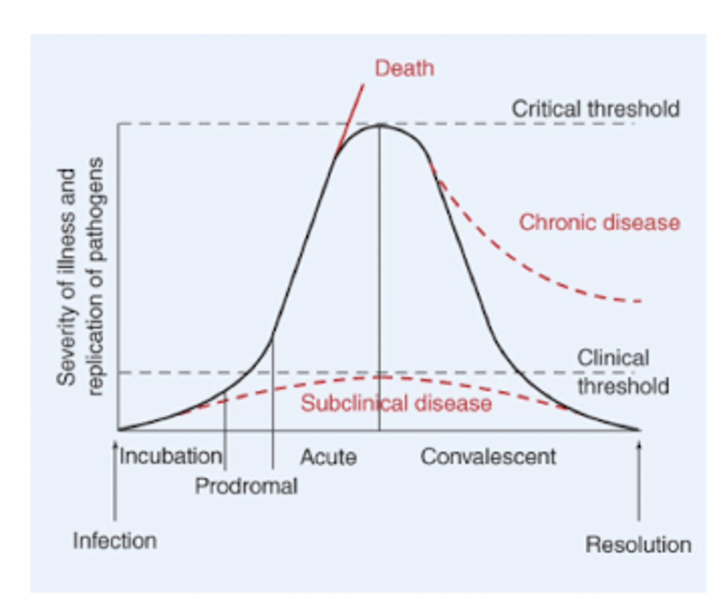
Explain the characteristics of the stages of the disease course
- incubation period: activate replication of a pathogen without recognizable symptoms
- prodromal stage: initial appearance of symptoms, mild fever, myalgia, headache, and fatigue (somewhat nonspecific)
- acute stage: maximum impact of the infectious process, inflammation and tissue damage (more specific)
- convalescent stage: progressive elimination of the pathogen
- resolution stage: total elimination of a pathogen
Distinguish the pathological functions of each class of virulence factors
- exotoxins: inactivate key cellular constituents and many are super antigens that induce excessive and nonspecific inflammatory responses
- endotoxins: can induce clotting, bleeding, inflammation, hypotension, and fever
- adhesion factors: bind to macromolecules on the surface of host cells, help to form potential mucous layer, and is critical for colonization of the pathogens
- evasive factors: inactivate host's immune system, some pathogens survive and reproduce within phagocytes after phagocytosis by neutralizing lysosomal contents with evasive factors
- invasive factors: facilitate the penetration of anatomical barriers
Commensalism, mutualism
- commensalism: the colonizing bacteria acquire nutrition, and the host gets neither benefit nor harm
- mutualism: both the microorganism and the host derive benefits from the interaction
Incidence, disease prevalence, endemic, epidemic, pandemic
- incidence: the number of new cases of an infectious disease that occur within a defined population over an established period of time
- disease prevalence: number of active cases at any given time in a population
- endemic: describes relatively stable incidence and prevalence in a particular geographic region
- epidemic: describes an abrupt and unexpected increase in the incidence of disease over endemic rates
- pandemic: refers to spread of disease beyond continental boundaries
Fomites, zoonoses, nosocomial infection, congenital infection
- fomites: inanimate objects contaminated with infected body fluids
- zoonoses: infectious diseases passed from other animal species to humans (rabies, HIV, plague flu)
- nosocomial infection: healthcare-associated infection
- congenital infection: infection of a child during gestation or birth from mother (vertical transmission)
Prodromal stage, acute stage, convalescent stage
- prodromal stage: initial appearance of symptoms (mild fever, myalgia, headache, and fatigue; somewhat nonspecific)
- acute stage: maximum impact of the infectious process (inflammation and tissue damage; more specific)
- convalescent stage: progressive elimination of the pathogen
Exotoxin, endotoxin, superantigen
- exotoxins: proteins released by pathogenic bacteria
- endotoxins: lipids and polysaccharides, not released
- superantigen: type of antigen that causes a massive and non-specific activation of T-cells in the immune system, leading to a potentially severe "cytokine storm"
Differentiate tissue regeneration and tissue repair
- tissue regeneration: replacement of injured tissue with cells of the same type and function
- tissue repair: occurs when extent or nature of damage cannot be reversed by regeneration alone
State the 4 stages of healing after tissue injury
1. hemostasis (minutes)
2. inflammation (hours)
3. proliferation (days)
4. remodeling (weeks-months)
Identify the determinants of regeneration versus repair after tissue injury
- nature of cells injured: some cells have margins renewal capacity
- extent of injury: the magnitude of injury may exceed regeneration capacity
- presence or absence of ongoing inflammation: due to concurrent infection or other factors, continued release of inflammatory mediators may disrupt balance toward repair
- underlying disease: may impair proliferative response or remodeling
State the common outcome of various signal transduction pathways of growth factors
Describe the two forms of ECM and their key components
- basement membrane: type IV collagen, laminin, and proteoglycan
- interstitial matrix: fibrillar collagens, elastin, proteoglycan and hyaluronan
Identify the three key growth factors that regulate fibrosis
- PDGF
- TGFB
- FGF-2
State the three key elements of shock
- life-threatening
- circulatory failure
- inadequate oxygenation
Name and describe the four main types of shock and their most common causes
- distributive shock: sepsis, anaphylaxis, neurogenic
- hypovolemic shock: hemorrhage, severe burns, severe vomitting, diarrhea
- cardiogenic shock: myocardial infractions, ventricular arrhythmia, cardiac myopathy, valvular disease
- obstructive shock: cardia tamponade, pulmonary embolism, pneumothorax
Identify the component of gram negative bacteria that mediates septic shock
lipopolysaccharide (LPS), also known as endotoxin
Describe the role of adrenomedullin in modulating vascular function
- enhances endothelial barrier
- induces VSMC relaxation
Define the term edema
accumulation of fluid in interstitial space
State the primary drivers of the movement of fluid into and out of vascular space
- hydrostatic pressure
- colloid osmotic pressure
Describe the mechanisms and common clinical causes of edema
- increased capillary permeability: cellulitis (local), sepsis, hypersensitivity reactions (systemic)
- increased capillary hydrostatic pressure: compartment syndrome, chronic venous insufficiency (local), heart failure, renal failure, pregnancy (systemic)
- decreased capillary oncotic pressure: protein deficient states; nephrotic syndrome, cirrhosis (systemic)
- lymphatic obstruction (lymphedema): tumor, trauma, infection (filariasis)
Explain the differences between monogenic and polygenic traits
- monogenic: single-gene traits; Mendel's law, only one pair of genes is involved (widow's peak, dimple, and a few more)
- polygenic: many genes are involved; traits have Gaussian distributions, the contribution of each gene to the traits may vary (height, skin, color, and many)
Describe how silent mutations, missense mutations, nonsense mutations, insertions and deletions impact protein function or expression
- silent mutation: no change in the amino acid sequence
- missense mutation: change in the amino acid sequence
- nonsense mutation: addition of a stop codon
- insertions/deletions: a multiple of three bases - insertions or deletions of one or more amino acids, not a multiple of three bases (frameshift)
Distinguish loss of function, gain of function, and dominant negative phenotypes
- complete loss of function: no expression, mutated proteins with no activity
- partial loss of function: reduced expression, mutated proteins with reduced activity
- gain of function: mutated proteins with abnormal activity (oncogenes)
- dominant negative: inhibition of the activity of the unmated protein expressed from the normal allele
Distinguish autosomal dominant, autosomal recessive, X-linked dominant, X-linked recessive, and mitochondria genetic disorders from inheritance patterns.
Autosomal Dominant: only one mutated allele for the disorder to manifest 50% chance of passing the trait to each child. Affects males and females equally. Unaffected relatives do not transmit the disorder.
Features: Often delayed onset. Example: Huntington disease, Marfan syndrome
Autosomal Recessive: Disorder appears only when both alleles are mutated.If both parents are carriers: 25% affected, 50% carriers, 25% normal. Affects males and females equally.
Features: early onset. Symptoms tend to be uniform. Example: Phenylketonuria, Tay-Sachs disease
X-Linked Dominant: single mutated allele on the X chromosome. Affected father: All daughters affected, sons normal. Affected mother: 50% chance for both sons and daughters.
Features: embryonic lethal for males. Example: Fragile X syndrome
X-Linked Recessive: Mutation on X chromosome; males affected, females usually carriers. Carrier mother: Sons: 50% affected, 50% normal. Daughters: 50% carriers, 50% normal. Affected father: Daughters: 100% carriers. Sons. 100% normal. Example: Hemophilia A, color blindness.
Mitochondrial Genetic Disorders: Inherited only from the mother. All children of an affected mother can inherit the mutation. Severity depends on ratio of normal to mutant mtDNA
Features: Affects organs with high energy demand. Example: Mitochondrial myopathies, encephalopathies.
Predict the chances of non-carrier, carrier, and affected children of genetic disorders based on the genotypes of the parents and the type of the disorders
- Autosomal Dominant
Genotypes: Aa × aa
Children:50% affected (Aa), 50% normal (aa)
No carriers.
- Autosomal Recessive
Genotypes: Aa × Aa
Children:25% normal (AA), 50% carrier (Aa), 25% affected (aa).
- X-Linked Recessive
Carrier mother (Xx) × Normal father (XY)
Sons: 50% affected, 50% normal
Daughters: 50% carriers, 50% normal.
- X-Linked Dominant
Affected father (xY) × Normal mother (XX)
Daughters: 100% affected
Sons: 100% normal.
- Mitochondrial Disorders
Affected mother: All children inherit mutation
Affected father: No children inherit mutation.
Explain the concepts of penetrance and expressivity
- penetrance: the percentage of individuals with a particular genotype who express the associated phenotype
- expressivity: the degree to which a genotype is expressed as a phenotype with an individual with the genotype
Explain the mechanisms that give rise abnormalities in the chromosome number
- nondisjunction during meiosis: gametes end up with an abnormal number of chromosomes; either 22 or 24 instead of 23
- fertilization of gametes with aneuploidy: the defects associated with monosomy of the autosomes are severe and often cause miscarriage in utero
- mosaicism: caused by nondisjunction occurring in the early stage of development
- prenatal testing: amniocentesis, prenatal cell-free DNA screening
Identify the genetic disorders occurred by abnormalities in the chromosome number
- down syndrome: trisomy 21
- turner syndrome: monosomy X (45, X/0)
- klinefelter syndrome: 47 XXXY or 48 XXXY
Explain how imprinting affects the inheritance patterns of genetic disorders
- imprinting introduces a parent-of-origin effect, making inheritance patterns more complex than standard Mendelian genetics
- the same mutation can cause disease only when inherited from a specific parent