1.1 Cell Structure
 Call Kai
 Learn
Learn Practice Test
 Spaced Repetition
 Match
Match Flashcards
 Knowt Play
1/58

There's no tags or description
Looks like no tags are added yet.
Name | Mastery | Learn | Test | Matching | Spaced | Call with Kai | Chat |
|---|
No analytics yet
Send a link to your students to track their progress
59 Terms

What is the definition of a eukaryotic cell?
Found in plants and animals
Contains nucleus

What is the definition of a prokaryotic cell?
Found in bacteria
No nucleus
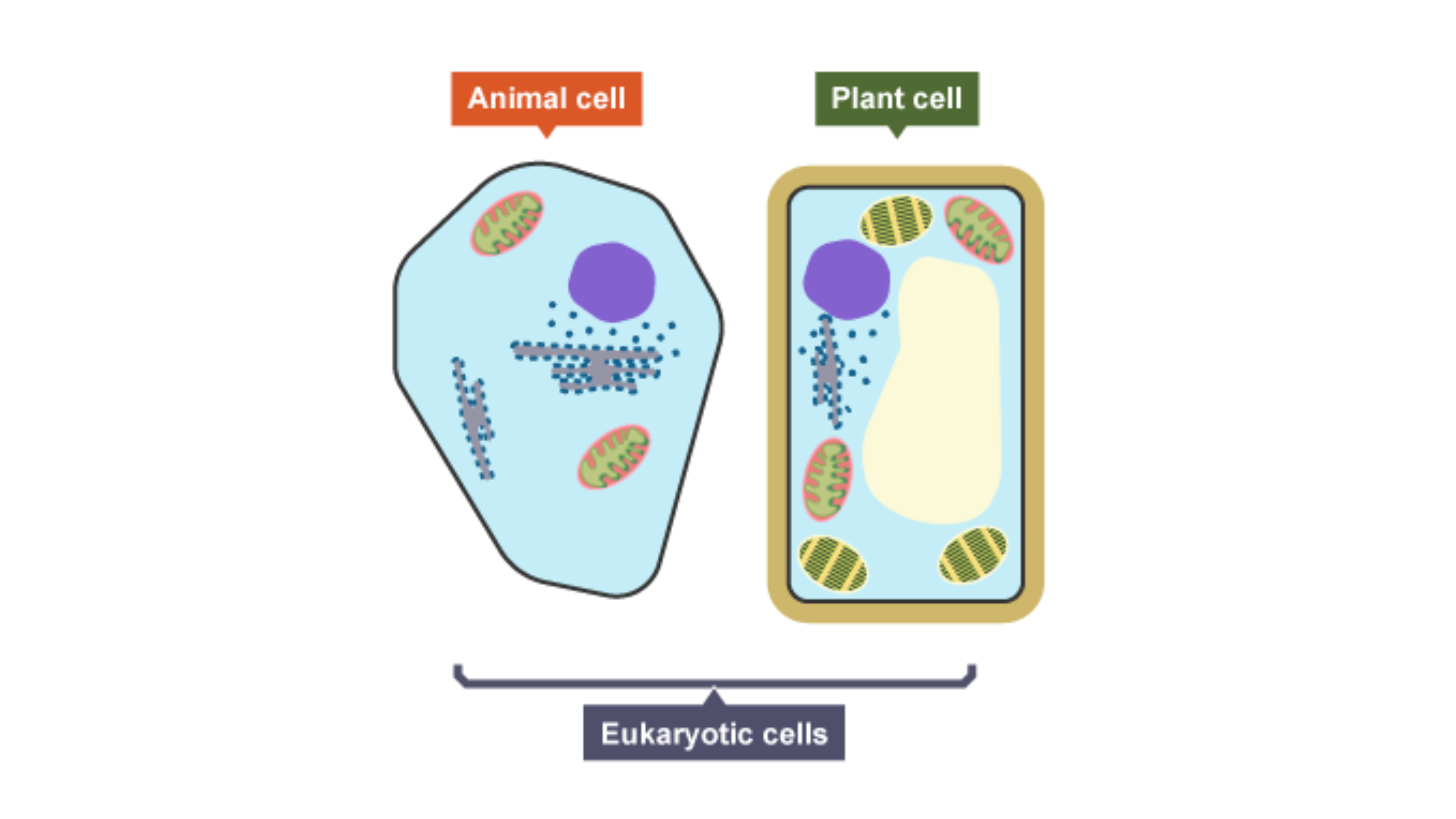
What do plant and animal cells (eukaryotic cells) have?
Cell membrane
Cytoplasm
Genetic material — enclosed in nucleus
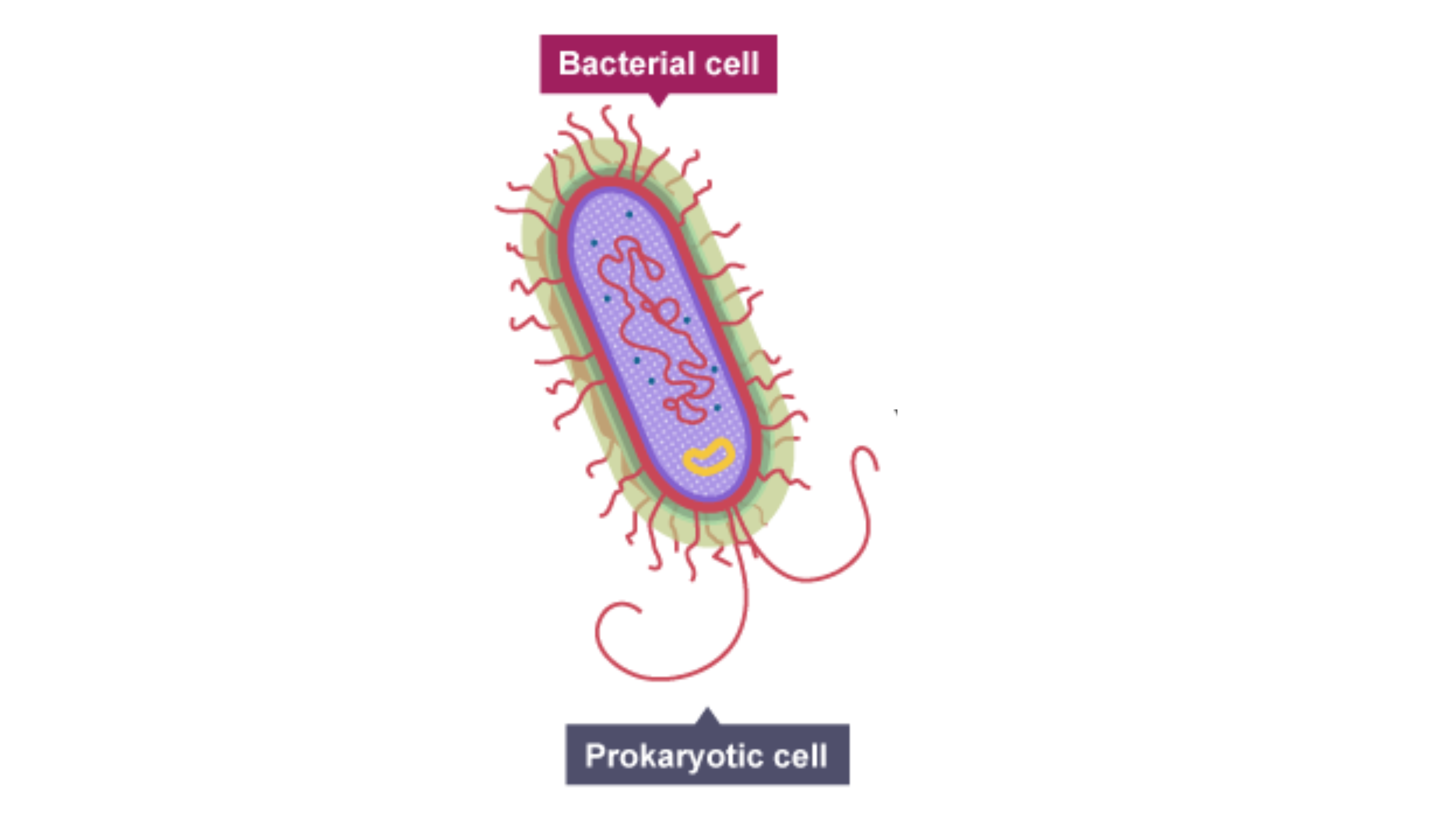
What do bacterial cells (prokaryotic cells) have?
Cytoplasm
Cell membrane
Surrounded by cell wall
Genetic material — not enclosed in nucleus
Single loop of DNA
Plasmids — small rings of DNA
Compare the size of eukaryotic cells and prokaryotic cells.
prokaryotic cells < eukaryotic cells
How much is a centimetre (cm) in metres?
1 × 10⁻² m
How much is a millimetre in metres?
1 × 10⁻³ m
How much is a micrometre in metres?
1 × 10⁻⁶ m
How much is a nanometre in metres?
1 × 10⁻⁹ m

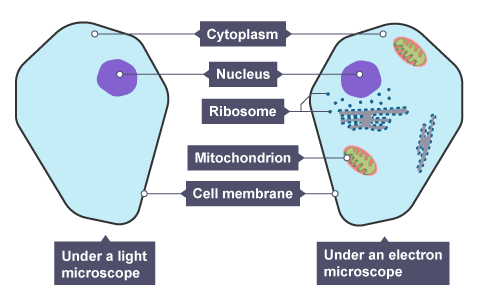
What parts do animal cells have?
Nucleus
Cytoplasm
Cell membrane
Mitochondria
Ribosomes

What parts do plant cells have?
Nucleus
Cytoplasm
Cell membrane
Mitochondria
Ribosomes
Chloroplasts
Cell wall — cellulose
Permanent vacuole — filled with cell sap
Plant and ________ cells have a cell wall made of ___________, which _____________ the cell.
algal
cellulose
strengthens

Label the animal cell.
A — Cytoplasm
B — Nucleus
C — Ribosomes
D — Mitochondria
E — Cell membrane

Label the plant cell.
A — Cytoplasm
B — Nucleus
C — Ribosomes
D — Vacuole
E — Mitochondria
F — Cell membrane
G — Cell wall
H — Chloroplasts
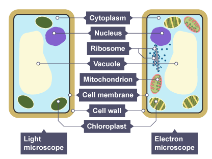
What is the function of the nucleus?
Controls activities of cell
Contains DNA
What is the function of the cell membrane?
Controls movement and transport of substances — in and out of cell
What is the function of the mitochondria?
Energy is released and transferred → aerobic respiration
What is the function of ribosomes?
Proteins are synthesised
What is the function of the cell wall?
Strengthens cell
Prevents bursting — too much water enters cell by osmosis
What is the function of chloroplasts?
Traps light → photosynthesis
What is the function of the permanent vacuole?
Contains cell sap — keeps cell turgid
What is the function of the cytoplasm?
Site of chemical reactions
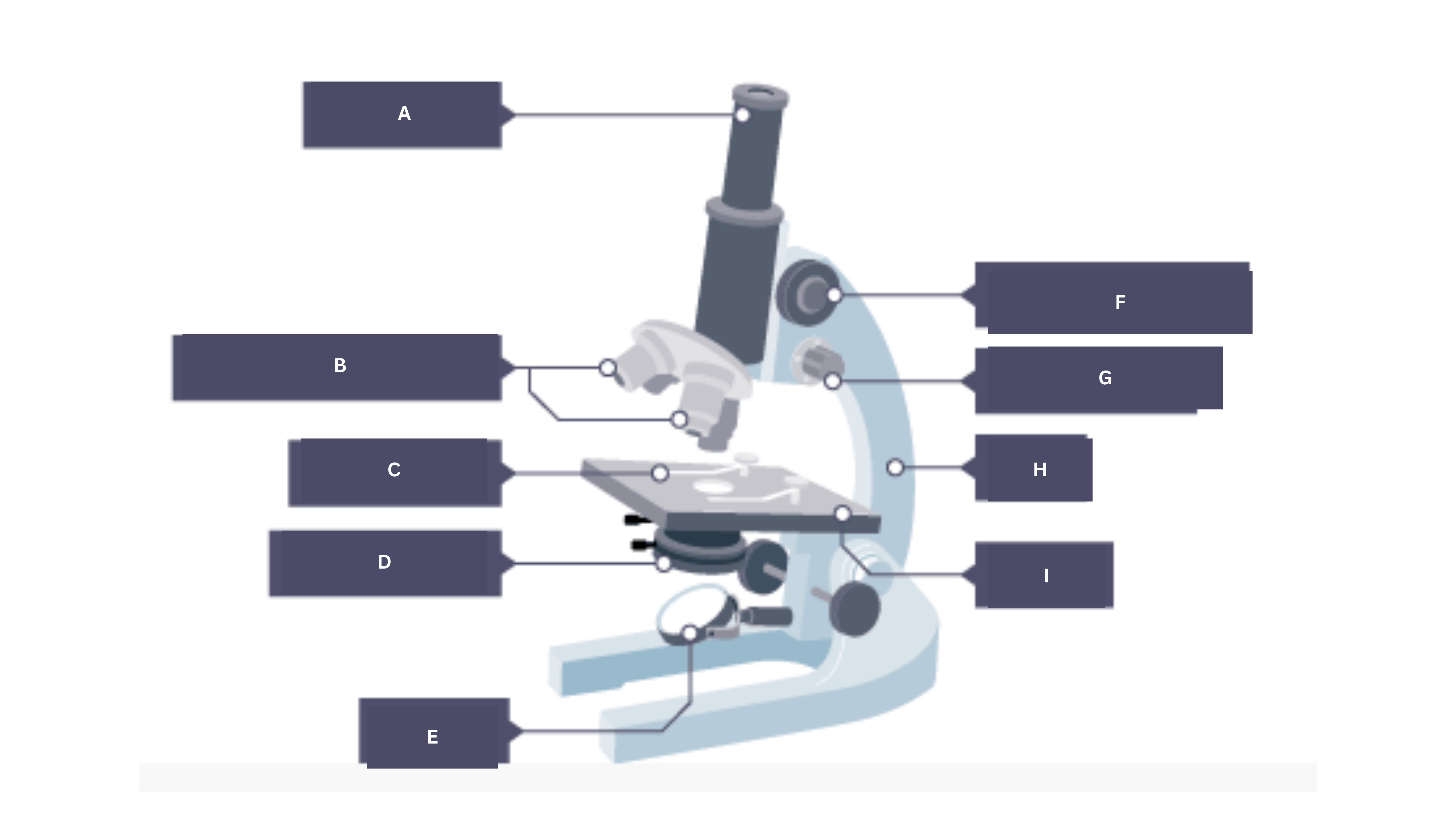
Label the microscope.
A — Eyepiece
B — Objective lenses
C — Stage clip
D — Condenser
E — Mirror
F — Coarse focus
G — Fine focus
H — Arm
I — Stage


Describe a method for Required Practical 1 — Microscopy.
Peel off thin epidermal layer on onion — forceps
Place sample flat onto drop of water on slide
Add few drops of iodine solution — stains cells
Slowly lower cover slip at an angle (mounting needle) — no air bubbles
Place slide on stage — select lowest power objective
Use coarse focus — brings cells into focus
Use fine focus — makes image clearer
![<p>A student prepared some animal cells to view using a microscope.</p><p><strong>Figure 2</strong> shows the student’s light microscope.</p><p>The student tried to look at the cells using the microscope.</p><p>Suggest one reason why the student could not see any cells when looking through part A. [1 mark]</p>](https://assets.knowt.com/user-attachments/9b67793c-76a0-4620-b395-5347a15a82e0.png)
A student prepared some animal cells to view using a microscope.
Figure 2 shows the student’s light microscope.
The student tried to look at the cells using the microscope.
Suggest one reason why the student could not see any cells when looking through part A. [1 mark]
No cells in the field of view
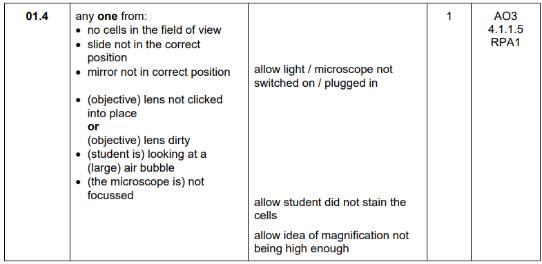
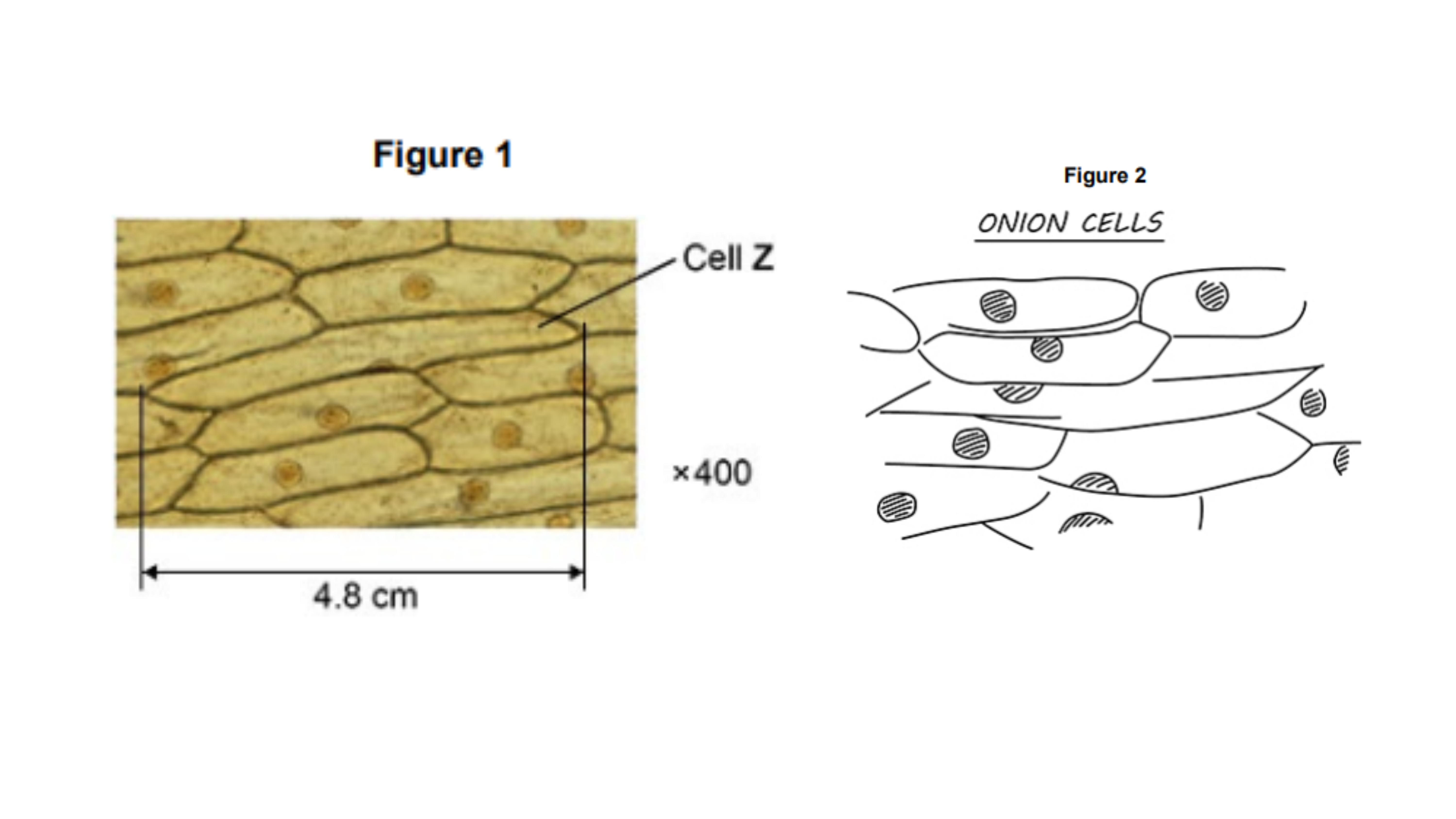
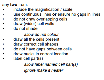
A student prepared some onion cells.
The student viewed the onion cells using a light microscope.
Figure 1 shows what the student saw under the microscope at a magnification of ×400.
Figure 2 shows the student’s drawing of Figure 1.
Give two ways the student could improve the drawing in Figure 2.
Include magnification
Use continuous lines
Why do you use a thin tissue layer in RP1?
Helps see individual cells clearly
Allows light to penetrate sample
Why do you use iodine in RP1?
Stains cells — makes sub-cellular structures visible
Why do you lower the cover slip at an angle in RP1?
Prevents air bubbles from getting trapped under slip
What are some hazards/risks in RP1 — how can they be minimised?
Iodine (irritant) — may cause rash/allergic reaction
Wear gloves
Wash hands after contact
Clean spills quickly
Sharp knives — can cut skin
Use chopping board
Cut away from body
Keep fingers clear of blade
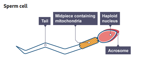
What are sperm cells specialised to do?
Carry male’s DNA to egg cell — reproduction

How does the structure of a sperm cell help it carry out its function?
Streamlined head and long tail — helps it swim to egg
Many mitochondria — site of respiration → energy for movement
Acrosome has digestive enzymes — breaks down outer layers of membrane of egg cell

What are nerve cells specialised to do?
Transmit electrical signals quickly — one place in body to another

How does the structure of a nerve cell help it carry out its function?
Long axon — impulses carried long distances
Dendrites — branched connections form with other nerve cells
Many mitochondria (nerve endings) — site of respiration → energy to make neurotransmitters
allows impulses to be passed from cell to cell

What are muscle cells specialised to do?
Contract and relax → causes movement in the body
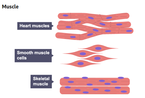
How does the structure of a muscle cell help it carry out its function?⁵
Proteins slide over each other → causes contraction
Many mitochondria — site of respiration → energy for contraction
Stores glycogen — used in respiration by mitochondria
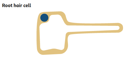
What are root hair cells specialised to do?
Take up:
water → osmosis
mineral ions → active transport — soil → cell

How does the structure of a root hair cell help it carry out its function?
Root hairs = large SA → more water moves in
Large permanent vacuole:
affects speed of movement of water — soil → cell
Mitochondria — site of respiration → energy for active transport

What are xylem cells specialised to do?
Transport water and mineral ions up plant — root → shoot

How does the structure of a xylem cell help it carry out its function?
Lignin = cell death — cells become hollow and join end-to-end → forms continuous tube
water and mineral ions move through
Lignin deposited in spirals — cells withstand pressure ← movement of water
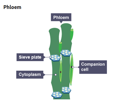
What are phloem cells specialised to do?
Carry products of photosynthesis to all parts of plant
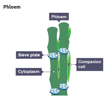
How does the structure of a phloem cell help it carry out its function?
Cell walls of each cell form sieve plates when broken down → movement of substances — cell → cell
Mitochondria in companion cells — energy for cells to stay alive
As an organism develops, cells _____________ to form different types of cells:
Most types of ___________ cell ______________ at an _______ stage.
Many types of ________ cells retain the ability to ______________ throughout life.
differentiate
animal
differentiate
early
plant
differentiate
What is cell division mainly restricted to in mature animals?
Repair
Replacement
What happens as a cell differentiates?
Acquires different sub-cellular structures — enables it to carry out specific function
→ Specialised cell
Why is cell differentiation important?
Allows production of different tissues and organs — perform vital functions
What is cell differentiation?
Process by which cells become specialised
An electron microscope has much higher _______________ and ___________ _________ than a light microscope.
This means that it can be used to study cells in much _________ _______.
magnification
resolving power
finer detail
What is the formula for magnification?
magnification = size of image ÷ size of real object
What process of cell division do bacteria multiply by?
Binary fission
Bacteria multiply by simple cell division (__________ _________) as often as once every ____ minutes if they have enough ___________ and a suitable _______________.
binary fission
20
nutrients
temperature
How can bacteria be grown?
Nutrient broth solution
Colonies on agar gel plate
What is required for investigating the action of disinfectants and antibiotics?
Uncontaminated cultures of microorganisms
You are preparing an uncontaminated culture using aseptic technique.
Explain why petri dishes and culture media must be sterilised before use.
Kills unwanted microorganisms already present — they could:
contaminate culture
compete with your bacteria for nutrients
You are preparing an uncontaminated culture using aseptic technique.
Explain why inoculating loops used to transfer microorganisms to the media must be sterilised by passing them through a flame.
Kills bacteria on loop — prevents transfer to agar
You are preparing an uncontaminated culture using aseptic technique.
Explain why the lid of the Petri dish should be secured with adhesive tape and stored upside down.
Loosely taping lid:
stops airborne microbes from entering — stills allows oxygen in → prevents growth of harmful anaerobic bacteria
Storing upside down:
stops condensation dripping onto colonies → would disrupt growth
You are preparing an uncontaminated culture using aseptic technique.
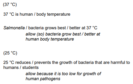
Explain why in school laboratories, cultures should generally be incubated at 25°C.
Reduces risk of growing human pathogens (37°C)
Required Practical 2 — Microbiology:
Describe a method for investigating the effect of antiseptics or antibiotics on bacterial growth using agar plates and measuring zones of inhibition.
Sterilise petri dish and agar → kills unwanted bacteria
Pass inoculating loop through flame → sterilises it
Use loop to spread bacteria onto agar
Open petri dish lid slightly → prevents microbes from air entering
Place filter paper discs soaked in different antiseptics onto agar — forceps
Seal lid with tape → prevents airborne microbes from entering
Incubate plate at 25°C → prevents growth of human pathogens
Measure zones of inhibition — ruler
compare effectiveness of each antiseptic
Explain why scientists use 37°C but students must use 25°C to incubate bacteria. [3 marks]
37°C is body temperature — bacteria grows better at 37°C
25°C prevents growth of bacteria that are harmful to humans