Normal Anatomy and Physiology of the Female Pelvis
 Call Kai
 Learn
Learn Practice Test
 Spaced Repetition
 Match
Match Flashcards
 Knowt Play
1/48

There's no tags or description
Looks like no tags are added yet.
Name | Mastery | Learn | Test | Matching | Spaced | Call with Kai | Chat |
|---|
No analytics yet
Send a link to your students to track their progress
49 Terms
Transabdominal Pelvic Ultrasonography
3.5 to 5 MHz transducer
wider field of view
requires distended urinary bladder
pushes uterus posteriorly
moves bowel out of pelvic cavity
Endovaginal Pelvic Ultrasonography
7.5 to 10 MHz
more detailed
limited field of view & depth of penetration
depends on age & sexual status
Bony Pelvis
composed of four bones:
two innominate (coxal) bones forming the anterior and lateral walls,
sacrum & coccyx forming the posterior wall.

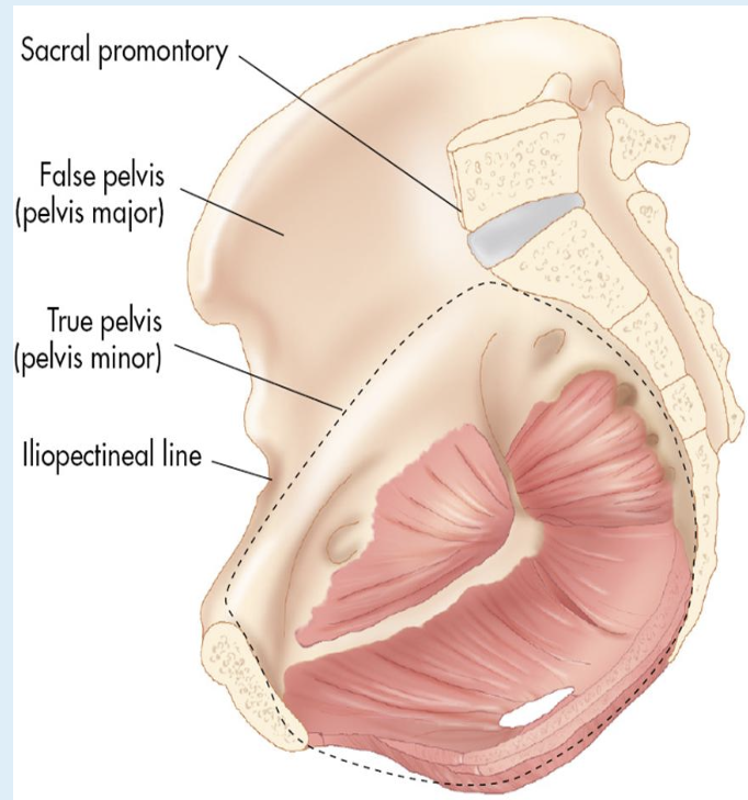
False Pelvis
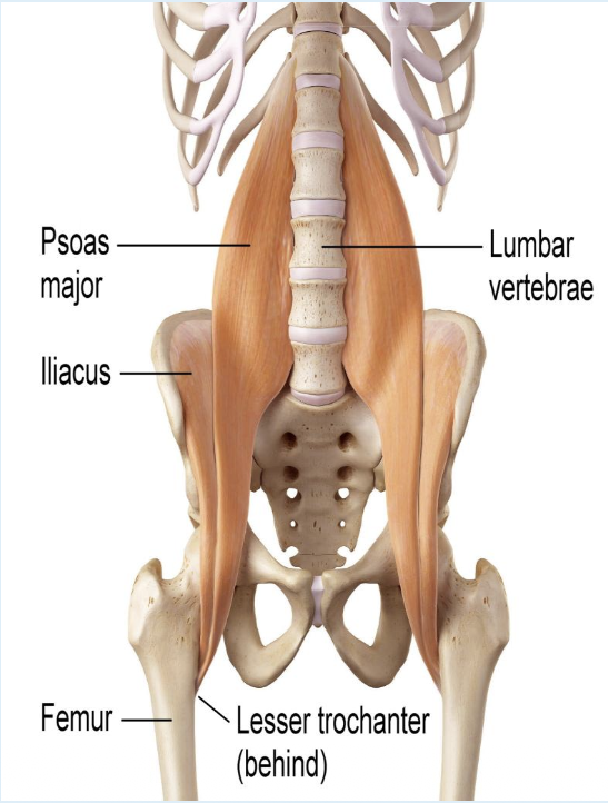
Muscles
psoas major
iliacus
create the iliopsoas muscle

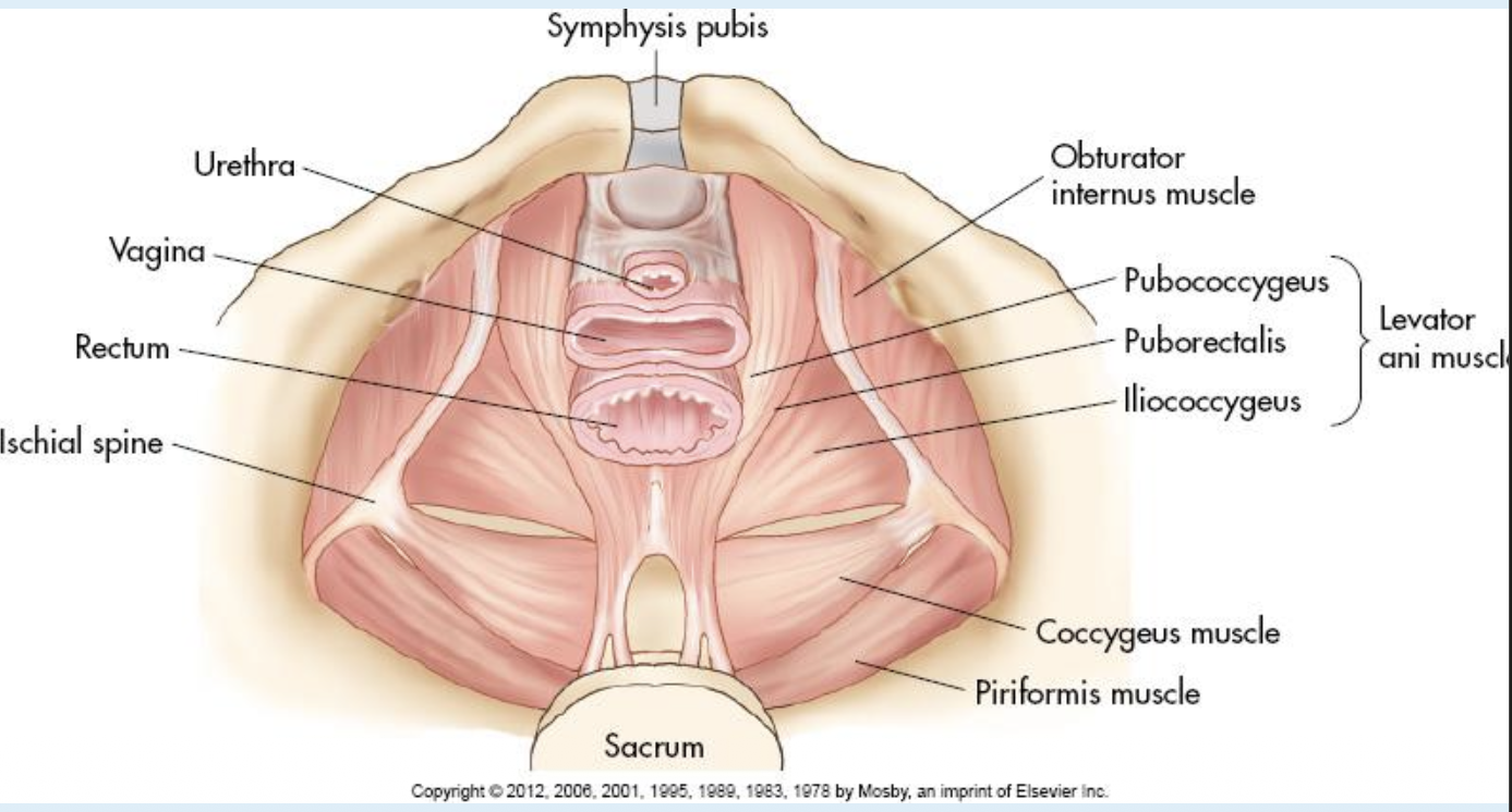
True Pelvis
piriformis muscles
obturator internus muscles
pelvic diaphram muscles
coccygeus muscles
levator ani (group of 3 closest to vaginal level)
pubococcygeus
iliococcygeus
puborectalis
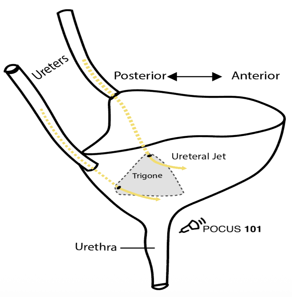
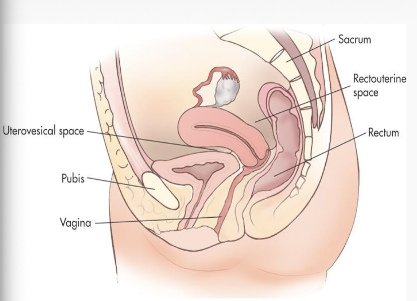
Bladder
apex = posterior to pubic bones
base = anterior to vagina
neck = rests on upper surface of urogenital diaphragm
Ureters
transport urine from kidneys to bladder
don’t typically see unless something is wrong w/ them
Vagina
collapsed muscular tube
approx. 9 cm in length
90-degree angle w/ the cervix
Cervix
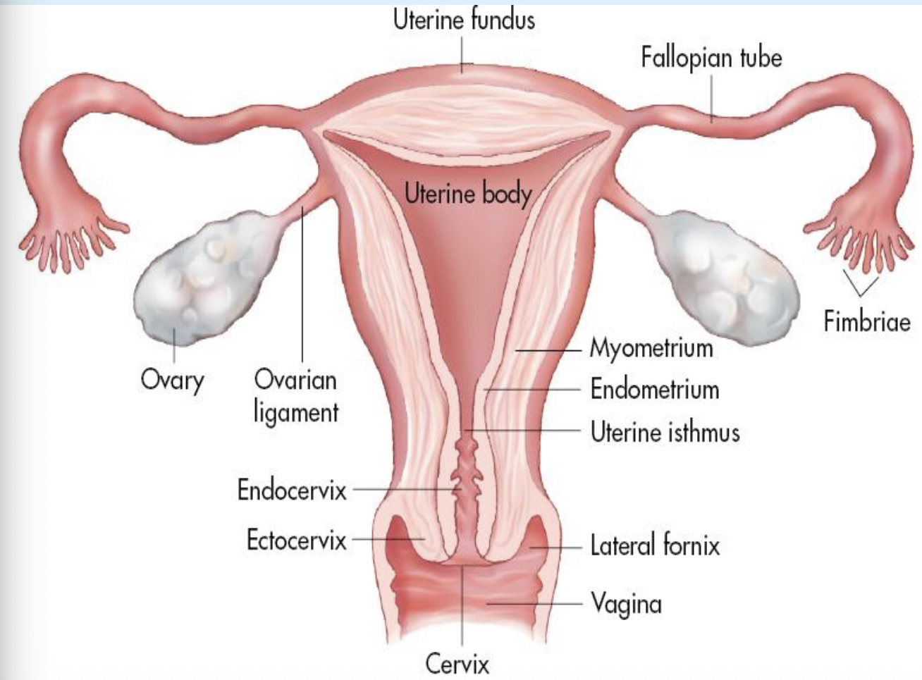
Endocervix
cervical canal
communicates w/ the uterine cavity
via internal os
vagina via the external os
Exocervix
outer portion of the cervix
protrudes into the upper vaginal canal
archlike recesses (fornices)

Uterus
hollow, pear shaped
Consists of:
fundus
widest (most superior)
body (corpus)
largest portion
Isthmus
point between body of uterus & cervix
bends anteriorly or posteriorly
Cervix
most inferior portion
Uterize Sizes
premenarchal
1.0 - 3.0 cm long by 0.5 - 1.0 cm wide
Menarchal
6.0 - 8.0 long by 3.0 - 5.0 cm wide
Multiparity
increases size by 1.0 - 2.0 cm
Postmenopausal
3.5 - 5.5 cm long by 2.0 - 3.0 cm wide
Perimetrium
serous outer layer of uterus (serosa)
Myometrium
thick middle layer of smooth muscle/connective tissue
bulk of the uterine wall
Endometrium
mucosal inner lining of the uterine body
Consists of:
superficial functional layer (zona functionalis)
sheds w/ menses
deep basal layer (zona basalis)
regenerates new endo. after menses
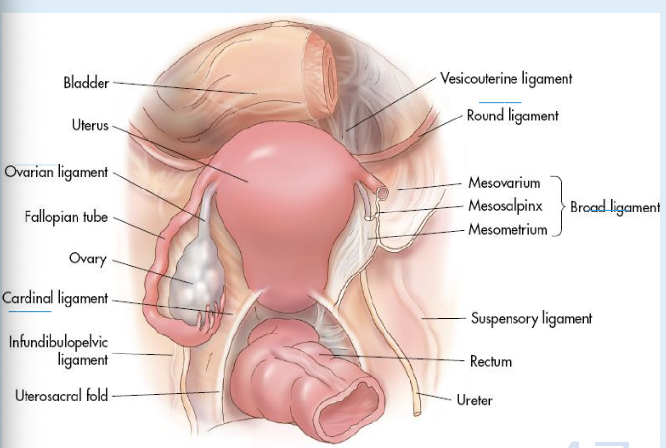
Broad Ligaments
Double folds of peritoneum
drapes over the fallopian tubes, uterus, and ovaries
lateral uterus to the pelvic sidewalls

Round Ligaments
Fibrous cords originating in front of and below the fallopian tubes
holding the uterine body in a forward position
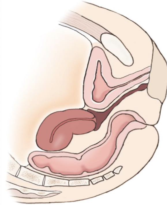
Anteverted Uterus
most common uterine position
fundus & body bent forward toward cervix

Anteflexed Uterus
body & fundus are curved forward on the cervix

Retroverted Uterus
entire uterus tilted posteriorly

Retroflexed Uterus
fundus & body bent backward toward the cervix
Uterine Prolapse
abnormal downward displacement of the uterus
caused by weakness in pelvic floor muscles & ligaments
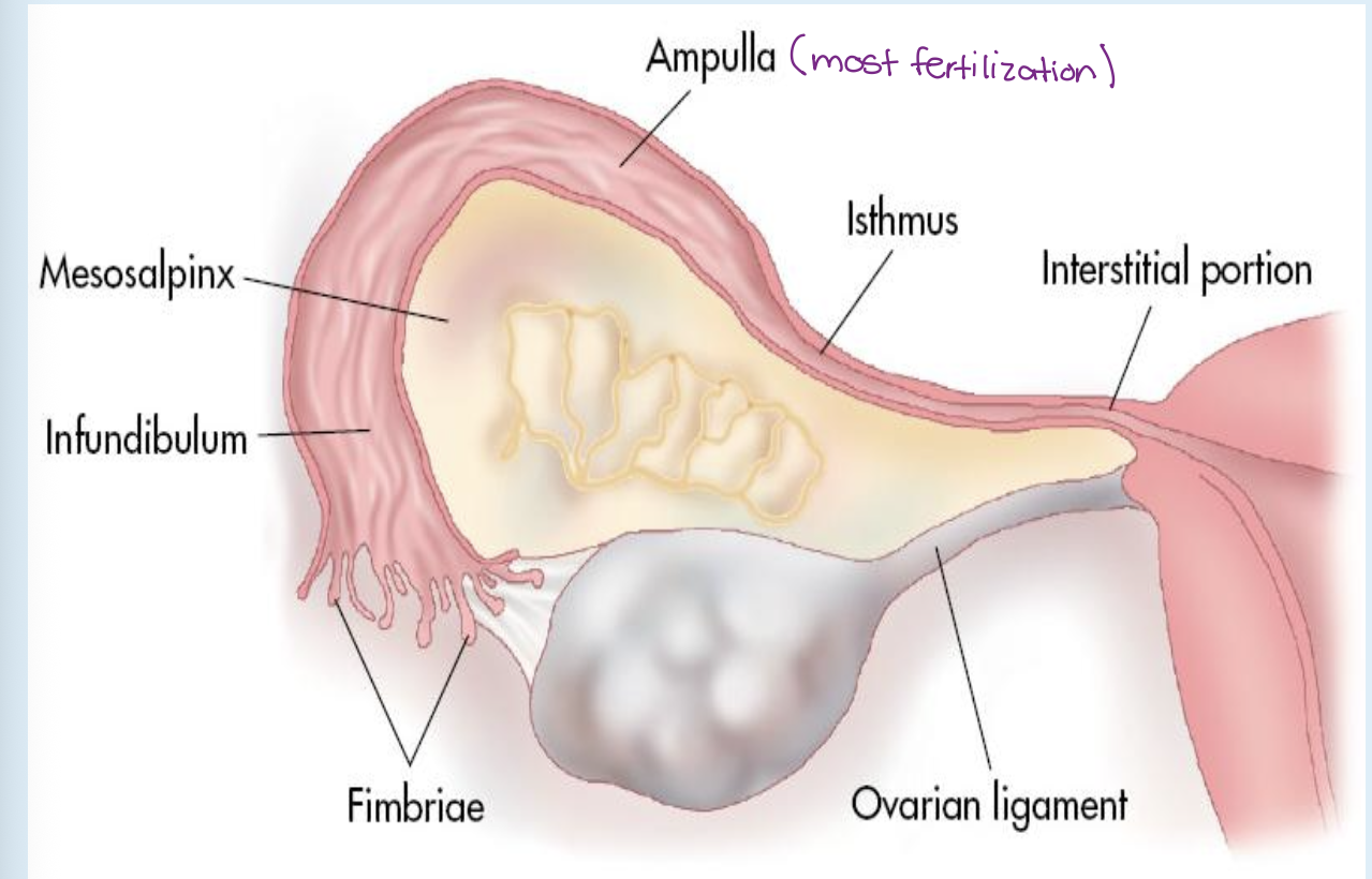
Fallopian tubes (oviducts)
coiled, muscular tubes
approx. 10-12 cm long & 1-4mm diameter
blood supply from ovarian arteries & veins
divided into 4 portions
infundibulum, ampulla, isthmus, interstitial portion
Infundibulum
lateral, widest portion
contains fimbriae
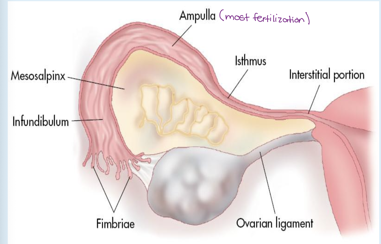
Ampulla
middle, longest, most coiled
most common site of ovum fertilization

Isthmus
located lateral to the uterus.
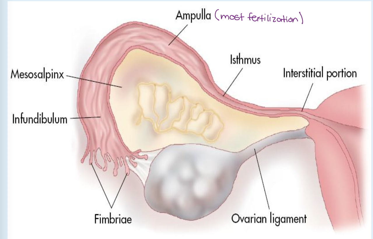
Interstitial Portion
narrowest portion
passes through uterine wall at the cornua
Ovary
almond-shaped organ
approx. 3 cm long in menarcheal women
responsible for ovum development
produces 2 hormones:
estrogen - secreted by follicles
progesterone - secreted by corpus luteum
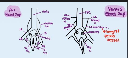
Dual blood supply
receives blood from ovarian artery & uterine artery
Blood drained
ovarian vein into IVC on right
renal vein on left

Blood Supply
Iliac arteries
anterior & medial to psoas muscles
bifurcate into
external iliac artery & internal iliac artery
Ovarian veins
left → drains into left renal vein
right → drains directly into IVC
Uterine Artery
Anastomose anteriorly & posteriorly within myometrium
form arcuate vessels that encircle uterus
Arcuate Arteries
Anechoic tubular structures in outer third of myometrium
Radial Arteries
arising from the arcuate vessels
penetrate deep into the myometrium to supply the base of the endometrium.
Spiral Arteries
Coiled terminal branches of radial arteries
supply functional layer of the endometrium
shed during menses
Polymenorrhea
irregular
less than 21 days
Oligomenorrhea
short & light menses
extending beyond 35 days
Amenorrhea
absence of menses
Dysmenorrhea
Painful menstruation
Premenarche
prepuberty (before onset of menses)
Menarche
regular menstrual cycles (approx. every 28 days)
Menopause
cessation of menses (typically occurring around age 50)
Ovulation
occurs at day 14 of a 28-day cycle
ovum released once a month by one of two ovaries
some will mature & be released while other degenerate
Follicular Phase
first phase of the ovarian cycle (days 1–14) driven by FSH
follicles grow/fill w/ fluid
secrete estrogen
select a dominant Graafian follicle
2 cm before ovulation
LH increases 24-36 hours before ovulation
Mittelschmerz = middle pain (dull ache on either side)
Luteal Phase
second phase (day 14 - 28)
FSH decreases due to LH surge
egg is either fertilized or degenerates
corpus luteum formation
progesterone secretion 9-11 days after
Menstrual Phase
1-5 days (1-5mm)
begins w/ declining progesteron levels
decreased blood flow → causes spiral arteries to shed
coincides with follicular phase of ovarian cycle (days 1-14)
Proliferative Phase
days 5-14
rising estrogen regenerates the functional layer
prepares endometrium for implantation
producing a characteristic 'three-line' (trilaminar) appearance
early = 4-8mm
late = 6-10mm
Secretory Phase
days 15–28
Corresponds with luteal phase of ovarian cycle
thick endometrium (7-14mm)
Menorrhagia
abnormally heavy or long periods (luteal phase affected)
Vesicouterine Pouch
anterior cul-de-sac
anterior to fundus between the bladder & uterus
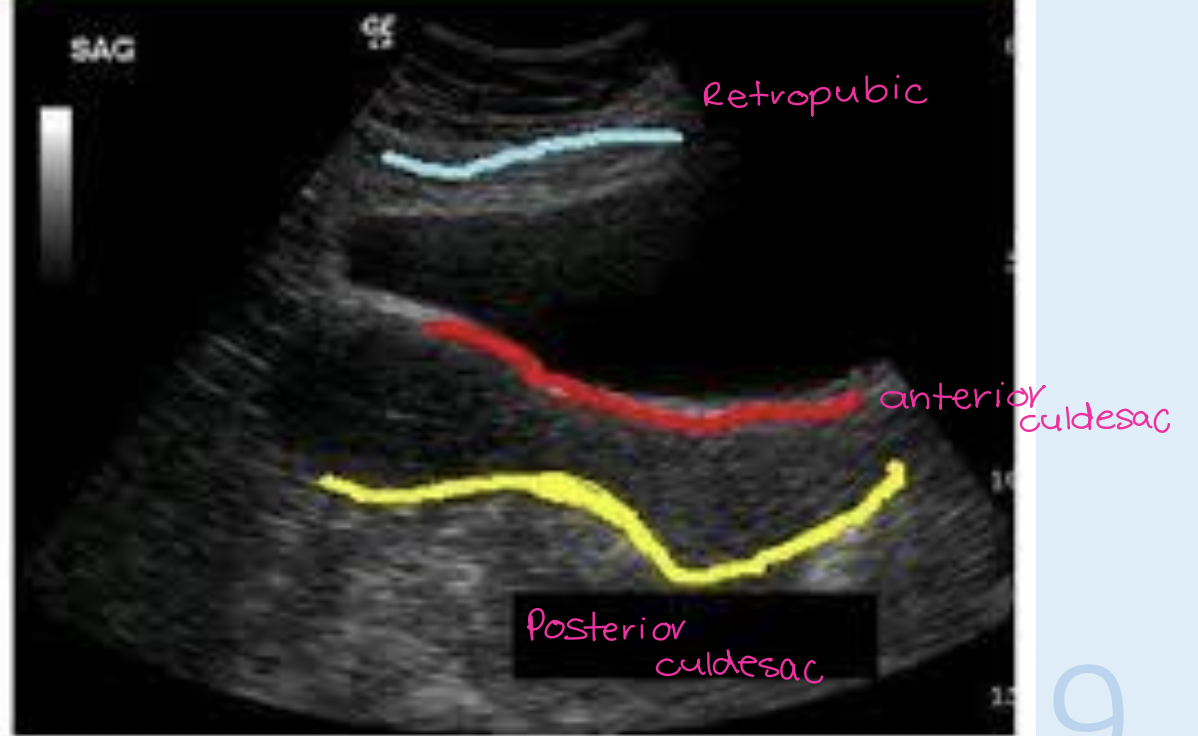
Rectouterine Pouch
posterior cul-de-sac or pouch of Douglas,
posterior to uterus between uterus & rectum
inferior & most posterior region of peritoneal cavity
most common for fluid

Retropubic
Space of Retziusan
anterior bladder wall & pubic symphysis
contain subcutaneous fat
