Cardiovascular/Blood vessels/Circulation
 Call Kai
 Learn
Learn Practice Test
 Spaced Repetition
 Match
Match Flashcards
 Knowt Play
1/77

There's no tags or description
Looks like no tags are added yet.
Name | Mastery | Learn | Test | Matching | Spaced | Call with Kai | Chat |
|---|
No analytics yet
Send a link to your students to track their progress
78 Terms
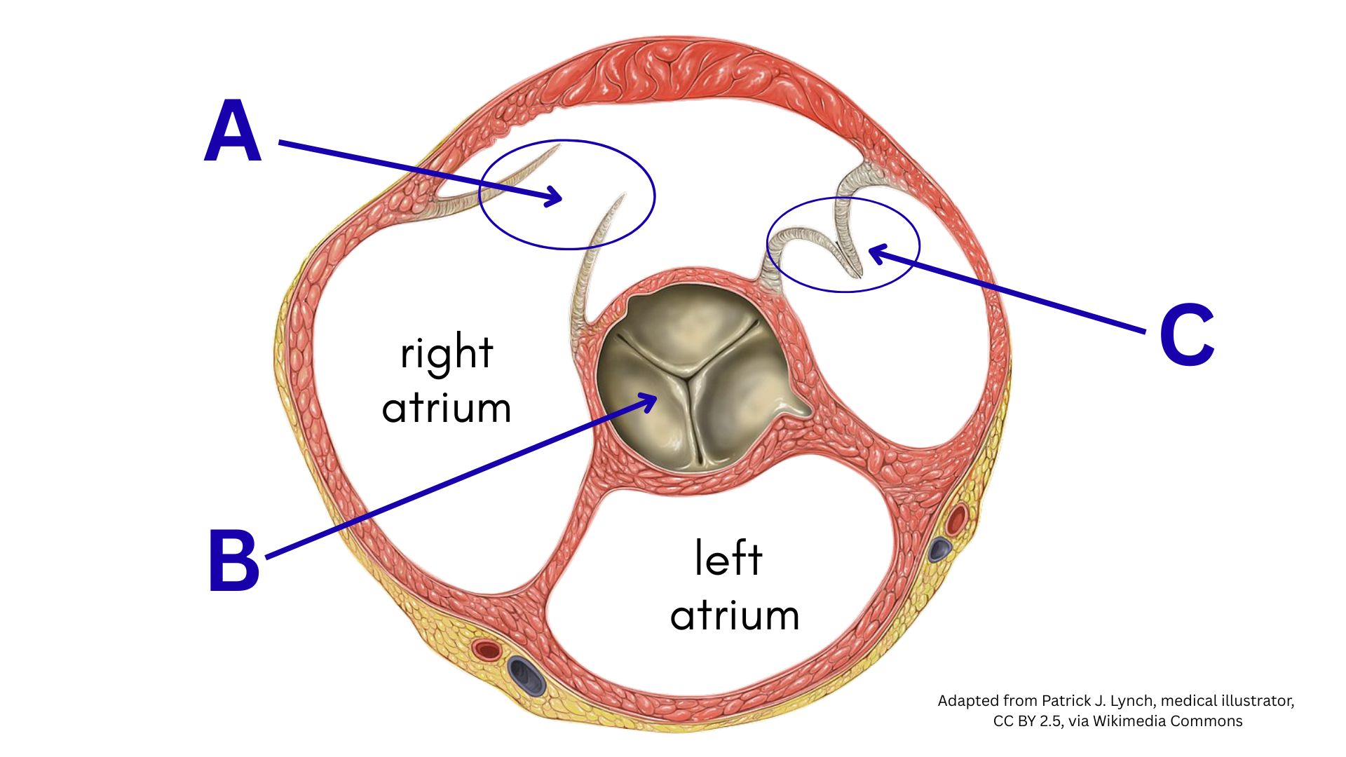
What is the structure labeled A on the heart model below?
tricuspid valve
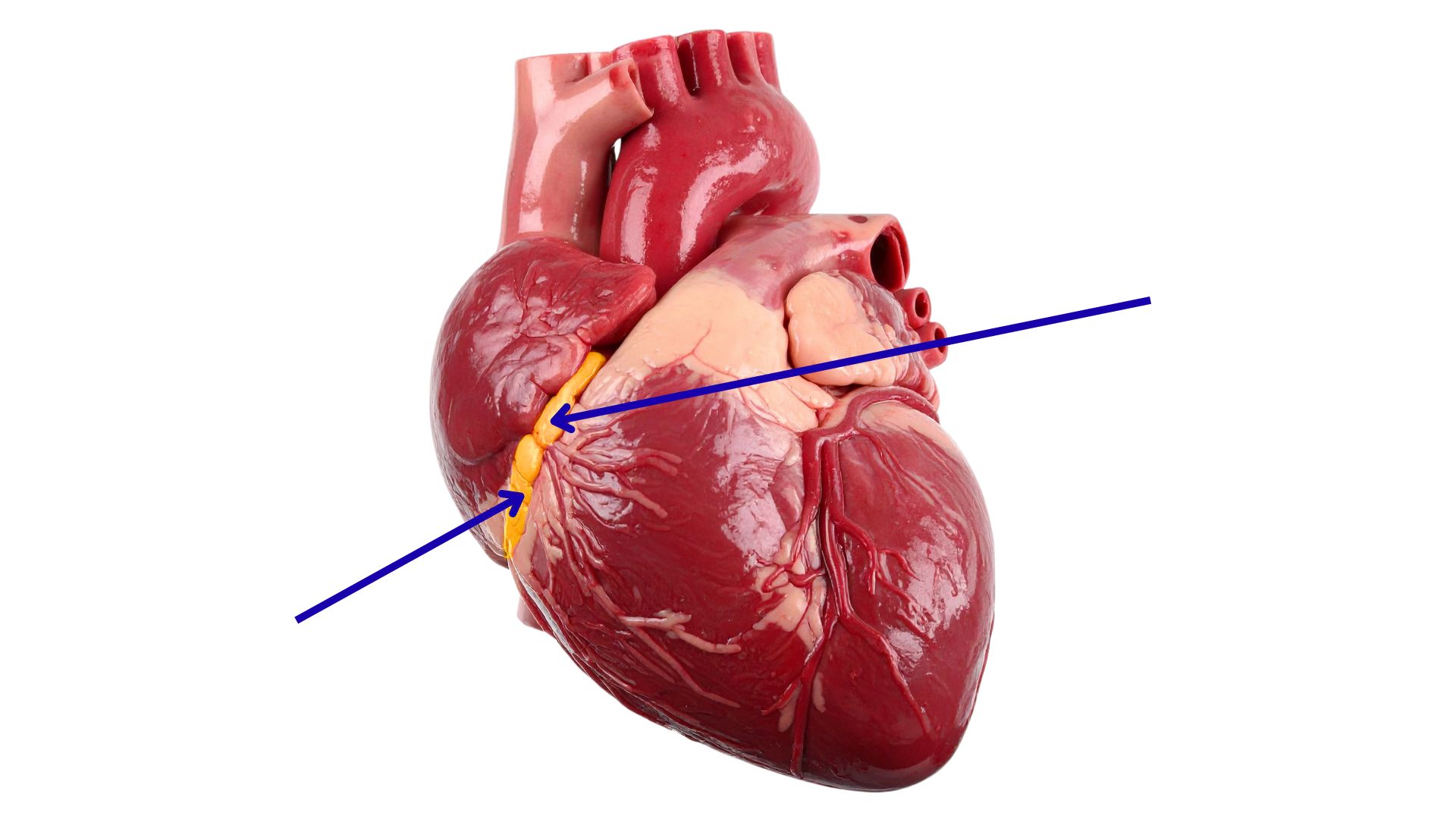
What is the highlighted structure on the heart model below?
Coronary sinus in coronary sulcus
Blood flows from the right atrium into the right ventricle through the [valve] valve.
Right AV valve
Tricuspid
Which pair of valves open during ventricular contraction?
pulmonary and aortic valves
The apex of the heart points where?
Downward and to the left
Which statement correctly describes the sulci on the surface of the heart
The posterior interventricular sulcus is located on the back of the heart.
The chordae tendineae and papillary muscles prevent what?
blood from flowing backward into the atria
Why is the left ventricular wall thicker than the right?
It must pump blood with greater force throughout the entire body.
![<p><span>What is the highlighted structure on the heart model below? [structure]</span></p>](https://assets.knowt.com/user-attachments/67379674-5e6e-49af-928a-f1f4cc1a32c9.jpg)
What is the highlighted structure on the heart model below? [structure]
Left ventricle
Blood returning from the lungs enters the [chamber].
Left atrium
The heart is located in the mediastinum, between which two organs?
Lungs
Which sequence shows the correct order of blood flow starting from the body?
Right atrium → Right ventricle → Lungs → Left atrium → Left ventricle → Body
When the ventricles contract, the [valves] close.
Atrioventricular (AV) valves
The left coronary artery mainly supplies [part].
the left atrium, left ventricle, and interventricular septum
The [vein] collects blood from the front of the heart and drains into the coronary sinus.
Great cardiac vein
What is the main function of the fibrous pericardium?
Anchors and protects the heart
![<p><span>What are the highlighted structures on the heart model below? [structure]</span></p>](https://assets.knowt.com/user-attachments/30285ccb-b895-469e-b098-ea9bb6dc8827.jpg)
What are the highlighted structures on the heart model below? [structure]
chordae tendineae
![<p>What is the highlighted structure on the heart model below? [structure]</p>](https://assets.knowt.com/user-attachments/ba541408-c6a6-4cac-b550-fe91684bd25f.jpg)
What is the highlighted structure on the heart model below? [structure]
Pulmonary trunk or artery
The [valve] prevents backflow of blood into the left ventricle.
Aortic semilunar valve
A patient has shortness of breath and blood backing up into the lungs. The echocardiogram shows mitral valve regurgitation. Which chamber is allowing blood to flow backward?
Left ventricle
What is the main function of the AV node in the conduction system?
delay the impulse so atria can finish contracting
Which heart sound corresponds to the closing of the atrioventricular valves?
S1 (“lub”)
During ventricular diastole, the atrioventricular valves open [when].
when atrial pressure exceeds ventricular pressure
During which phase does most ventricular filling occur passively?
ventricular diastole
What is the main reason inhalation amplifies right-sided heart murmurs?
it increases blood flow into the right heart
What happens to stroke volume (SV) when heart rate becomes very high?
SV decreases because diastole is shortened
Arrange these conduction system components from fastest to slowest intrinsic firing rate: SA node, AV node, Bundle of His, Purkinje fibers.
SA node → AV node → Bundle of His → Purkinje fibers
What percentage of ventricular filling is contributed by atrial contraction (“atrial kick”)?
20 to 30%
Which part of the nervous system slows the heart rate at rest?
parasympathetic ardioinhibitory
[ion] is essential for the plateau phase of a cardiac contractile cell action potential.
Calcium
Which property allows the heart to generate its own electrical impulses without nervous system input?
Autorhythmicity
Which ECG wave represents ventricular depolarization?
QRS complex
How does high calcium influence the heart?
Makes the heart beat stronger
During isovolumic contraction, [valves] are closed.
All valves
In the conduction system, the [what] acts as the natural pacemaker of the heart.
SA node
Which of the following conditions would likely decrease heart rate?
Rising blood pressure
![<p><span>What is the highlighted structure on the heart model below? [structure]</span></p>](https://assets.knowt.com/user-attachments/b7000bb2-742b-4ab3-bc9c-ba5b079ee862.jpg)
What is the highlighted structure on the heart model below? [structure]
Aortic arch
Aorta
Which valve does NOT have chordae tendineae or papillary muscles?
Pulmonary semilunar valve
Aortic valve
Which pair of valves open during ventricular contraction?
Pulmonary and aortic valves
The space between the parietal and visceral layers of the pericardium is called what?
Pericardial cavity
Which statement correctly describes the sulci on the surface of the heart?
The posterior interventricular sulcus is located on the back of the heart.
A 50-year-old patient develops cardiac tamponade after chest trauma. Which finding would the nurse expect?
Distended neck veins
Which heart wall layer is made mostly of cardiac muscle tissue?
Myocardium
Blood supply to the brain comes mainly from the
Internal carotid
Vertebral arteries
Baroreceptors detect changes in
Blood pressure
The ___ veins join to form the inferior vena cava.
Common iliac veins
The brachial vein is formed by the union of the ___
Radial vein and ulnar vein
When does local tissue autoregulation cause vasodilation?
Oxygen is low or carbon dioxide is high
The Circle of Willis ensures that there is [what].
constant blood supply to the brain
RAAS (Renin-Angiotensin-Aldosterone System) increases blood pressure by [what].
retaining salt and water and constricting vessels
The external iliac artery becomes the [artery] after passing under the inguinal ligament.
femoral artery
The radial artery is cut in a wrist laceration. Which structure is directly affected?
Lateral forearm and hand
A patient has a deep vein thrombosis (DVT) in the femoral vein. What is the immediate concern?
Blood pooling in leg, risk of clot moving to lungs
About 3 to 4 liters of fluid per day enter lymphatic capillaries. Why?
to prevent edema and maintain blood volume
During surgery, the internal jugular vein is clamped. What is a likely immediate effect?
Impaired blood drainage from brain and deep face
A patient has a compression fracture at L4, affecting the common iliac veins. What is an immediate downstream consequence?
Blood pooling in pelvis and lower limbs
A patient is standing still for a long time and feels faint. Which factor is most responsible?
Skeletal muscle pump not active
Arterioles are called resistance vessels because they [what].
Slow down blood flow and regulate blood pressure
Injury to the hepatic veins will most directly impair:
Blood leaving the liver to the IVC
A compression injury occurs to the popliteal artery behind the knee. Which area will suffer reduced blood flow?
Foot and lower leg
A nurse notes that the patient’s large veins in the legs appear flattened. Why?
Lower pressure and thin walls
A patient’s renal artery is blocked. Which organ will be immediately affected?
Kidney
Which type of blood vessel carries blood away from the heart?
Arteries
A patient is losing a lot of blood. Which blood vessels act as a reserve to help maintain circulation?
Veins
A compression injury occurs to the popliteal artery behind the knee. Which area will suffer reduced blood flow?
Foot and lower leg
A superficial laceration of the cephalic vein on the arm will cause
Minor superficial bleeding, little oxygen deprivation
ADH helps maintain blood pressure by
keeping water in body and constricting vessels
A patient has very thick blood due to high red blood cell count. What will probably happen?
Slow blood flow and increase resistance
A patient has external iliac artery blockage. Which body part is most affected?
Anterior thigh and lower limb
Which pressure pushes fluid out of capillaries into tissues?
Capillary hydrostatic pressure
During surgery, the internal jugular vein is clamped. What is a likely immediate effect?
Impaired blood drainage from brain and deep face
During exercise, which tissues receive more blood?
Muscles, heart, and lungs
A patient has low blood volume due to dehydration. Which effect is expected?
Blood pressure falls
The ___ supplies the stomach, spleen, and liver.
Celiac trunk
Which of the following veins is superficial and has no matching artery?
Great saphenous vein
Epinephrine and norepinephrine affect the cardiovascular system by [what].
Raising heart rate, constricting vessels
What does the azygos system do?
Drains thoracic wall and empties into SVC
The pulmonary trunk splits into the
right and left pulmonary arteries