Interactive echocardiography
 Call Kai
 Learn
Learn Practice Test
 Spaced Repetition
 Match
Match Flashcards
 Knowt Play
1/49

There's no tags or description
Looks like no tags are added yet.
Name | Mastery | Learn | Test | Matching | Spaced | Call with Kai | Chat |
|---|
No analytics yet
Send a link to your students to track their progress
50 Terms
What are the different views on an ultrasound?
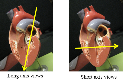
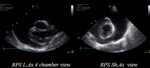
Describe the view shown below?
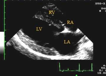
Through the right chest wall
Right para sternal long axis 4 chamber view
Most cardiac problems are evident on this view
When would the end of diastole be on the ECG?
Start of QRS complex on ECG
When would the end of systole be on an ultrasound and an ECG?
Smallest LV dimension / end of T wave
How do you achieve RPS short axis views?
Rotate the ultrasound probe (thumb would go from sternal to the caudle part of the animal)
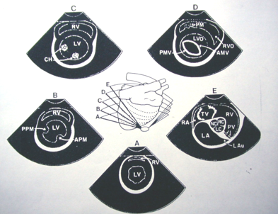
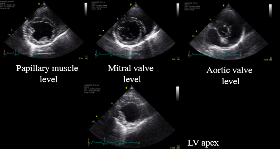
How do we measure the heart on an ultrasound?
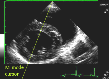
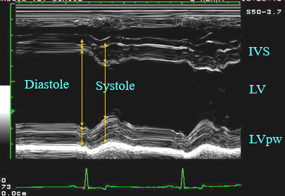
What is fractional shortening?
Simplest, most commonly used measurement of systolic function (pump function) - only looking at radial movement
Normal > 25%(breed dependent)
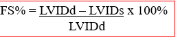
When is fractional shortening not accurate to assess systolic function?
Significant mitral regurgitation
Wall motion abnormalities
Right sided heart disease associated with pressure overload
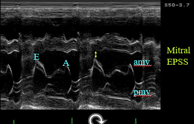
How does the mitral valve pressent in M-mode?
Mitral E point to septal separation (EPSS) normal <7 mm (any dog breed / size)
INCREASED EPSS in DCM / myocardial failure
How do you assess the RPSLA view on an ultrasound?
Chamber sizes relative to each other (right should be 1/3 of left)
Wall thickness relative to chambers (wall in left should be 1/3 of lumen)
Systolic function
What is abnormal here?
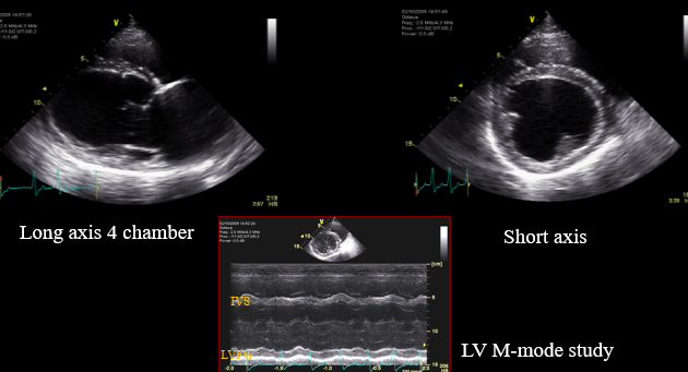
The left ventricle looks dilated, rounded, thin walls, not pumping very well (hypokinetic) (seen on m-node)
Dilated cardiomyopathy
it was also not pumping very well
Describe the m-node in this canine DCM case
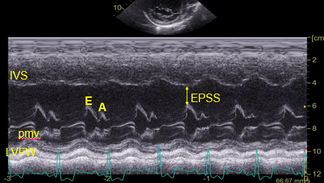
Great distance from the E point and the septum
How do you calculate the ejection fraction and what is normal?
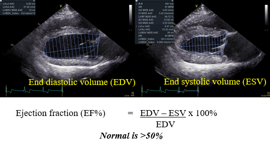
(Simpson's method of discs)
What is the index of sphericity?
Attempt to quantify subjective assessment of rounding of LV
Normal > 1.7

What is abnormal here?
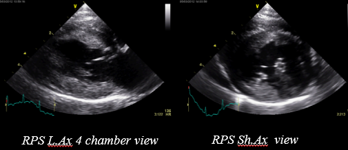
Thick walls and left atrium big (rounded)
Concentric hypertrophy of the left ventricle —> systemic hypertension?
Hypertrophic cardiomyopathy —> pumps well but L ventricle has trouble filling
L sided congestive HF
What else could cause concentric hypertrophy of the left ventricle?
Aortic stenosis
Systemic hypertension (measure blood pressure to rule out)
What is abnormal in this image?
Pericardial effusion
May show right sided congestive heart failure (due to this being a lower pressure system)
Pericadial pressure is higher than R atrial pressure —> collapses during diastole —> cardiac tamponade (drain pericardial effusion)
What is another type of ultrasound?
Spectral Doppler (velocity time graph)
Pulsed wave (PW) & Continuous wave (CW)
Colour flow Doppler
Tissue Doppler (for myocardial motion)
What are the different effects of the doppler on the RBCs?
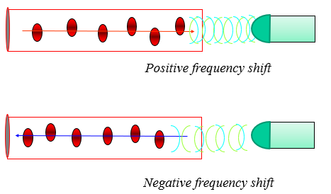
Can work out the velocity of blood flow if you are parallel to it
Velocity of RBCs (estimate) is proportional to frequency shift between reflected and transmitted ultrasound
Must be as parallel to flow as poss for accuracy
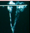
What would the doppler show if you put it at the apex vs base of the heart?
Apex:
Blood flowing away from the probe (will look like below with laminar flow)
Pulse wave doppler
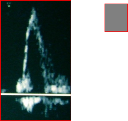
Base:
blood flowing towards probe
What are the features of a pulse wave doppler?
spatially specific, just sample RBC velocity within the sample volume
limit to the depth you can sample and limit to the peak velocity you can record
What are the features of a continuous wave doppler?
samples all along the cursor line – peak velocities displayed (not spatially specific)
Can record at depth and high peak velocities
How will turbulent vs laminar flow look on a doppler?
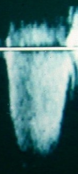
Turbulent:
Whirls of RBCs in multiple directions, multiple velocities
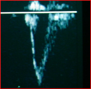
Laminar:
Sheets (laminae) of RBCs in similar directions
What is the colour flow doppler?
Colours associated with movement
BART map – blue away, red towards (transducer)
The faster it is the brighter
What is the velocity limit in the colour flow doppler known as?
Nyquist limit - if it is faster than the limit it shows aliasing
What view is being shown here?
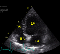
Left apical 4 chamber view
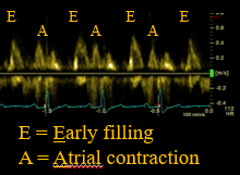
What would the m-node look like on a left apical 4 chamber view?
What view is being shown here and what does the colour represent?
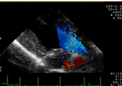
Left apical 3 chamber view
Blue: away from transducer represents aortic outflow
What does the green indicate on this doppler?
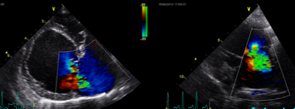
Turbulent flow
This patient had mitral valve regurgitation
What are the abnormalities here?
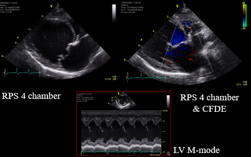
The mitral valve shows nodular thickening (also mitral prolapse into the atria)
Atrial septum pushed away form midline and atria rounded
Severe mitral regurgitation shown in the video with green
What is this m-node depicting?
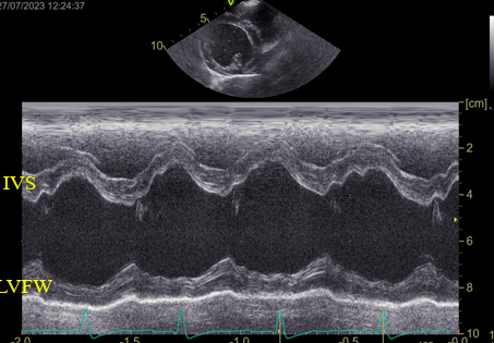
Hyperkinetic left ventricle
Reduced afterload on LV due to severe MR
What is being shown here?
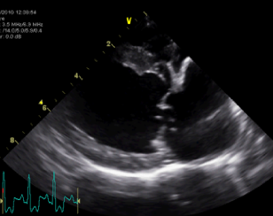
Flail MV due to rupture of Chordae Tendinae (watch video)
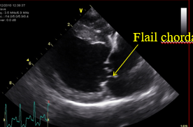
What would you see with mitral vegetative endocarditis?
Lesion on the valve that moves independently of the valve (will not always have a murmur)
Animal will be pyrexic
What measurements can you do at short axis views at level of aorta?
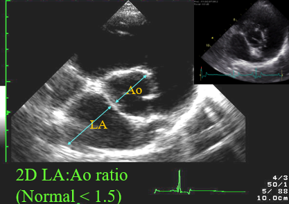
What is being shown in this LPS cranial view for LAA?
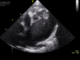
Thrombus in the top right
The 'smoke' below this tells you the blood is no longer acting like fluid (swirls around in the video)
What view is being shown here?
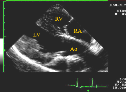
Right parasternal long axis five chamber view
What is being shown here?
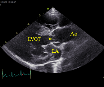
Fibrous muscular lesion where the star is which narrows before the valve
Sub-aortic stenosis
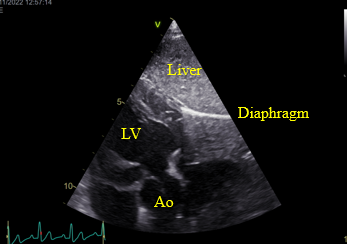
Why is the sub-costal view useful?
Optimal alignment with aortic outflow
Probe sits beneath the sternum (will have to press hard)
Will see the liver and the diaphragm
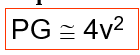
How can we identify the severity of the aortic stenosis?
Pressure gradient (PG) between LV and Ao
Modified Bernouilli Equation (higher worse the pressure gradient)
Would use the doppler to get the velocity
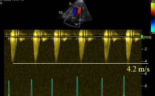
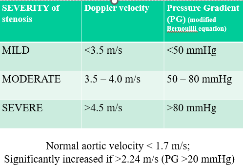
How do we grade the Severity of Aortic and Pulmonic Stenosis?
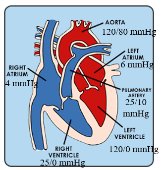
What are the normal pressures in the chambers and the great vessels of the heart?
What is abnormal in this Right parasternal long axis 4 chamber view?
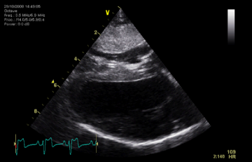
Myocardial infiltrate? (would occur if there are thick walls) - ddx
There is a right ventricular concentric hypertrophy (left ventricle is underfilled so the walls also look thick)
The velocity below is 7.2 on the PA spectral doppler, using the Bernouilli equation what is the RV-PA pressure gradient?
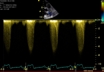
This is the case with right ventricular concentric hypertrophy
207mmHg - SEVERE stenosis
How would we calculate RV pressure if the patient has a normal PA pressure in systole?
Add PG to PA
What is being shown in this m-mode?
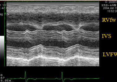
Paradoxical septal motion due to severe pulmonic stenosis
What is abnormal here? (the top is right)
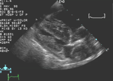
Pressure overload of the right ventricle (dilated) and abnormal structure in the right atrium
Thrombus within the pulmonary artery - bottom right (pulmonary hypertension)
Left ventricle looks underfilled (left ventricular hypertrophy)
Pericardial effusion
Dirofilariasis
What are the results of dirofilariasis?
Pulmonary hypertension and pulmonary thromboembolism causes pressure overload on RV
Right sided CHF results (also pericardial effusion)
How do you deal with dirofilariasis?
Extract worms via jugular vein
What is being shown here?
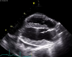
Pleural effusion - on the left you can see the mediastinum
What is being shown here?
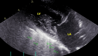
B-lines with large left atrium
Indicative of cardiogenic pulmonary oedema