Medical Terminology Unit 2 Part 1
1/43
There's no tags or description
Looks like no tags are added yet.
Name | Mastery | Learn | Test | Matching | Spaced | Call with Kai |
|---|
No analytics yet
Send a link to your students to track their progress
44 Terms
Pulmonology
Is the medical specialty that studies the anatomy and physiology of the respiratory system.
Pulmonologists use diagnostic tests, medical and surgical procedures, and drugs to treat respiratory diseases.
pulmon/o-: means lung
-logy: means study of
Nasal Cavity
The nose is the first part of the upper respiratory tract
It contains the nasal cavity, which is divided by the septum
nas/o = nose
-al = pertaining to
Three turbinates in the nasal cavity slow the flow of air so it can be warmed and moistened.
Nasal cavity and turbinates are lined with mucosa.
The mucous membrane humidifies air and produces mucus.
This is especially important in cold or dry areas.
Mucus and hair in the nose trap particles and prevent them from reaching the lungs.
Pharynx
The posterior part of the nasal cavity merges with the pharynx
The pharynx is a common passage for inhaled air, exhaled air, and food.
The pharynx is composed of:
- Nasopharynx
-Oropharynx
- Laryngopharynx
Larynx
The larynx (voice box) is the first part of the lower respiratory tract.
The larynx remains open during speech and respiration to allow air into the trachea.
During swallowing, the larynx pulls up to the epiglottis, forcing food into the esophagus.
Trachea
Below the vocal cords, the larynx merges into the trachea.
The trachea aka windpipe, is a passageway for inhaled and exhaled air.
It has a column of C-shaped cartilage.
-The anterior side is rigid due to the cartilage to prevent collapse.
- The posterior side has no cartilage and is soft.
The Bronchi
The trachea divides into right and left bronchi (plural of bronchus).
Bronchi are supported by rings of cartilage, similar to the trachea.
The primary (right and left) bronchi enter the lungs, where they divide into smaller bronchioles.
Bronchioles
Bronchioles are the smallest bronchi with a diameter of 1 millimeter or less.
Their walls contain smooth muscle and no cartilage.
The smooth muscle can contract or relax, to narrow or widen the bronchial lumen.
Anatomy of the Lungs
The lungs are spongy, air-filled structures.
The right lung contains three lobes (divisions) and the left lung contains two.
-Right upper lobe (RUL)
-Right middle lobe (RML)
-Right lower lobe (RLL)
-Left upper lobe (LUL)
-Left lower lobe (LLL)
The top of the lung is the apex.
The base rests on the diaphragm
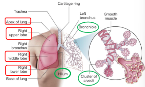
Alveoli
-Exchange oxygen and carbon dioxide with nearby capillaries.
- Air sacs that are functional units of the lung.
-They secrete surfactant that keeps their walls from collapsing.
-The alveoli form the pulmonary parenchyma.
Thorax
The thorax is the bony area between the neck and the diaphragm.
-The rib cage (sternum, ribs, and spine) protects the lungs and thoracic cavity.
-The lungs fill most of the thoracic cavity.
-The mediastinum lies between the lungs and contains the trachea, heart, and esophagus.
-The diaphragm makes up the inferior border of the thoracic cavity.
Pleural cavity
Within the thoracic cavity, each lung lies in a pleural cavity.
This space is surrounded by a double-layered serous membrane called pleura.
The pleura secretes pleural fluid that allows the layers to smoothly slide past each other.
Respiration
A two-way path: Inhalation/inspiration and Exhalation/expiration
Eupnea
Having a normal rate and depth of respiration is Eupnea
Inhalation
Diaphragm contracts and moves downward, intercostals pull ribs up and out creating negative pressure, causing air to flow into the lungs.
Exhalation
Relaxation of diaphragm and intercostals, air flows out.
Respiration involves five processes:
Ventilation is the movement of air in and out of the lungs.
External respiration is the exchange of oxygen and carbon dioxide between the blood and the lungs at the alveoli.
Gas transport is the movement of oxygen and carbon dioxide by the blood.
Internal respiration is the movement of oxygen and carbon dioxide from blood into cells.
Cellular respiration is the use of oxygen by the cells to produce energy and waste (carbon dioxide) through metabolism.
Cardiology
The medical specialty that studies the anatomy and physiology of the cardiovascular system.
Cardiologists use diagnostic tests, medical and surgical procedures, and drugs to treat cardiovascular diseases.
- cardi/o- = heart
- -logy = study of
Anatomy of the heart
The heart is located in the thoracic cavity, behind the sternum and between the lungs.
It is roughly the size of a fist and contrasts at least once per second.
Mounds
Mounds reflect the locations of the heart’s internal chambers. (external surface of heart
Grooves
Grooves contain fat, blood vessels, and nerves. (external surface of heart)
Pericardium
The pericardium is a two-layered membrane that forms the U-shaped pericardial sac.
Outer layer is the parietal pericardium.
Inner layer is the epicardium.
Pericardial fluid fills the space between the layers.
Myocardium
The muscular myocardium lies beneath the epicardium.
Endocardium
The endocardium is the innermost layer that lines the heart’s chambers and valves.
4 chambers of heart
The heart has four chambers: right atrium, right ventricle, left atrium, and left ventricle.
The interior of the heart contains four chambers: two on top and two on bottom.
Each small upper chamber is an atrium.
Each large lower chamber is a ventricle.
All four chambers are filled with blood, lined with endocardium, and surrounded by myocardium.
Septum
The septum is the central wall that divides into the right and left sides.
Four valves control blood flow through the heart.
Two valves are on the right side:
The tricuspid valve has three cusps and lies between the right atrium and right ventricle.
The pulmonary valve has three cusps and lies between the right ventricle and the pulmonary arteries.
Four valves control blood flow through the heart.
Two valves are on the left side:
The mitral valve has two cusps and is located between the left atrium and left ventricle.
The aortic valve has three cusps and is located between the left ventricle and the aorta.
Chordae tendineae
The tricuspid and mitral valves both have chordae tendineae.
These ropelike strands extend from each valve’s leaflets to small muscles in the ventricular walls.
Chordae tendineae help keep the leaflets closed during ventricular contraction.
Mediastinum
The mediastinum is an irregularly shaped central area between the lungs that holds the heart and parts of the great vessels.
The thoracic cavity contains the lungs and mediastinum.
Other structures in the mediastinum include the thymus, trachea, and esophagus.
Blood vessels
All blood vessels:
Have a central opening or lumen.
Are lined with a smooth layer of endothelium, also called the intima.
3 kinds of blood vessels:
Arteries
Capillaries
Veins
Arteries
Carry blood away from the heart.
Are large blood vessels that branch into smaller vessels called arterioles.
Arteries and arterioles share some important characteristics:
- All carry blood away from the heart.
-Most carry bright red oxygen-rich blood.
-Most lie deep beneath the skin.
-All have smooth muscle in their walls that allows for vasoconstriction and vasodilation.
-Vasoconstriction and vasodilation of the arteries are important ways in which the body regulates blood pressure.
Capillaries
Connect arteries and veins
Smallest blood vessels.
Their lumen is so narrow that blood cells must pass through in single file.
Capillaries lie between arterioles and venules and deliver blood to each cell in the body.
Veins
Carry blood towards the heart
From capillaries, blood moves into venules.
Multiple venules converge to form veins.
Veins and venules share some important characteristics:
-All carry blood to the heart.
-Many are near the surface of the body and can be seen.
-The largest veins have valves that ensure one-way blood flow.
Aorta
Largest artery in the body.
It receives oxygenated blood from the left ventricle and carries it to the systemic circulation
Coronary arteries
First vessels to branch off the aorta.
These small arteries provide the heart muscle with the oxygen necessary for contraction.
Aortic arch
The aortic arch then gives rise to other arteries that serve the rest of the body.
Two major veins
The two major veins of the body are the superior and inferior venae cavae:
-Both carry deoxygenated blood to the right atrium of the heart.
The inferior vena cava serves the abdomen, pelvis, and legs.
The superior vena cava serves the head, neck, arms, and chest.
Systemic circulation
Systemic circulation includes the vessels everywhere in the body except in the lungs.
Pulmonary circulation
Pulmonary circulation includes the vessels going to, within, and coming from the lungs.
Sinoatrial (SA) node
Located in the upper right atrium, serves as the heart’s pacemaker.
It initiates the electrical impulse that begins each heartbeat with the contraction of the two atria.
Systole and diastole
A normal heartbeat has two phases:
-Systole is the period when the heart contracts.
-Diastole is the resting period between contractions.
Normal Sinus Rhythm (NSR)
When the SA node controls heart rate, the heart is in normal sinus rhythm (NSR).
Ectopic sites
When other areas (ectopic sites) produce impulses, an abnormal rhythm may result