OSPE Pathology (medlibrary)
1/68
There's no tags or description
Looks like no tags are added yet.
Name | Mastery | Learn | Test | Matching | Spaced | Call with Kai | Chat |
|---|
No analytics yet
Send a link to your students to track their progress
69 Terms
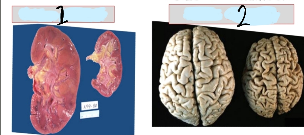
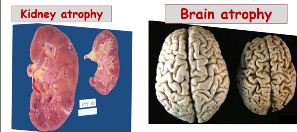
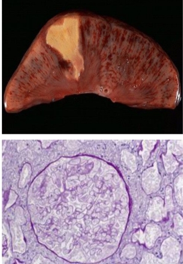
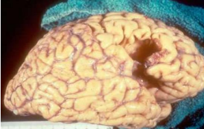
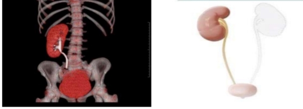
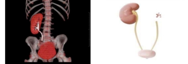
What is depicted in this image and what is the definition of this condition?
1- kidney atrophy
2-brain atrophy
Atrophy: decrease in size of mature organ due to decrease in cell size (and ,or) number
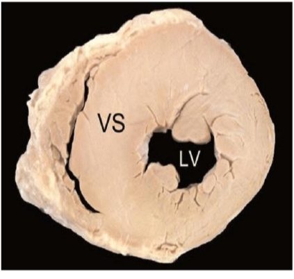
What happened?and what is the reason?
And is it reversible or irreversible ?
left ventricular hypertrophy due to hypertension
all adaptation are reversible
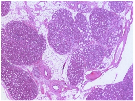
Identify then define
breast hyperplasia
Hyperplasia: increase in size of mature organ due to increase in cell number
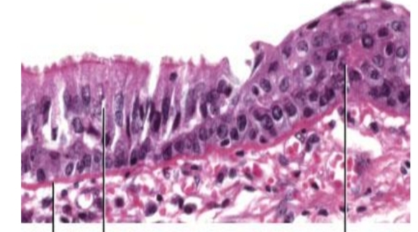
Identify then define
squamous metaplasia of respiratory epithelium in smokers
Metaplasia:change of one type of tissue to another type of same category.
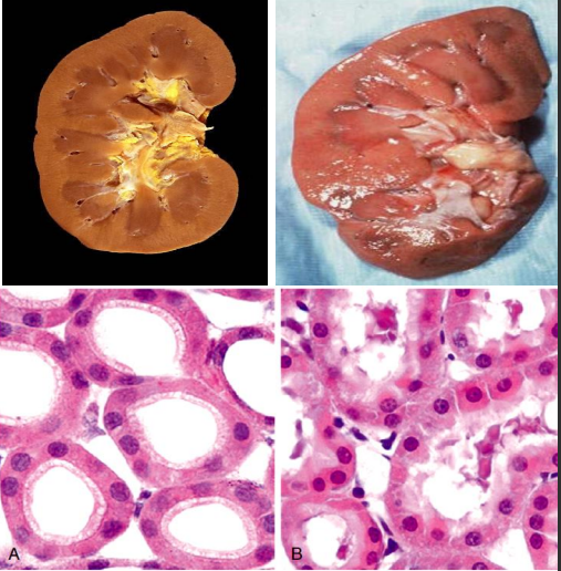
What type of injury and specific condition is depicted? What is seen microscopically?
Reversible injury, Cloudy Swelling of kidney. Microscopically, the proximal convoluted tubules are most affected.
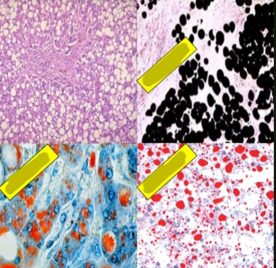
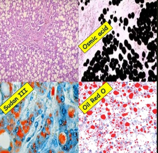
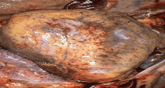
What condition is shown and how does it appear with H&E stain and other special stains”mention thier names”?
Fatty liver.
H&E stain shows a clear vacuole with a signet ring appearance.
Fresh and frozen sections stained with Osmic acid appear black, Sudan III appears orange, and Oil Red O appears red.
What type of necrosis is this and where is it commonly seen?
Coagulative necrosis of kidney.
ده شكل ال kidney
محدش يغلط و يكتب liver 🙂
This is the commonest type of necrosis, seen in acute ischemia of any organ except the brain (heart, kidney & spleen).
identify ? other examples ?
liquefactive necrosis of the brain
pyogenic abscess
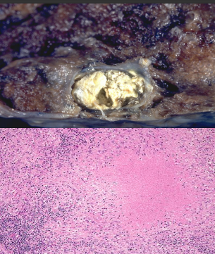
identify ?
examples ?
caseation necrosis
examples are (TB,Syphilis )
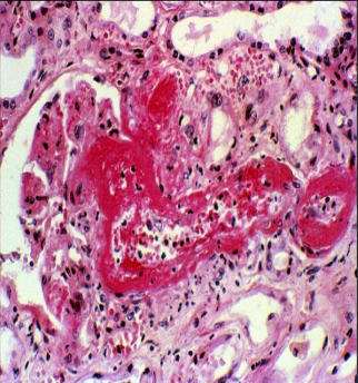
identify ?
occurs when ?
fibrinoid necrosis
Occurs in vasculitis and hypertension
identify ?
What is the reason and looks like what?
mucoid carcinoma due to mucin accumulation
Looks like mucin lakes
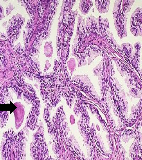
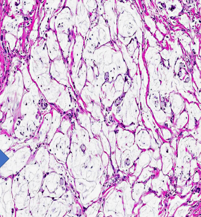
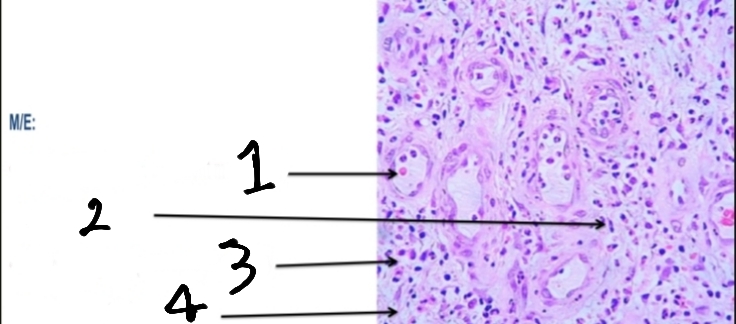
What is the term for this accumulation shown in the image?and arrow point to what?
Hyalinosis
Arrow point to Corpora amylacia in senile prostatic hyperplasia
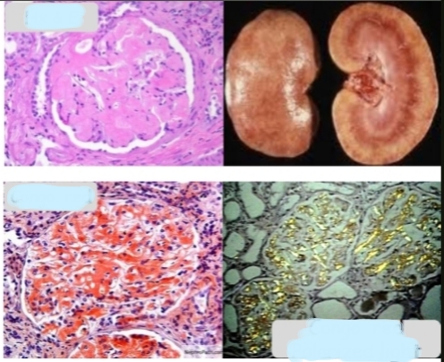
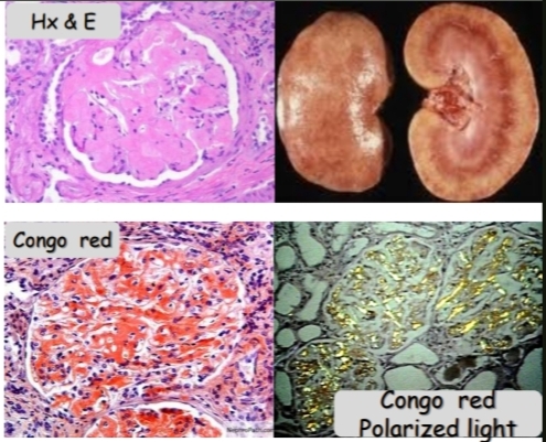
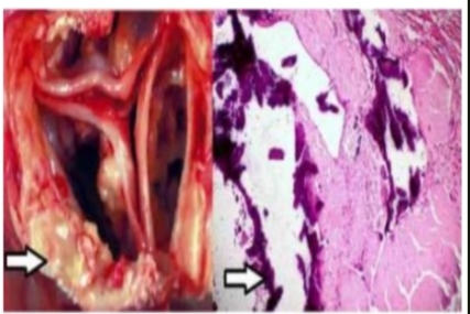
What condition is shown ? When examining a tissue biopsy for this condition, what stains or methods are typically used? Describe what is seen with each stain or method mentioned.
Amyloidosis of the kidney
The stains used are:
Hx & E stain: Eosinophilic material.
Congo red stain: Orange-red color.
Polarized light (after Congo red): Apple green birefringence.
What type of tissue accumulation is shown and what are its macroscopic and microscopic appearances?
Tissue Accumulations; Calcium.
Dystrophic calcification of heart valve. N/E (naked eye) is chalky white
M/E (microscopic picture) is deeply basophilic.
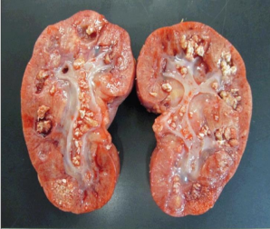
What is this condition and other sites?
Metastatic calcification of kidney (Nephrocalcinosis).
Sites include renal tubular epithelium (Nephrocalcinosis), wall of arteries, mucosa of the stomach, and lung alveoli.
في نيمونيك لل sites كنت عملاها و فادتني شخصيا و هي: كلمة KASL قريبة من نطق كسل
K for kidney
A for arteries
S for stomach
L for lung alveoli
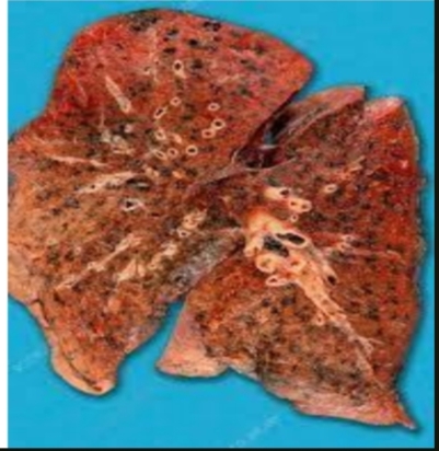
Identify and show the reason
Anthracosis due to accumulation of carbon particles in lung tissue.
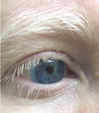
identify and show its reason
albinism due to melanin hypopigmentation
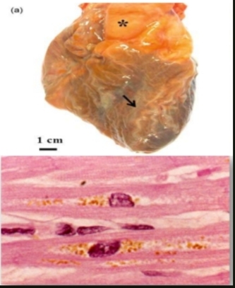
What condition is shown and what pigment is deposited? What are the causes?
Brown atrophy of the heart.
Para-nuclear deposition of Lipochrome (Lipofuscin) pigment in myocardial cells.
Causes include aging, wasting diseases, and cancer.
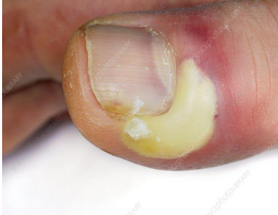
identify ? And what are its cardinal signs?
abscess ( Acute Localized suppurative inflammation).
Cardinal signs are redness, hotness, swelling, pain, and loss of function.
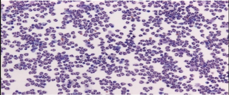
What is this and What are the main cells seen in it?
Suppurative inflammation.
The main cells are Neutrophils & pus cells.
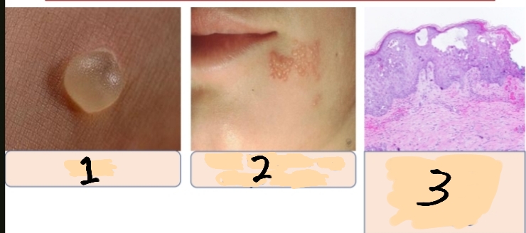
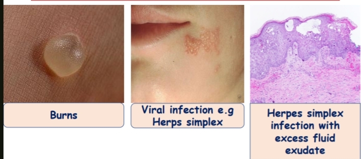
Describe the type of inflammation by providing its name and characteristic feature, its listed causes that are shown in the image 1,2
And 3 refer to what ?
Name of Inflammation: Serous inflammation
Characteristic Feature: Excess fluid exudation
Listed Causes: 1-Burns
2-Viral infection (e.g., Herpes simplex)
3→Herpes simplex infection with excess fluid exudate

identify?and What is the key feature of this?
fibrinous inflammation
Key feature: excess fibrin
Identify and What is the main characteristic of it? +Give an example?
Catarrhal inflammation
Main characteristic:Excess mucous secretion.
Example: Common cold (catarrhal rhinitis).
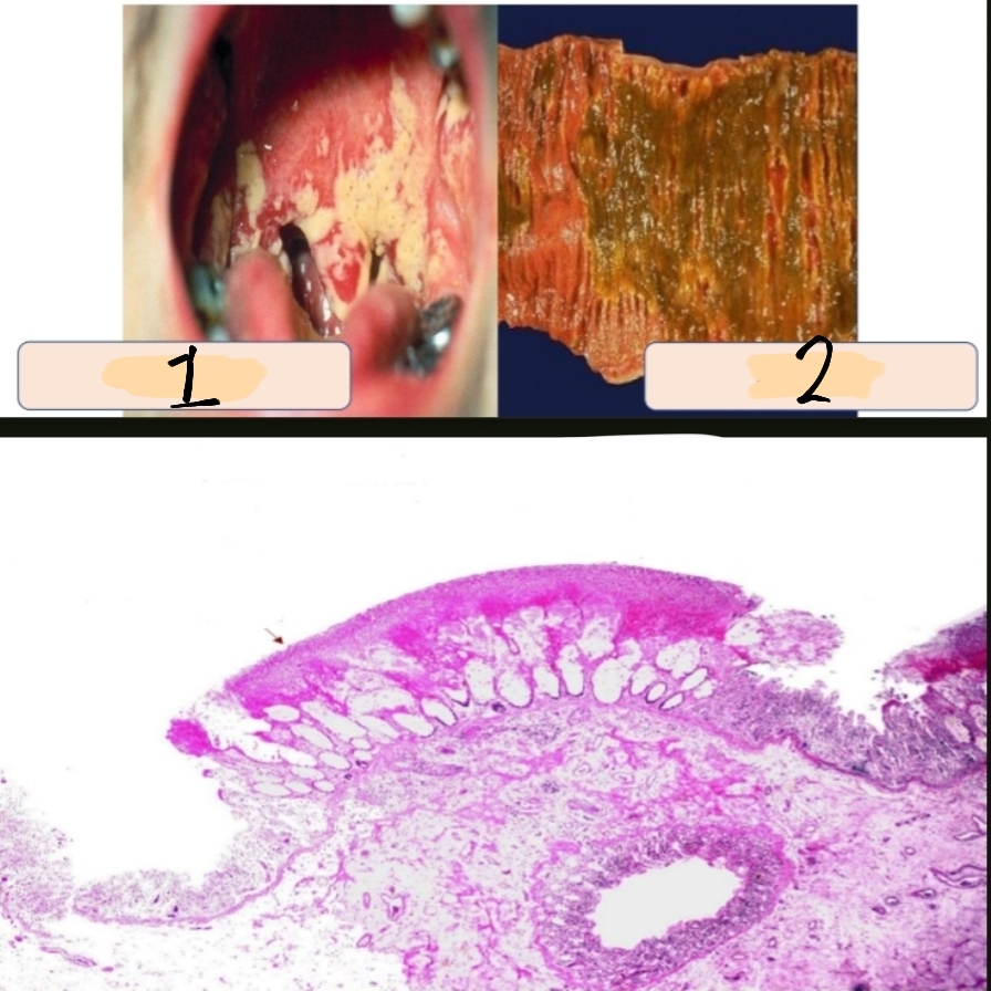
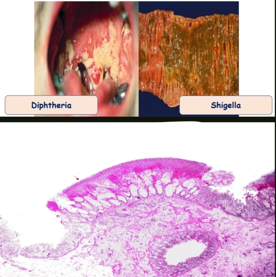
What is this and composed of what?
Enumerate its examples
1 refer to ?
2 refer to?
pseudomembranous inflammation(adherent false membrane).
The membrane is formed of necrotic mucosa + fibrin + inflammatory exudate.
1-Diphtheria
2-Shigella.
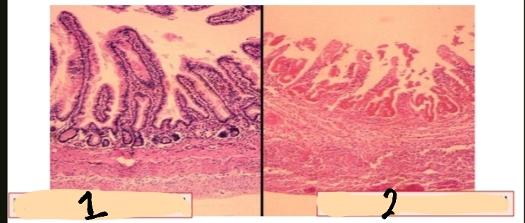
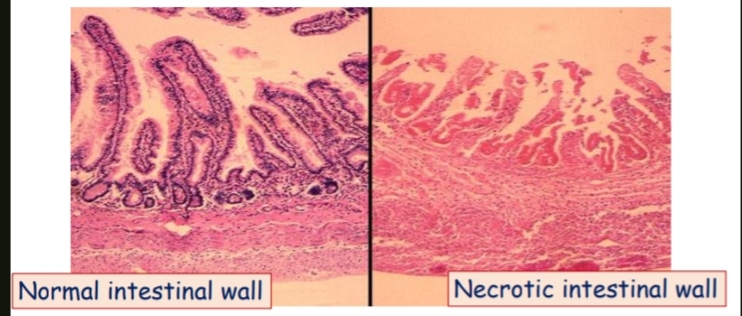
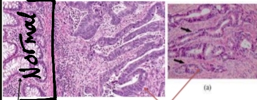
What is this and What is the main feature of it?
Based on your observation,1 or 2 depicts a normal state of the intestinal wall?
necrotizing inflammation
Main feature:excess tissue necrosis
1 normal
2 necrotic
) مش هيجيلي صورة لل normal في الامتحان و لكن خليكم عارفين الفرق بس )
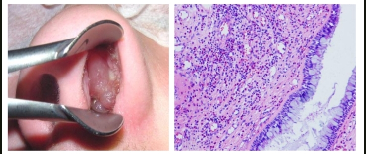
identify ?
composed of ?
allergic nasal polyp with excess eosinophils
(Allergic inflammation)
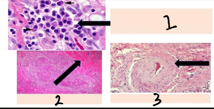
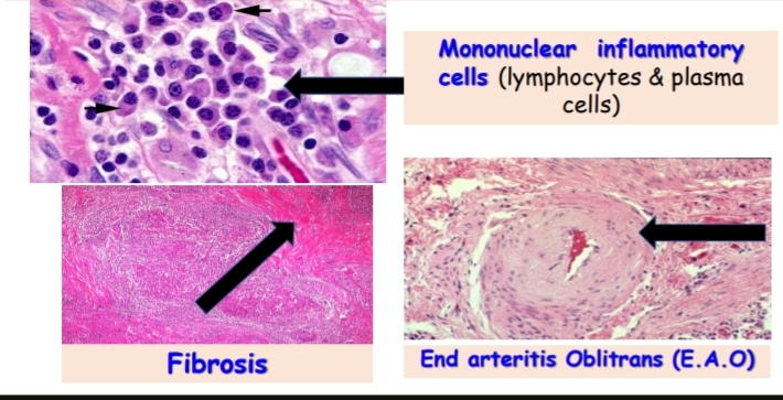
what are considered the classic microscopic features of chronic inflammation?
1-Mononuclear inflammatory cells (lymphocytes & plasma cells)
2-fibrosis
3-End arteritis Oblitrans (Ε.Α.Ο)
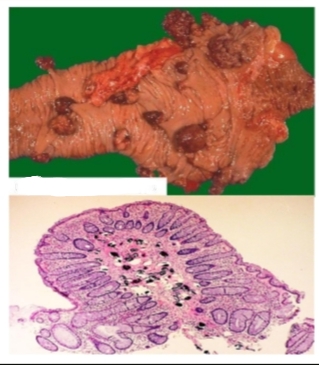
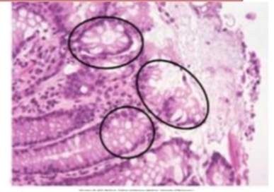
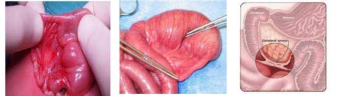
Identify then Describe the macroscopic and microscopic features
bilharzial colonic polyp.
Macroscopically: they are multiple,sessile or pedunculated, and simple (unbranched) or compound (branched).
Microscopically: there is a central core of connective tissue containing bilharzial granuloma with excess eosinophils. The covering mucosa may be intact, hyperplastic, or ulcerated.
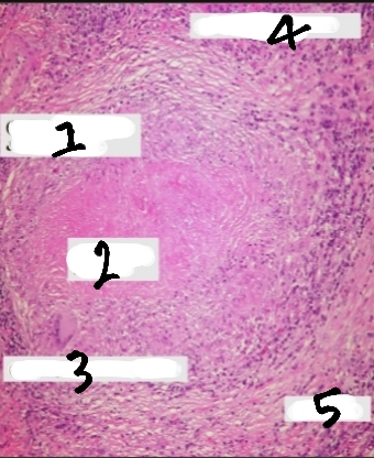
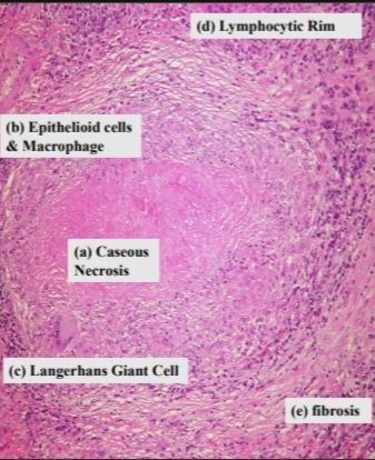
Identify and What are the microscopic components ?
Tubercle
1. Epithelioid cells and macrophages.
2.caseous necrosis
3.Langhan's giant cells.
4. Lymphocytes.
5.Peripheral fibroblastic reaction (fibrosis).
Identify and What are the components of it?
+Arrows point to what?
Primary Pulmonary TB
1. Ghon's focus (located subpleural).(Arrows point to it)
2. Tuberculous Lymphangitis.
3. Tuberculous lymphadenitis.
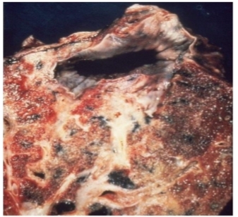
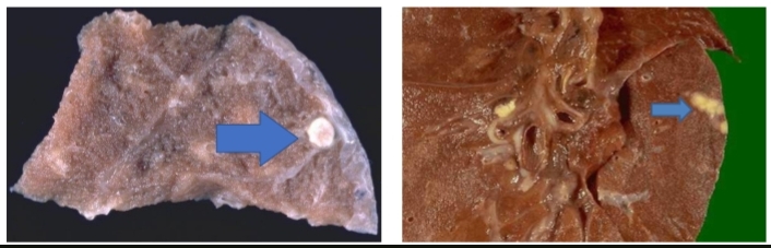
Identify and What is a characteristic apical lesion ? What often occurs in most cases?
Secondary pulmonary tuberculosis
An apical lesion (Assmann focus).
In most cases:cavitations occur.
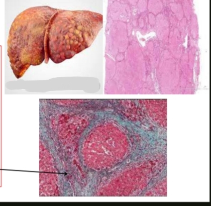
What condition is shown and what causes it? What special stain is used and what does it highlight”arrow refer to it”?
Liver cirrhosis due to improper regeneration admixed with fibrosis.
Special stain used is Masson trichrome, which stains the fibrous tissue blue(the arrow refer to it)
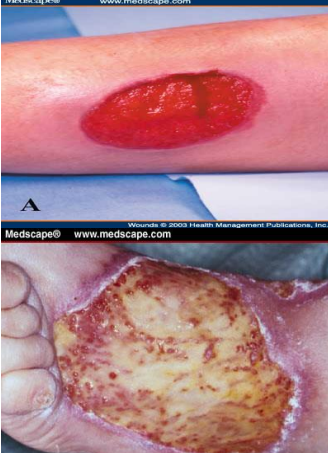
identify and What is it (give definition)+
What is its naked eye appearance?
granulation tissue
It is a transitory tissue formed during the process of repair.
N/E: Red, granular, moist, fragile (bleeds easily), resistant to infection, and insensitive.
For the previous question, mention the microscopic features according to numbers on photo.
1-Capillaries(leaky; no basement membrane)
2-Fibroblasts
3-Inflammatory cells(macrophages mainly)
4-Edema in the background

What type of tissue repair outcome is shown?
Scar hypertrophy
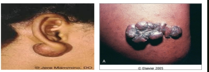
identify ?
keloid
identify ?
mention other types ?
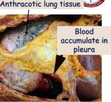
Number 1,2 refer to what?
Internal hemorrhage,hemothorax
hemoperitonium , haemopericardium , hemoarthrosis
1-Anthracotic lung tissue
2-Blood accumulate in pleura
identify ?
other types ?
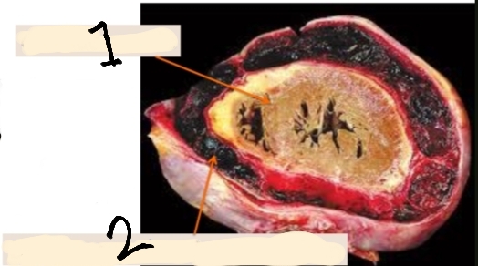
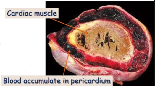
Number 1,2 refer to what?
Internal hemorrhage,hemopericardium
hemothorax , hemoperitonium
1-Cardiac muscle
2-Blood accumulate in pericardium
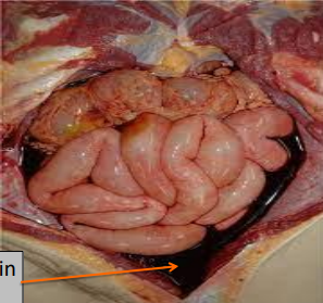
Identify.
Arrow point to what?
و زي الاثنين اللي فاتوا في حتة ذكر الامثلة
Internal hemorrhage,hemoperitonium
Arrow for blood accumulate in peritoneum
identify.
Its size look like what ?
petechial hemorrhage (interstitial hemorrhage)
pinhead-sized
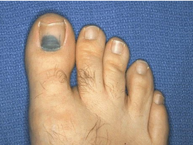
identify ?
ecchymosis (interstitial hemorrhage)
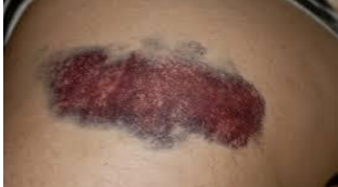
identify .
hematoma ( interstitial hemorrhage)
Large amount of blood causing swelling
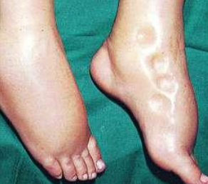
What type of edema is shown and what are its causes?
Pitting edema.
Caused by renal edema, cardiac edema, and nutritional edema.
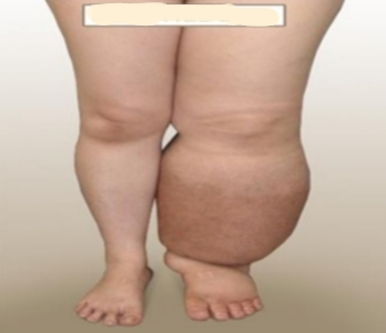
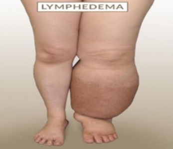
What type of edema is shown and what are its causes?
Non-Pitting edema
Caused by inflammatory edema and lymphedema.
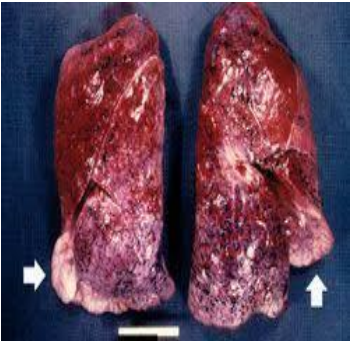
Identify
and what causes the brown color and induration?
Chronic venous congestion of the lung(Brown induration of the lung).
Brown color is due to hemosiderin, and induration is due to fibrosis.
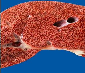
identify ?
And look like what?
what causes the dark red foci and yellow background?
chronic venous congestion of the liver
Nut Meg Liver.
Dark red foci are due to congestion, and the yellow background is due to fatty change
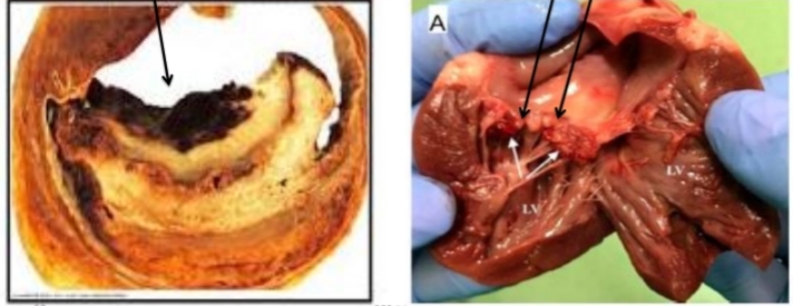
What types of thrombi are shown?
1- Thrombi in the wall of the cardiac chamber (Mural thrombi).
2-Valvular thrombi (vegetation)

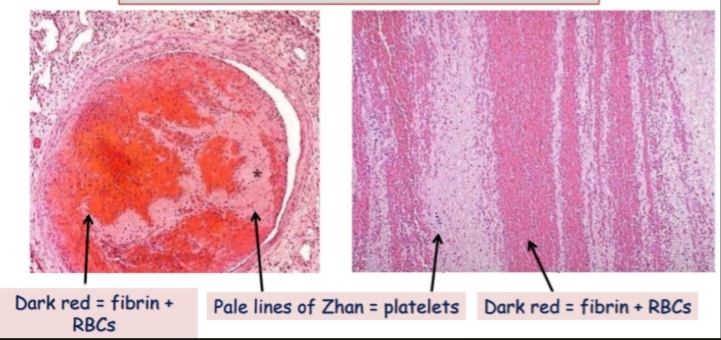
identify ?
composition of arrows ?
microscopic picture of thrombus
1,3-Dark red areas represent fibrin + RBCs.
2- Pale lines represent lines of Zahn (platelets).
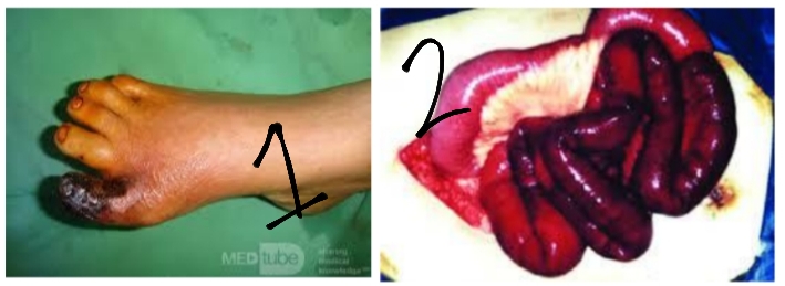
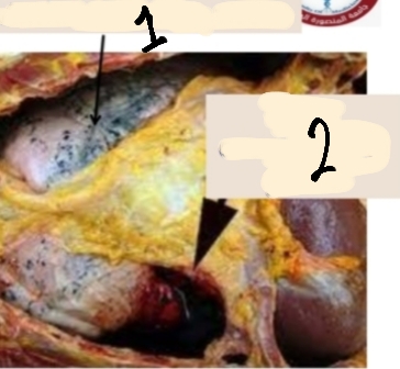
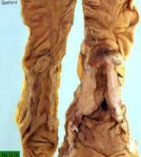
What types of gangrene are shown?
1-dry gangrene of foot
2-wet gangrene of intestine
identify then define
agenesis:Congenital absence of an organ due to complete lack of development.
identify then define
aplasia:The organ is represented by rudimentary structure.
identify then define
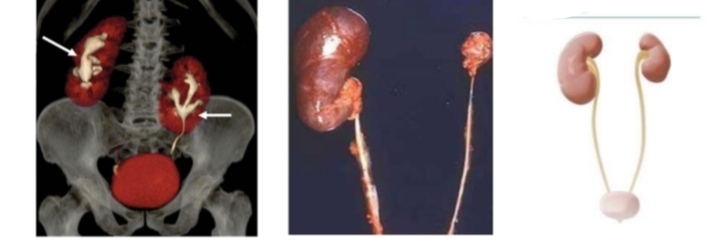
hypoplasia:The organ is normal structure but fails to reach to adult size
identify then define
atrasia:Absence of a normal opening or failure of canalization of hollow organ.
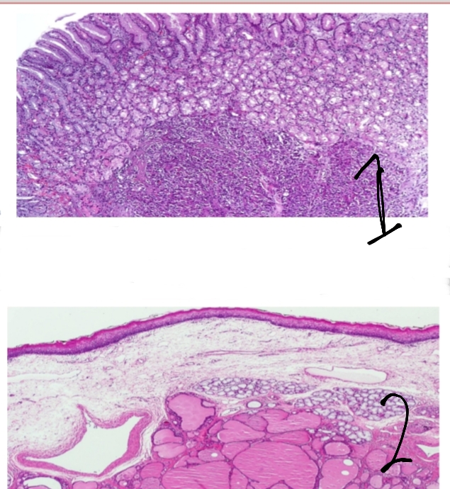
identify(المجمل العام لاسم الحالة) then define
1,2 refer to what ?
Heterotropia (Choristoma) : the presence of tissue in an abnormal location.
1-pancreatic heterotropia in the stomach
2-lingual thyroid (thyroid heterotopia in the base of the tongue).
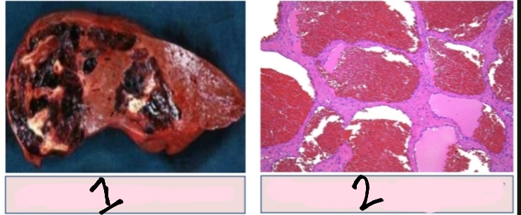
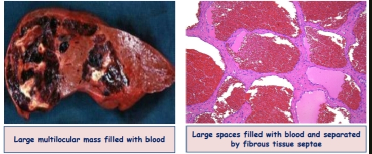
Identify الحالة دي اسمها ايه
Then
Identify 1 and 2
Most common organ
Hamartoma (cavernous hemangioma)
1-Large multilocular mass filled with blood
2-Large spaces filled with blood and separated by fibrous tissue septae
Most common organ is liver
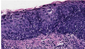
identify .
dysplasia of squamous epithelium
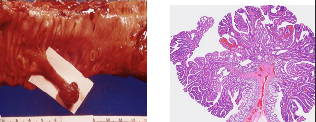
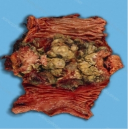
What is shown in these images (N/E and M/E)?
Colon adenoma (Adenomatous polyp of the colon).
identify .
colonic adenocarcinoma infeltrating mass
identify ?
colonic adenocarcinoma fungating mass
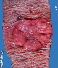
identify .
colonic adenocarcinoma ulcerating mass
Identify then describe
colonic adenocarcinoma
Malignant acini lined by malignant epithelial cells and separated by desmoplastic stroma.
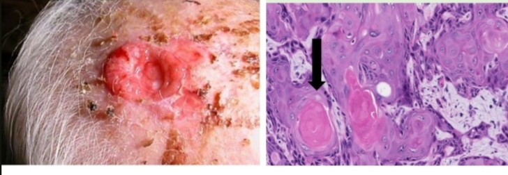
identify then describe microscopically and macroscopically
+Arrow refer to?
squamous cell carcinoma
N/E: Ulcer with raised everted edge,necrotic floor, and fixed base.
M/E: Sheets of malignant squamous cells containing central keratin pearls (Cell nests) separated by desmoplastic stroma.
Arrow refer to cell nests
identify.
What are its microscopic and macroscopic features?
basal cell carcinoma (locally malignant tumor). Microscopic features include sheets and groups of atypical basaloid cells with peripheral pallisading separated by fibrous tissue stroma.
Macroscopically, it appears as an ulcer with a raised inverted edge and necrotic beaded floor.
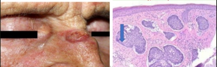
identify then Describe the macroscopic and microscopic appearance
lipoma
Macroscopically, it is a well-circumscribed soft yellowish mass with a greasy cut section. Microscopically, it shows lobules of mature fat cells separated by scanty fibrous tissue stroma.
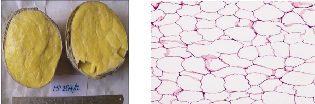
Identify.
Acute diffuse suppurative appendicitis.
بعرف الشريحه من بره انها دائرتين و شريطه
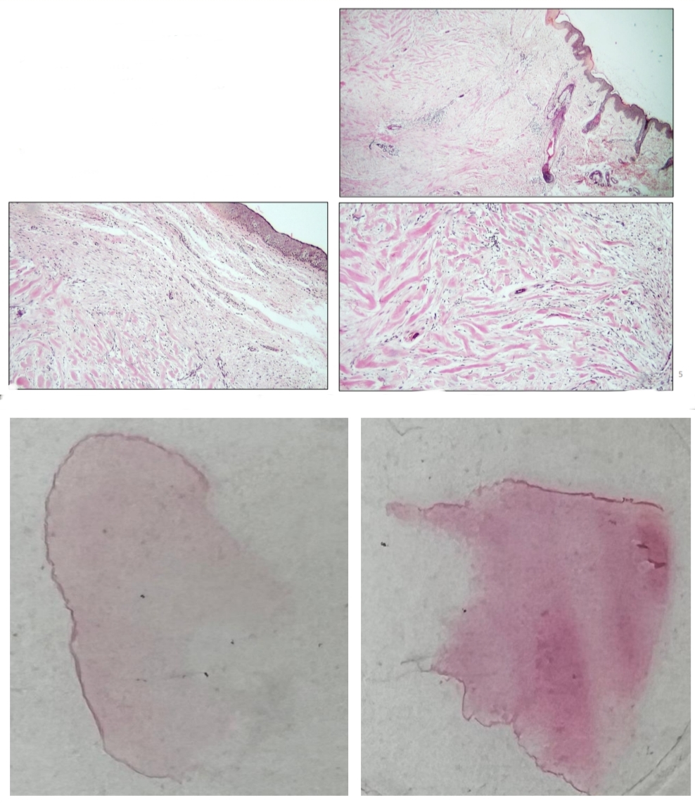
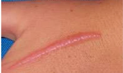
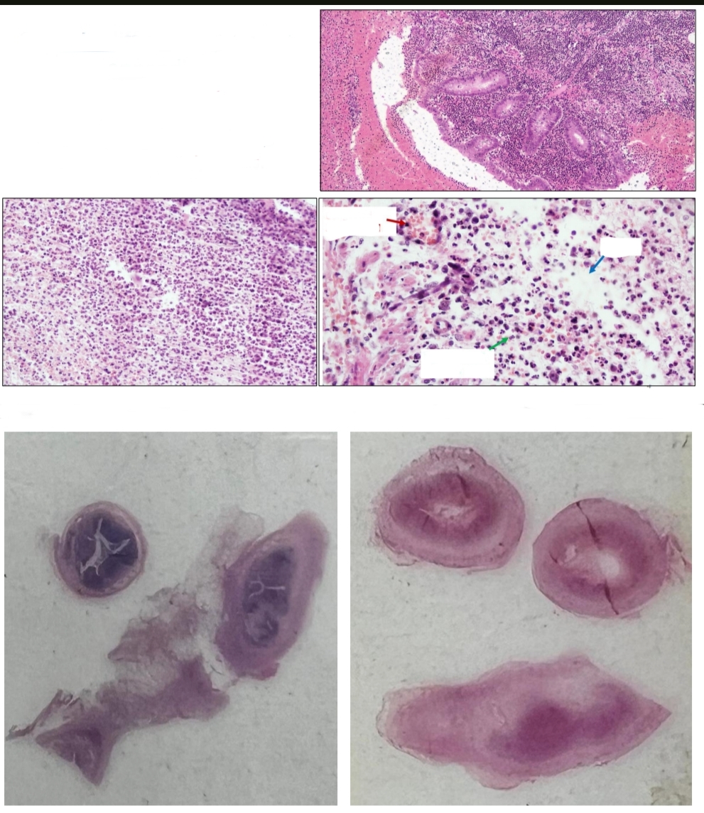
Identify .
Skin scar
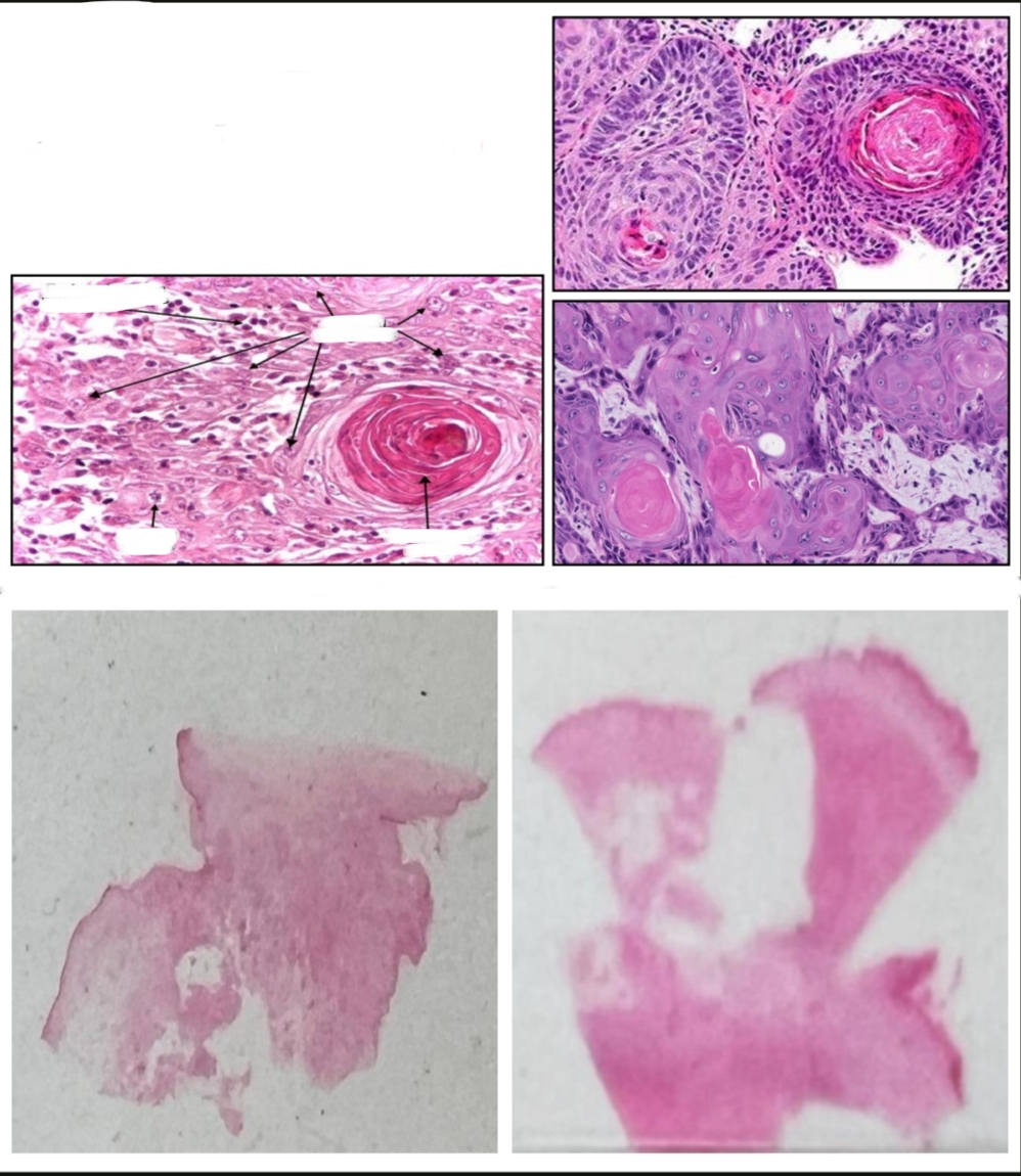
Identify.
Squamous cell carcinoma
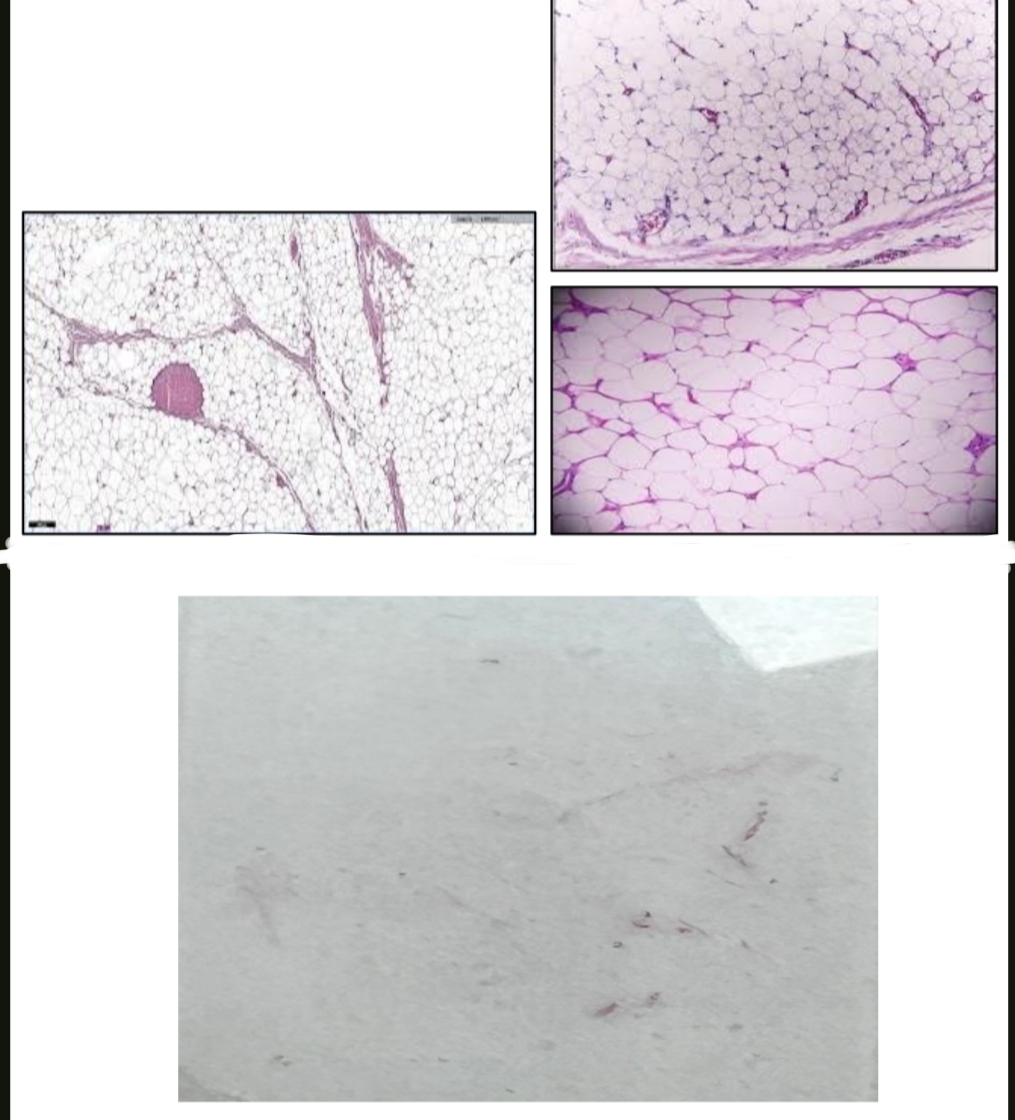
Identify.
Lipoma.
الشريحه بتجيلي فاضية بسبب ان lipids حصلها dissolve during staining

For more