Cardiovascular Lab 1 & 2
 Call Kai
 Learn
Learn Practice Test
 Spaced Repetition
 Match
Match Flashcards
 Knowt Play
1/71

There's no tags or description
Looks like no tags are added yet.
Name | Mastery | Learn | Test | Matching | Spaced | Call with Kai | Chat |
|---|
No analytics yet
Send a link to your students to track their progress
72 Terms
Congenital heart defect & myocarditis (viral, bacterial, protozoal, fungal, algae)
Thor, a 9-month-old male intact Boxer, presented to the TAMU ER for respiratory distress at home. The owner noticed that 2 days ago, his abdomen appeared bloated, and he thought that Thor had eaten too much.
Yesterday, Thor began to have trouble breathing. Physical exam reveals muffled heart sounds, harsh lung sounds, and a fluid wave on abdominal palpation. Jugular pulses were obvious. Due to poor prognosis and financial concerns, the owner elected humane euthanasia.
What are differential diagnoses for heart disease in a dog at this age?
Increased intravascular hydrostatic pressure
At necropsy, 5 liters of clear red fluid is in the abdominal cavity. With cardiac disease, what is the mechanism for the development of ascites?
chronic passive congestion
What causes "nutmeg liver"?
B
Ascites and chronic passive congestion of the liver indicate a:
A. Left-sided heart failure
B. Right-sided heart failure
C. Biventricular heart failure
right
______-sided congestive heart failure results in increased pressure in the vessels delivering blood to the right atrium
left
______-sided congestive heart failure, signs are associated with a backup of pressure in the vessels delivering blood to the left ventricle
C
Here is the thorax with the pericardial sac opened.
What are the visible lesions?
A. Pericarditis
B. Lymphangiectasia
C. Pale areas in the myocardium (either inflammation or degeneration/necrosis)
D. Serous atrophy of fat

F
In dogs (any breed or any age), which disease process does not produce pale tan foci/streaks in the heart?
A. Canine parvovirus type 2 necrotizing myocarditis (puppies)
B. Aspergillus terreus pyogranulomatous myocarditis (German Shepherds)
C. Prototheca zopfii (algae) granulomatous myocarditis
D. Brain-heart syndrome in a dog with brain trauma (myocardial degeneration/necrosis)
E. Doxorubicin toxicity (myocardial degeneration/necrosis)
F. Hemangiosarcoma in the right auricle
G. Lymphoma infiltrating the myocardium
H. Arrhythmogenic right ventricular cardiomyopathy in Boxers
I. Coccidioides immitis or Coccidioides posadasii granulomatous myocarditis
right
The cardiomegaly is due to ventricular dilation. Which ventricle is dilated?

A
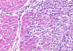
The pale foci of the heart correspond to these microscopic areas. What is the primary process?
A. Inflammatory infiltrate
B. Neoplastic infiltrate
C. Degeneration/necrosis

D
Do you see an infectious agent? What is it?
A. Histoplasma capsulatum
B. Toxoplasma gondii
C. Leishmania donovani
D. Trypanosoma cruzi

triatoma sp. (kissing bugs)
What is the vector for Chagas disease?
B
A 24-month-old Brahman bull is found acutely down and unable to rise. In 24 hours, it developed "bloody urine". The
next day, the bull was dead. You are called in to do a necropsy. At necropsy, areas of pallor were present in skeletal muscles.
Your most likely interpretation for this lesion is:
A. Necro-hemorrhagic myositis
B. Degeneration/necrosis

lymphoma (HULAS sites)
Most common neoplasia in the heart of cattle?
B
Is this lesion?
A. Inflammatory
B. Degenerative
horses
Which species is most susceptible to monensin toxicosis?
C
Which toxic plant should NOT be considered as a potential cause for skeletal and cardiac muscle degeneration/necrosis?
A. Coyotillo (Karwinskia humboldtiana)
B. Coffee senna (Senna occidentalis)
C. Locoweed (Astragalus sp.)
D. White snakeroot (Ageratina altissima)
C
Gabby is an 11-year-old, female, dog who presented to TAMU ER for a swollen abdomen. Gabby had gotten loose from the yard a couple of months ago and was gone for a few hours before returning. About a month and a half ago, Gabby's abdomen begin to swell. Due to this event, the owner was suspicious that she may have gotten pregnant when she got out a couple of weeks before. Over the past few days, she was gasping for breath, not walking, and not eating.
Physical examination revealed an oral mass, a right-sided heart murmur (tricuspid insufficiency) and a fluid wave on abdominal palpation.
The oral mass is confirmed during autopsy. What is the most likely diagnosis?
A) Squamous cell carcinoma
B) Fibrosarcoma
C) Malignant melanoma
D) Hemangiosarcoma

B
The analysis of the peritoneal fluid revealed a straw-colored to slightly red transparent fluid with total protein of > 2.5 g/dL and nucleated cells = 4.0x10³/ul, mostly composed of macrophages with occasional erythrophagocytosis.
This fluid can be classified as:
A) Transudate
B) Modified transudate
C) Exudate
D) Chylous effusion

C
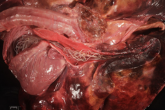
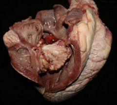
Here is the right side of the heart and pulmonary artery opened up. Name the parasite:
A) Onchocerca lupi
B) Spirocerca lupi
C) Dirofilaria immitis
D) Dirofilaria repens
D
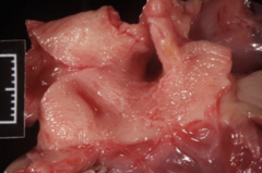
The intima of the pulmonary artery has a shaggy and roughened appearance. What is the name of this lesion?
A) Arteritis
B) Aneurysm
C) Atherosclerosis
D) Fibroelastosis
D
In this heartworm case, the clinical signs and the autopsy lesions are consistent with:
A. Disseminated intravascular coagulopathy
B. Pulmonary thrombosis
C. Protein-losing enteropathy
D. Right-sided heart failure

systemic venous congestion -> generalized edema & chronic passive liver congestion -> ascites & nutmeg liver
What are the pathologies associated with right-sided congestive heart failure?
pulmonary venous congestion -> pulmonary edema -> intra-alveolar macrophage with phagocytosed erythrocytes -> iron in alveolar macrophages (heart failure cells)
What are the pathologies associated with left-sided congestive heart failure?
C
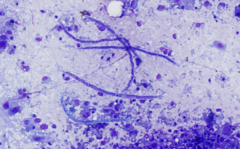
Besides neoplastic melanocytes, the impression smear of the oral mass revealed microfilariae.
Microfilariae are infective to:
A) Dog
B) Human
C) Mosquito
L3
What is the infective stage of Dirofilaria immitis to dogs?
C
Dogs with dirofilariasis may develop coughing and increased of respiratory rate. The cause is the presence of adult Dirofilaria immitis in the branches of the pulmonary arteries leading to:
A. Cranioventral bronchopneumonia
B. Interstitial pneumonia
C. Multifocal pulmonary thrombosis and infarct
A
Dogs with heartworm disease may also develop sudden onset of severe lethargy and weakness accompanied by hemoglobinemia and hemoglobinuria. This condition is called:
A) Caval syndrome
B) Immune-mediated glomerulonephritis
C) Pulmonary vascular disease
D) Right-sided heart failure
A
You were called to a sheep farm where out of 76 sheep, 5 have died over the last few weeks, 4 were sick, and 4 recovered. The age of the sheep varied from 8 months to 4 years old. The clinical course ranged from three to seven days in sheep that died (with an average of five days) and approximately 10 days in sheep that recovered. Clinical signs included anorexia, fever, lethargy, loss of condition, and facial swelling, mainly involving the lips.
Pressing a finger into the facial skin, an indentation remains for several seconds after removing your finger. In this case, the facial swelling is secondary to:
A) Subcutaneous edema
B) Osteosarcoma
C) Pyogranulomatous myositis
moderate, multifocal to coalescing, acute, ulcerative and hemorrhagic stomatitis
You were called to a sheep farm where out of 76 sheep, 5 have died over the last few weeks, 4 were sick, and 4 recovered. The age of the sheep varied from 8 months to 4 years old. The clinical course ranged from three to seven days in sheep that died (with an average of five days) and approximately 10 days in sheep that recovered. Clinical signs included anorexia, fever, lethargy, loss of condition, and facial swelling, mainly involving the lips.
Other frequent clinical signs included respiratory distress, tachypnea, cough, pulmonary rattle, hyperemic or cyanotic oral mucosa, mainly involving the hard palate.
Please, provide a morphologic diagnosis:

B
Goats: Additionally, these lesions were seen in the pulmonary artery and rumen. Based on these findings, what is your main differential diagnosis?
A) Foot-and-mouth disease
B) Bluetongue
C) Contagious ecthyma

C
What is the etiology and mode of transmission of bluetongue?
A) Parapoxvirus; transmission by direct contact
B) Picornavirus; direct and indirect contact (saliva, aerosol etc)
C) Orbivirus; transmission by biting midges (Culicoides sp.)
all of the above but B is best
What are the best samples for testing for bluetongue (select all that apply)?
A) Semen (live animal)
B) EDTA whole blood (live animal)
C) Spleen (dead animal)
C
What is the primary histologic lesion which could explain the lesions and clinical signs? (bottle jaw, anorexia, lethargy, loss of condition, greenish discharge from nares)
A) Edema
B) Hemorrhage
C) Vasculitis

Epizootic hemorrhagic disease
What disease in deer is similar to bluetongue in goats?

C
Which classic clinical sign or lesion indicates that a deer survived hemorrhagic disease?
A) edema in hindlimbs
B) pulmonary artery stenosis
C) hoof sloughing/cracking or growth (stress) rings
foramen ovale & ductus arteriosus
Case: Dystocia foal
What are the two shunts we can still see from the fetal circulation?
ventricular septal defect with secondary congestive heart failure
volume & pressure overload: eccentric hypertrophy of left ventricle, concentric hypertrophy of right ventricle
Case: Billy, 1-yo, male, Nigerian Dwarf goat
Presented for doing poorly
Diagnosis?
What type of cardiac overload can be observed? Which chamber?

patent ductus arteriosus
pulmonary hypertension; eccentric hypertrophy of left ventricle, concentric hypertrophy of right ventricle
Case: Athena, 3-yo, FS, Labrador Retriever dog
Presented with a 5/6 heart murmur. Radiographs show biventricular enlargement and pulmonary edema.
Diagnosis?
What are secondary cardiac findings?

tricuspid valve dysplasia; volume overload
Case: Leslie, 1-yo, M, Boxer dog
Severe right sided heart enlargement and supraventricular tachycardia that is unresponsive to anti-arrhythmic medication.
Diagnosis?
How does this cause right-sided enlargement?

pulmonic stenosis -> pressure overload in right ventricle -> right ventricular hypertrophy, right-sided congestive heart failure and ascites
subaortic stenosis -> pressure overload in left ventricle -> left ventricle hypertrophy, left-sided congestive heart failure and pulmonary hypertension
Case: Mel, 2-yo, FS, Chihuahua dog
Severe hypoxemia, respiratory distress, pleural effusion, scant pericardial effusion.
Diagnoses?
What type of ventricular overload is expected?
What secondary findings can be seen?
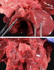
dirofilariasis & pulmonary artery fibroelastosis
pulmonary thromboembolism & proliferative endarteritis (tunica intima)
Case: Jax, MC, Mixed breed dog
History of exercise intolerance and reluctance to move.
Morphologic diagnoses? Possible consequences?
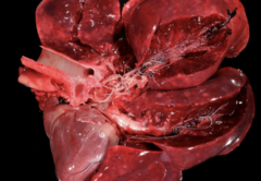
proliferative & obstructive pulmonary endarteritis (pulmonary vascular disease), pulmonary hypertension
right sided heart failure
caval syndrome due to occlusion of vena cava leading to hemolytic anemia
What are possible clinical outcomes of dirofilariasis in dogs?
Severe, locally extensive, acute, endarteritis with thrombosis with intralesional nematode larvae
Larval migration of Strongylus vulgaris
Case: Holly, 1-yo, female, Quarter horse
Presented for severe colic. Laparotomy reveled infarcted cecum apex.
Diagnosis? Etiology?

cranial mesenteric artery & L4-L5
What vessel does larval migration of Strongylus vulgaris migrate to? What stages are present?
atherosclerosis of coronary arteries; hypothyroidism
Raven, 10-yo, MC, Rottweiller
Presented for humane euthanasia after not responding well to treatment.
Total T4: 0.4 ug/dL (Reference 1-4 ug/dL)
Free T4: 0.35 ng/dL (Reference 0.8-2.5 ng/dL)
TSH: 0.85 ng/mL (Reference 0.05-0.42 ng/mL)
Diagnosis?
Underlying endocrine disease?

lipid-laden macrophages and cholesterol clefts within the tunica media
Pictured: Atherosclerosis of coronary artery in a rottweiller with underlying hypothyroidism.
What is observed histologically?

Phlebitis with thromboembolism; repeated venipuncture & endotoxemia
Case: Seabiscuit, 11-yo, MC horse • Presented for colic with secondary hypoproteinemia, suspect DIC.
Diagnosis?
Please state two reasons for this to occur.
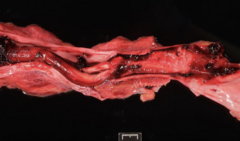
endocardial and intimal mineralization
Case: Luca, 8 yo, MC, German Shepherd
Presented for euthanasia due to progressive decline after a previous diagnosis of lymphoma.
Diagnosis?

Lymphomas can trigger hypercalcemia, which can deposit on tissue (metastatic mineralization)
process called hypercalcemia of malignancy
How is lymphoma related to mineralization?
AGASACA/mammary carcinomas
pulmonary carcinoma
chronic lymphocytic leukemia
parathyroid carcinoma
thymoma
multiple myeloma
What neoplasms can trigger hypercalcemia of malignancy?
Chronic foot root abscess - long-standing bacteremia - endocarditis of the tricuspid valve - embolic pneumonia
Case: 75-day-old Boer goat wether
Lethargy, hyporexia, increased respiratory effort. Patient died spontaneously. A foot abscess was found in the right forelimb during necropsy.
Draft a possible pathogenesis for the heart and lung changes

Ingestion of metal (nail, wire) → reticulum → puncture the wall of the reticulum → enter the diaphragm → pericardial sac → pericarditis → right sided congestive heart failure (CHF)
What is the pathogenesis of hardware disease (traumatic reticuloperitonitis)?
ruminal magnets
How can hardware disease be prevented?
liver, spleen, right auricle
What are the common primary sites for hemangiosarcoma?
maine coone, ragdoll, sphynx
Predisposed cat breeds for hypertrophic cardiomyopathy?
large breeds, such as doberman pinschers, great danes
Commonly affected dog breeds for dilated cardiomyopathy?
genetic basis, doxorubicin toxicity, infectious (Chagas, parvovirus myocarditis)
What are the potential causes of DCM in dogs?
taurine deficiency
What are the potential causes of DCM in cats?
Chemodectoma
Lymphoma
Thymoma
Ectopic thyroid carcinoma
Differentials for a heart base mass?

boston terriers & boxers
What breeds are predisposed to heart base masses?
secondary to local tumor growth
What causes clinical signs seen with chemodectomas?
Myxomatous valve degeneration with mitral valve prolapse and chordae tendineae rupture
Case: Mixed breed dog, 12-year-old, male castrated Presented for sudden death
Morphologic diagnosis?
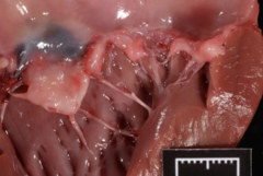
old toy & medium-sized breeds (cavalier king charles spaniels, chihuahuas, dachshunds)
What signalment (& breeds) is commonly seen with myxomatous valve degeneration?
Marked, multifocal to coalescing, acute (subacute), suppurative (vegetative) valvular endocarditis
Case: Boxer dog, 6-year-old, intact male
Weak, febrile, tachypneic, tachycardia
Morphologic diagnosis?

subacute renal infarct
Case: Boxer dog, 6-year-old, intact male
Weak, febrile, tachypneic, tachycardia
Morphologic diagnosis?

Marked, locally extensive, acute, suppurative valvular tricuspid endocarditis
Trueperella pyogenes
(Dogs: Streptococcus spp., Staphylococcus aureus, Escherichia coli, Bartonella sp.)
Case: Simmental bull, 2-year-old
Bull had a large preputial abscess. Weak, tachycardic with arrhythmia, febrile
Morphologic diagnosis? Marked, locally extensive, acute, suppurative valvular tricuspid endocarditis ▪ Common pathogens?
left-sided valves: mitral & aortic; usually with pre-existing lesions in the valves
What is the preferred site of valvular endocarditis in dogs?
tricuspid valve; often without prior valvular damage
What is the preferred site of valvular endocarditis in dogs?
Chagas disease; Trypanosoma cruzi
Severe necrotizing myocarditis with infiltration of lymphocytes, macrophages, and plasma cells.
pulmonary congestion: left-sided heart failure
peritoneal effusion: right-sided heart failure
Case: Great Pyrenees littermates
One presented for acute lethargy, and the other one presented for sudden death. Owner noticed the dog eating kissing bugs.
Name the disease and cause?
Why the heart is pale?
What caused pulmonary congestion and edema and peritoneal effusion?

vitamin D toxicosis; calcinogenic toxic plants (Solanum malacoxylon, Cestrum diurnum, Trisetum flavescens)
What can cause endocardial & intimal mineralization in horses?
Johne's disease or paratuberculosis caused by Mycobacterium avium subsp. paratuberculosis
What can cause endocardial & intimal mineralization in cattle?