Medical Exam
 Call Kai
 Learn
Learn Practice Test
 Spaced Repetition
 Match
Match Flashcards
 Knowt Play
1/215

There's no tags or description
Looks like no tags are added yet.
Name | Mastery | Learn | Test | Matching | Spaced | Call with Kai | Chat |
|---|
No analytics yet
Send a link to your students to track their progress
216 Terms
What is the order for history taking?
PC (Presenting complaint)
HxPC (History of presenting complaint)
DDx (Differential diagnosis) - ideally, obs are being taken at same time
SAMPLE (Signs and symptoms [Including SOCRATES], Allergies, Medications, Past Medical History, Last meal, Events/Evaluate all said)
If not already covered in SAMPLE, should ensure you have:
DHx (Medications and allergies)
FHx (family history)
SHx (Social history)
RoS (Review of Systems)
Now you should have an IMP (Impression)
Now move onto TxT
How full does a sharps box need to be before disposing of it?
2/3
What is the capacity of the suction bag?
1L
What is the presenting complaint (PC), and what questions could you ask as part of this?
Objective statement of what the patient thinks is wrong with them. Should be documented verbatim.
What has happened today?
What have you called us for?
What is going on?
What are the Differential Diagnoses (DDx)?
What are all the things it could be?
Describe the SAMPLE process and questions you could ask for each element of it
S - Signs and symptoms
Ask questions pertaining to the signs (what you see) and symptoms (what pat is experiencing) to rule out DDx. Use SOCRATES to evaluate
A - Allergies
Do you have any allergies?
M - Medications
What prescribed medication are you taking?
Are you on any prescribed medication?
Have you taken any recreational drugs?
Do you have a prescription list?
Do you mind if I access your medical records?
P - Past medical history
Do you have any medical conditions, have you had any medical conditions?
L - Last meal
When was your last meal and what was it? When was your last bowl movement and was it normal? Have you been passing urine okay and what has it looked like?
E - Evaluate/Events
What led up to you experiencing this problem?
What made you call?
So just to confirm… XYZ
Describe SOCRATES and questions you could ask in each section
S - Site
Can you point to the pain/feeling?
O - Onset
When did it start?
Was it gradual or sudden?
What were you doing at the time?
C - Character
What does it feel like?
R - Radiation
Does it move?
Does it go anywhere else?
A - Association
Does it make you feel anything else?
T - Timescale
How long has it been going on for?
Is it there all the time? Or does it come and go?
E - Exacerbating factors
Does anything make it worse?
Does anything maker it better?
S - Severity
How bad is it on a scale of one to ten? One being a slight annoyance and 10 being the worst pain you could ever imagine?
Describe the Review of Systems (RoS) and questions you could ask
Review of bodily systems based on presenting complaint and your history taking, relating to your impression and differential diagnosis.
e.g. if sounds like breathing and or cardiac problem, ask questions pertaining to the cardiovascular and respiratory system.
Does it hurt when you reach up?
Do you have any swelling on your feet or ankles? When did that start?
On one lung or both lung?
Why do observations need to be repeated?
Baseline observations pain a picture of your patient’s health on arrival
Continued monitoring indicates whether your treatment is working or whether you’ve missed something
How do you measure body temperature, what is the normal range, and what are some challenges that may need to be addressed?
Normal Range
35.8 - 38.2*C (37 is good average)
Approach
Using thermoscan tympanic (can detect changes to core body temperature quickest measuring tympanic membrane), and less invasive
Attach single use cover on end of tympanic device
Pull er backwards and upwards and insert in ear
Press button and wait for beep, discard single use cover in clinical waste
Note temperature and report findings
Clean probe
Always use same ear for repeat readings
Challenges
Hearing aid: can generate heat for up to 30mins, remove before
How do you measure levels of consciousness, what is the normal values, and what are some challenges that may need to be addressed?
Normal range
A on AVPU
GCS 15
Approach
Use AVPU to assess (check eyes tracking, responding with voice, trap squeeze)
Challenges
How do you measure severity of pain and what are some challenges that may need to be addressed?
Approach
SOCRATES
Severity - 0-10
Timescale - 5/60 5mins, 5/24 5 hours, 5/7 5 days
Character and radiation - description of crushing, squeezing, stabbing
Challenges
Pain is subjective, can be exacerbating and alleviating factors
How do you attach ECG nodes for a 4 lead and 12 lead, what is the normal values, and what are some challenges that may need to be addressed?
Approach
Consent, chaperone, dry the skin, shave hairy skin, move metal out of way of dots, position comfortably, ensure electrodes sealed and in date
4 lead (Lead 2) electrode placement - (limb leads)
Ride Your Green Bike (clockwise on anatomical model for limb placement)
Red on right arm, Yellow on left arm, Green on left leg, Black on right leg
12 lead electrode placement
V1, V2, V3, V4, V5, V6 along chest in straight, down, up pattern
V7, V8, V9 tracking along back under scapula
V4 can be moves to other side of chest to look at inferior view
Challenges
Sweaty chest, chest hair, jewellery and clothing
How do you measure pupils, what is the normal values, and what are some challenges that may need to be addressed?
Normal value
Pupils equal and reactive to light (PEARL)
3-4mm
Approach
Prior to pen torch visually check pupils to check for equal size and or abnormal changes in shape
Use pupil gauge on pen torch to determine size
Shine pen touch on eye for three seconds - Prompt constriction should be observed - repeat on other eye
Document pupil size and reactivity
Challenges
False eyes
How do you measure respiratory rate, sounds and rise/fall, what are the normal values, and what are some challenges that may need to be addressed?
Normal values
12-20 for adults
Equal chest rise and fall
Normal breath sounds
Approach
Respiratory rate: count over 30 seconds while taking pulse
Breath sounds: Listen with stethoscope 6 points back and front, ladder pattern left to right
Chest rise and fall: Back of hands or palms spread butterfly while taking deep breath
Challenges
Heavy clothing
Heavy tissue
How do you measure blood glucose, what are the normal values, and what are some challenges that may need to be addressed?
Normal values
Non diabetic
3.0-5.6 mmol/L
Diabetics
4.0-7.0 mmol/L before meals
Less than 10 mmol/L 1.5 hours after meal
Around 8 mmol/L at end of day towards sleep
How do you measure capnogrpahy (end tidal CO2), what are the normal values, and what are some challenges that may need to be addressed?
Normal values:
Measures asthma, bronchospasm, COPD CO2 retention
4.67 - 6.00 kPa (35-45 mmHg)
Good box waveform - (A-B insipratory baseline, B-C expiratory upstroke, C-V alveolar plateau, D end-tidal amount of CO2 [end of expiration], D-E waveform starts as inspiration starts
Bronchospasm can show show ‘shark fin’ shape sharper higher C-D incline
Application:
Must be used if iGel, intubated, receiving assisted ventilation, to ascertain quality of CPR. Advised to be used in acute/severe resp distress, acutely unwell patients
How do you measure blood oxygen saturations (SPO2), what are the normal values, and what are some challenges that may need to be addressed?
Normal values:
94-98% in normal patients
88-92% in COPD patients
Application:
Ensure finger is clean, free of heavily applied nail polish, ensure patient sitting still
Place says probe on patient’s finger
Challenges
Heavily applied nail varnish
Cold skin/poor circulation
Moving patient
How do you measure pulse rate, what are the normal values, and what are some challenges that may need to be addressed?
Normal values:
60-100bpm in normal adults (higher in children)
Regular, strong pulse in normal adults
Application:
Pulse can be felt at:
Carotid
Brachial (crook of elbow)
Radial
Femoral
Posterior tibial (between ankle and Achilles)
Dorsalis Pedis (tendon in line with large toe)
Count for 30 secondS
Challenges
Recent exercise
Anxiety
Cardiac arrhythmias
How do you measure capillary refill, what are the normal values, and what are some challenges that may need to be addressed?
Normal values:
Return to normal skin colour in under 2 seconds
Application:
Apply pressure to patient’s finger for 5 seconds, then release
Time to refill
Challenges:
Cold
Poor circulation
How do you measure capillary refill, what are the normal values, and what are some challenges that may need to be addressed?
Normal values:
Return to normal skin colour in under 2 seconds
Application:
Apply pressure to patient’s finger for 5 seconds, then release
Time to refill
Challenges:
Cold
Poor circulation
Describe the stepwise approach to airway management
Remember the PMSASIF
Positioning
Manouevers
Suction
Adjuncts
Supraglotic Airway
Intubation
Front of neck access
What is a patent airway?
Open
Maintained
Protected
All three must be established in primary survey before moving to breathing
What are some signs of an obstructed airway?
Snoring, coughing, cyanosis, reduced chest rise and fall, wheeze, stridor
Explain the triple airway manoeuvre and when elements of it would not be indicated
Head-tilt Chin-lift Jaw-thrust
Head-tilt Chin-lift: palm on head, two fingers on chin, slightly tilt head back to open airway and inspect. Do not hyperextend. NOT indicated for ?c-spine injuries.
Jaw thrust: palms on cheeks, forefingers on angle of jaw, thumbs on chin and push up ensuring mouth is open.
Explain neutral alignment
When managing airway you should aim to create a neutral alignment position, with the tragus in line with the sternal notch. This can be achieved with putting something under the head, neck or shoulders (ramping)
What PPE might you need to manage airway?
Mask, goggle, gloves, sharps box, clinical waste bag, spare equipment, kit dump - all checked in date and sealed
What are the two types of foreign body airway obstructions and how are they differentiated in times of signs and symptoms?
Complete and Partial
Partial: patient able to speak, cough, breathe - although may be with some difficulty. Strider may be present on auscultation. Some or one sided chest rise and fall.
Complete: complete obstruction - pat not able to speak, cough or breathe at all. Silent chest on auscultation. No chest rise or fall. May be unconscious.
What are common causes of airway obstruction?
Food, teeth, toys, blood, vomit, secretions, coins
Describe the management of airway obstructions in an adult
if effective cough, encourage coughing (likely partial obstruction)
If no effective cough and conscious, consider critical care, check airway for any obvious obstruction, 5 back blows followed by 5 abdominal thrusts, then back to 5 back blows and so on until obstruction relieved
If no effective cough and unconscious, consider critical care, open airway and check for any obvious obstruction, and start CPR BLS.
Describe the management of airway obstructions in a paediatric patient
if effective cough, encourage coughing (likely partial obstruction)
If no effective cough and conscious, consider critical care, check airway for any obvious obstruction. If child, 5 back blows followed by 5 abdominal thrusts, then back to 5 back blows and so on until obstruction relieved. If infant (under 1) 5 back blows (place child face down) followed by 5 chest thrusts (hold child on back supporting head), then 5 back blows and so on until obstruction relieved.
If no effective cough and unconscious, consider critical care, open airway and try rescue breaths, and start paediatric BLS.
Describe how to use an OP tube including indications and contraindications
- Measure sizing from lips to angle of jaw
- insert curved up twist down
- should fit flush with lips
- single use
- reassess pat breathing after insertion
- have one size up and down ready Paeds are sized per age. Do NOT twist, use a tongue depressed as palette is soft
Indications
- no gag reflex
- unconscious and no gage reflect
- cardiac arrest
Contraindications
- gag reflex
Describe how to use NP tubes
Sized 6 and 7 Must be lubed first Not for pat under 12yo Right nostril first Curve down insertion, should sit flush with nostril Don't force in
Indications:
- unconscious no gag reflex
- trismus
- unable to insert OP tube
Contraindications:
- under 12
- gag reflect
Cautions:
- facial/ base of skull trauma
Describe how to use a suction
- Oxygenate patient as suction removed O2
- Patient position laterally
- Attach suction tubing and yankauer wand (adult or paed)
- Suction on the way out
- Figure of 8, be gentle
- Max of 15 seconds, ideally 10
- Only suction as far as you can see
In cases where fogey suction, there is a single use hand held bulb suction in BVM bag
Describe how to use a BVM
- Attach O2 tube to bag spigot, NOT mask
- Ensure bag inflates
- CE grip on mask with fingers performing slight jaw thrust to open mouth and bring jaw up to meet mask and seal
- Gently squeeze bag, squeeze over one second enough to see chest wall movement
- Removed for shocks or suction
- ventilation rate per age in JRCALC
- can be done with one person, but two person technique where one holds mask and jaw together in jaw thrust position and one person bags
What are common causes of cardiac arrest?
Cardiac disease
Arrhythmia
Electrolyte disorder
Poisoning
Drowning
Hypothermia
Hyperthermia
Asthma
Anaphylaxis
Trauma
Electrocution
What are key tasks on arrival to recognise and manage cardiac arrest?
D+R
A - check clear airway, manoeuvre and pulse check same time
B - look listen and feel for breath
C- Call for backup,CPR and shock
What are CPR essentials
Lower half of sternum
Depth of 5-6cm
Rhythm of 100-120bpm
Allow full recoil
LP 1000 and shocking essentials
Apply pads and turn on [ANALYSE]
Follow audible prompts
Continue CPR when charging
Shock safety - say ‘STAND CLEAR’ , look at pat top to toe to ensure stood clear, look at patient then shock
Straight back to CPR
Swap CPR every 2 mins
What are some post ROSC care essentials
Use checklist - DONT RUSH
‘Challenge and response’ → ask question and wait for an answer
Full set of OBS
Titrate oxygen
Think about destination → ED or HAC?
Plan extrication → Trolley bed out, facing right way
Needs to be at least 10 mins on scene to make sure sustained ROSC
Explain the Pit Crew model to manage cardiac arrest
Everyone knows their role and that of the others team members
One person on airway management with airway kit dump next to them (at head)
One person doing chest compressions with defib next to them (one side of patient)
One person waiting to take over from chest compressions
One person doing paramedic ALS drugs (other side of patient)
One person doing team leader checklist

Explain the handover from ILS to BLS (you are first and paramedic arrives).
Try to use SBAR
Time arrest confirmed/start of BLS
Presenting rhythm
Number of shocks
Medical Hx
Any Hs and Ts covered
Stage of checklist (if used)
Where and when should we check for pulses in a cardiac arrest scenario?
To confirm arrest or ongoing arrest, pulse check at end of 5×30:2 cycle
Should check in 2 places for accuracy, either:
Carotid
Radial
Femoral
Check for pulses in
VT
PEA
Describe Ventricular Fibrillation and actions during a cardiac arrest
Waveform is chaotic with no discernible pattern
AED will recognise this and charge shock. You should shock.
Do NOT need to pulse check this rhythm, it is incompatible with life

Describe Pulseless Electrical Activity (PEA) and actions during a cardiac arrest
Waveform may look like a recognisable, regular rhythm
AED will not charge and will not advise shock. Do not shock.
DO need to check for a pulse as waveform is possibly compatible with life

Describe Asystole and actions during a cardiac arrest
Typical “flatline” waveform
AED will not charge shocks and will not advise shock. You should not shock.
You do NOT need to check for a pulse, incompatible with life

Describe VT and actions during a cardiac arrest
High, peaking, rhythmic waveform in a ‘shark tooth’ pattern
AED will charge a shock and will advise to shock
You DO need to check for a pulse. If a pulse is present, do NOT chock even if AED advises to. If no pulse, Shock.

What are the 4 H’s and 4 T’s
These are the reversible causes of cardiac arrest that you should consider and treat differently
Hypo/Hyperthermia (warm up/cool down)
Hypoglycaemia (give glucose)
Hypovolemia (staunch bleeding/give fluid)
Hypoxia (rescue breaths and oxygenate)
Tamponade (hospital/hems)
Toxins (narcan)
Tension pneumothorax (needle decompression)
Thrombus (PE/MI - convey quickly)
What should you consider for pregnant patients in cardiac arrest?
Hypoxia/hypovolemia are common causes
Manual uterine displacement
Displace to patient’s left side so as to not compress inferior vena cava when doing compressions
Plan for time critical transfer to hospital
What should you consider for drowned patients in cardiac arrests?
As soon as patient has been rescued:
clear the airway
Provide 5 initial ventilations (rescue breaths)
Commence CPR
What is PACE?
A mnemonic for escalation of assertiveness when calling out something you see is wrong
P - probe
A - Alert
C - Concern
E - escalate
Describe ATMIST and what it is used for?
Mnemonic for handovers during traumatic cardiac arrests
A - Age
T - Time of incident
M - Mechanism of injury
I - Injuries
S - Signs and symptoms
T - Treatment given/immediate needs
Describe the differences between Hypoxia, Hypoxaemia and Hypercapnia
Hypoxia → inadequate O2 at cellular level
Hypoxaemia → abnormal deficiency in concentration of O2 in arterial blood
Hypercapnia → greater than normal CO2 in blood
Describe the implications and benefits of oxygen therapy
- O2 is a medication and should be treated as such
- Should only be given to those with a clinical need
- Oxygen treats hypoxaemia and NOT breathlessness
- Oxygen treatment should be titrated to achieve normal saturations in healthy adults (94-98%)
By increasing oxygen content in the lungs you can force the haemoglobin to carry more O2 thereby reducing affects of Hypoxia
What are some basic health and safety principles for using medical gases such as oxygen or entonox?
- Cylinders should be stored in well ventilated special cupboards, not used for any other purpose, empty and full cylinders stored separately, properly supported off the floor
- No smoking, naked flames, sources of heat, oil or grease near cylinders and particularly no oil or grease near valves or outlets
- No dropping or rolling cylinders on ground
- Always check label
Describe the process of setting up oxygen for use
- Check label
- Check contraindications
- Ensure cannula securely attached
- Open valve fully (black knob)
- Use flow regulator for appropriate litres
- Watch contents gauge
What are the types of masks and cannulas that can be used with oxygen?
- Bag Valve Mask (BVM) 15L
- Non-rebreather mask 10-15L
- Simple face mask 5-10L
- Venturi mask 4L
- Nasal cannula 2-6L
- Nebuliser 6-8L
Describe some challenges of recording an accurate oxygen saturation
Heavily applied nail varnish, poor circulation due to cold or illness
Describe the properties of entonox
- 50% nitrous oxide, 50% oxygen
- self administered and inhaled gas that is quick to act but quick to dissipate in effect
Explain the effect of temperature on entonox
Gases can separate at -6*. Before use invert canister a few times.
Describe the process of setting up Entonox for use
- Ensure demand valve clean and ready for use
- Fit new filter and mouthpiece
- Push hose firmly into Schrader valve and hear click
- Slowly turn cylinder on (black knob)
- Purge test demand valve
- Check for leaks, instruct pat on use
What are the contraindications for Entonox?
- Chest injury and/or suspected pneumothorax
- Air embolism
- Severe head injury with impaired consciousness (possible air embolism)
- Decompression sickness (consider risk if anyone has been diving last 24hrs)
- Violently disturbed psychiatric patients
- Severe bollous emphysema
- Abdominal pain with gross distension
- Intraocular injection of gas in last 8 weeks
What are some indications for entonox use?
- Cardiac chest pain
- Moderate to severe pain
- Labour pains
Define Medicine
Substance taken or applied to body that affects function or composition of body or microbes within it
Define pharmacology
Study of drugs and effects on body
Define Pharmacodynamics, and describe agonists and antagonists
What the drug does to the body (mechanism of action, effect on cells/tissues)
Drugs usually act by attaching to specific receptor proteins in body
Agonists activate receptors to produce a response ‘unlock’ effects
Antagonists block receptors to stop a response, they ‘block’ effects
Define Pharmacokinetics
What the body does to the drug
Absorption (how enters body)
Parenteral routes
Physical breach of skin e.g. injection, intravenous, sub-cutaneous, I/O
Non-parenteral routes
Drug absorbed passively e.g. by mouth, buccaneers/sub-lingual, inhalation/aerosol, rectal
Distribution (how spreads through body, i.e. bloodstream)
Metabolism (drug is chemically modified, i.e. in liver)
Excretion (drug and metabolites removed from body, i.e. urine)
What are GSL, P and POM classifications of medicines?
Will find these on side of medicines:
GSL (general sales list) - ‘Over the counter’ medications
P (pharmacy only medicines) - can only be prescribed by a pharmacist
POM (prescription only medicines) - can only be prescribed by a prescribing HCP (GP, consultant)
What do you need to remember for documenting medicine administration?
Indication for giving or contraindication for not giving
Indication for taking it away e.g. JRCALC guideline to remove for COPD pats)
What checks should you do as part of your drug checks before administering?
Is patient allergic?
Correct drug, presentation (form) and dosage?
Is intact?
If fluid is it clear?
Is it within expiry?
What is the AAP scope of practice for ECGs?
Interpret Lead 2 ECGs
Attach leads and dots for both 2 Lead and 12 Lead ECGs
Print ECG strips
What are important elements to remember about Lead 2 ECGs specifically?
Inadequate for diagnosis, useful as a monitoring tool
Equidistant leads
Avoid crossing wires
Can either attach on shoulders and hips or wrists and ankles (RYGB, clockwise facing patient)
Use fresh dots out of a sealed packet
Prepare the skin
Dry
Free from loose skin
Avoid hairy areas
Ideal to obtain on trolley bed or bed with patient semi-recumbent
What does an ECG measure?
The path of electrical conductivity through the heart
What should you keep in mind about interpreting ECGs and their printed results?
Many patients may have severe cardiac disease present despite a normal ECG. It is only an investigative tools and must be interpreted in light of the clinical findings.
Ensure has calibrated starting box and 25mm/sec on bottom
History and clinical examination is essential
Remember to attach a copy of the ECG (with Pat name and DOB on ECG) with the PRF (if on paper), or photograph for ePCR.
Keep a copy of the ECG with name and DOB physically when hand over
How does depolarisation towards/away from electrodes show as a deflection on the ECG?
Any energy moving towards the positive electrode (depolarisation away from negative) is going to be shown as a positive (upwards) deflection
Any energy moving away from the positive electrode (depolarisation towards negative electrode) is going to be shown as negative (downwards) deflection

In what direction does the 2 Lead ECG measure depolarisation between each of the electrodes?

How does the grid pattern on ECG paper measure time and amplitude?
Time across X axis, amplitude across Y axis
Time
Every small box along x axis is 0.04 seconds (40 milliseconds)
Therefore, every large square (5 small boxes) is 0.2 seconds (200 milliseconds)
And so on, 5 large squares is a second
And so on, 30 of the large squares is 6 seconds
Amplitude
Every small box along the Y axis is 0.1 mV amplitude
Therefore, every large square (5 small boxes) is 0.5 mV amplitude
What are the elements of NSR on an ECG?
P wave
PR interval
Q wave
R wave
S wave
ST segment
T wave
Describe the P wave
Depolarisation of the Atria on an ECG
Should be small and smooth
Measures electrical activity going around the atria from the SA node over the top of the atria
If absent or abnormal, may be an indication of heart block

Describe PR interval
Delay at AV junction
Includes the P wave and the short straight line afterwards (this is the delay part)
Normal interval in adults is 120-200Msec (3-5 small boxes)

Describe the Q wave
Septal depolarisation (depolarisation from left to right across heart wall)
May not be present in all patients
If wider than 40 Msec or taller than 2mm, may indicate previous MI
May be seen as a slight negative deflection
Part of the QRS complex
Normal QRS interval in adults is 80-120Msec (2-3 small boxes)

Describe the R wave
Ventricular depolarisation (energy going down bundle of his) [also repolarisation of ventricles not picked up]
First upward deflection
Part of the QRS complex
Normal QRS interval in adults is 80-120Msec (2-3 small boxes)
![<p>Ventricular depolarisation (energy going down bundle of his) [also repolarisation of ventricles not picked up]</p><ul><li><p>First upward deflection</p></li><li><p>Part of the QRS complex</p></li><li><p>Normal QRS interval in adults is 80-120Msec (2-3 small boxes)</p></li></ul><p></p>](https://assets.knowt.com/user-attachments/d9af33d4-78d7-46ca-8b0d-4216781e6f35.jpg)
Describe the S wave
Late stage depolarisation (energy going all around ventricles back up) [also atrial repolarisation not picked up]
Downward inflection after R wave
Part of the QRS complex
Normal QRS interval in adults is 80-120Msec (2-3 small boxes)
![<p>Late stage depolarisation (energy going all around ventricles back up) [also atrial repolarisation not picked up]</p><ul><li><p>Downward inflection after R wave</p></li><li><p>Part of the QRS complex</p></li><li><p>Normal QRS interval in adults is 80-120Msec (2-3 small boxes)</p></li></ul><p></p>](https://assets.knowt.com/user-attachments/9001d5d7-4506-44db-9acc-7817e2e84402.jpg)
Describe the ST segment
Isoelectric segment (should be flat, relaxation phase between ventricular depolarisation and depolarisation)
elevation or depression may indicate an MI

Describe the T wave
Repolarisation of ventricles (resting phase)
Wave after the ST segment
Part of the QT interval
QTc interval should be less than 500Msec (2.5 large boxes)
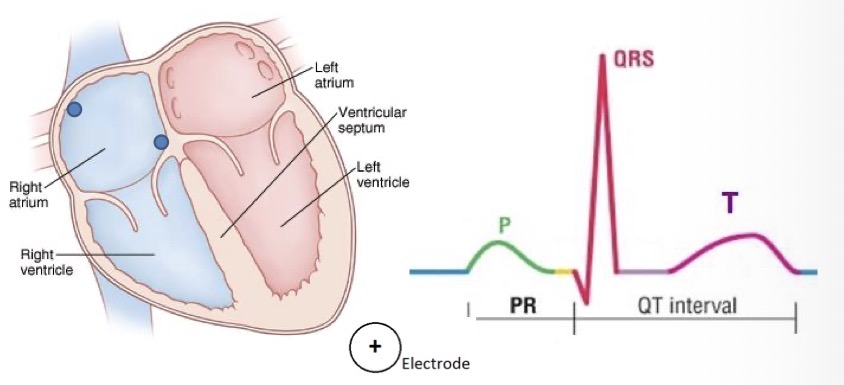
Describe the TP interval
The interval between the end of the T wave and the start of the next P wave (End of repolarisation of ventricles and start of depolarisation of atria)

Describe the RR interval
The interval between the top of the QRS and the top of the next QRS
RR interval can give you a heart rate
Count over 30 large boxes (6 seconds)
Count RR intervals over these 30 boxes
Multiply by 10 to give over 60 seconds
=HR/M

What are some questions your need to ask to interpret Lead 2 ECG?
Is there electrical activity?
What is the rhythm
If have PRST = sinus rhythm
What is the rate
P waves
Shape
Before every QRS
QRS after every P
What is the P-R interval
Duration (should be 120-200 Msec (3-5 small boxes))
QRS complex
Shape
Duration (should be 80-120 Msec (2-3 small boxes))
How can you tell if the rhythm is regular or irregular, or regularly irregular.
If RR intervals spaced out evenly, regular
If an RR interval is skipped/elongated/shortened randomly, irregular
If an RR interval is skipped/elongated/shortened in a pattern, regularly irregular
What is artefact?
Interference that can disrupt the ECG
Muscular or electrical artefact can occur
Fix by
Moving electrode form large muscle mass
Turn off electrical equipment if necessary

What is the standard 12 lead ECG electrode placement?
V1: 4th intercostal space (ICS), right sternal border
V2: 4th ICS along left sternal border
V4: 5th ICS, mid-clavicular line (MCL) [find end of shoulder, beginning of clavicle, in half]
V3: Midway between V2 and V4
V5: 5th ICS, Anterior axillary line (AAL) [same level as V4]
V6: 5th ICS, Mid axillary line (MAL) [same levels as V4]
![<ul><li><p>V1: 4th intercostal space (ICS), right sternal border</p></li><li><p>V2: 4th ICS along left sternal border</p></li><li><p>V4: 5th ICS, mid-clavicular line (MCL) [find end of shoulder, beginning of clavicle, in half]</p></li><li><p>V3: Midway between V2 and V4</p></li><li><p>V5: 5th ICS, Anterior axillary line (AAL) [same level as V4]</p></li><li><p>V6: 5th ICS, Mid axillary line (MAL) [same levels as V4]</p></li></ul><p></p>](https://assets.knowt.com/user-attachments/dc910e1c-680c-4082-8ee4-4f14e201e50a.jpg)
What is the 12 lead ECG electrode placement for a V4R (looking at right side of heart) trace?
Remove V4 from the chesT
Put a new electrode on the lead
Place lead on 5th ICS, MCL, on left
Reprint the 12 lead
Label the ECG (V4R)

What is the 12 lead ECG electrode placement for a posterior ECG?
Remove V4, V5, V6
Put new electrodes on these leads (These now become V7, V8, V9)
Place V8 under the angle of the scapula
Place V7 on its left and V9 on its right
V7, V8 and V9 should be continuous with the dots of V4,V5, V6 in a straight line
Print off another 12 lead ECG annotating ‘posterior ECG’ at the top
Cross out V4, V5, and V6 and re-label it ‘V7, V8, V9’
Define Infection
Invasion of the body by a harmful organism or infections agent such as a virus, parasite, bacterium or fungus
Define Colonisation
Presence of microorganisms on a body surface (such as skin, mouth, intestines or airway) that does not cause disease in the person or signs of infection
Describe the chain of infection
Infectious Agents → Susceptible Host → Portal of Entry → Mode of Transmission → Portal of Exit → Reservoir → Infectious Agents
Describe patient placement in the context of IPC
Clear handover of information including symptoms of infection or known infections
May prompt instigation of isolation facilities
Describe the 5 moments of Hand Hygiene in the context of IPC
Clean hands before touching patient
Clean hands before any ascetic procedure
Clean hands after exposure risk to bodily fluids
Clean hands before touching patient and patient surroundings
Clean hands after touching patient and patient surroundings
Which coloured buckets are used for which areas?
Red - Toilets and showers
Blue - General areas
Green- Kitchen and dining areas
Yellow - Ambulances
Describe the three steps of decontaminating environments and equipment
Cleaning
Involves fluid and friction, usually detergent and water to physically remove organic matter
Disinfection
Process of eliminating or redacting harmful micro-organisms from surface, e.g. Clinel wipes
Sterilisation
Killing all microorganisms through physical or chemical means. Not undertaken by LAS.