6.2 The blood system
1/9
There's no tags or description
Looks like no tags are added yet.
Name | Mastery | Learn | Test | Matching | Spaced |
|---|
No study sessions yet.
10 Terms
Outline the role, structural adaptations, drawn structure, and examples of arteries.
Arteries
Role: Transports oxygenated blood (except pulmonary artery) from heart ventricles → Body tissues
SA:
Elastic fibres & thick muscle wall: Elastic stretch & recoil + Muscle copes, accommodate & maintain high pressure blood flow between pump cycles
Arterioles: connect arteries to capillaries
No valves: high pressure, no backflow
Thicker diameter: >10mm
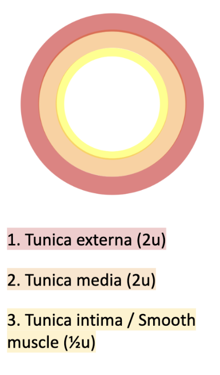
Drawn structure
Red: Tunica externa (2u)
Orange: Tunica media (2u)
Yellow: Tunica intima / Smooth muscle (½u)
Examples:
Aorta: Oxygenated blood from heart → organs through aorta
Pulmonary artery: Deoxygenated blood from heart → lungs through P.A.
Renal artery: Blood arrives at the kidney through this artery to be filtered
Outline the role, structural adaptations, drawn structure, and examples of veins.
Veins
Role: Transports deoxygenated blood (except pulmonary vein) from heart ventricles → Body tissues
SA:
Thinner walls than artery: lower pressure of blood
Larger lumen: lower pressure of blood hinders flow, thicker lumen to facilitates blood flow
Valves: ensure circulation of blood by preventing backflow
Venules: connect capillaries to veins
Thicker diameter: >10mm
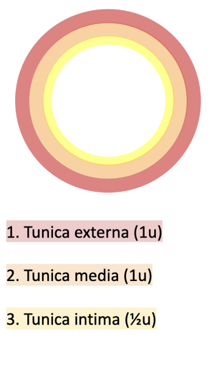
Drawn structure
Red: Tunica externa (1u)
Orange: Tunica media (1u)
Yellow: Tunica intima (½u)
Examples:
Vena cava: Deoxygenated blood from organs → heart through this vein
Pulmonary vein: Oxygenated blood from lungs → heart through this vein
Renal vein: Filtered blood leaves kidney through this vein
Outline the role, structural adaptations, and drawn structure of capillaries.
Capillaries
Role: Transports…
Connect arteries → veins
Food & oxygen from blood → cells
Waste (e.g. CO2) from cells → blood
SA:
Thin (1 cell-thick) & permeable walls: Facilitate diffusion of materials between cells (in tissue) and blood (in capillary)
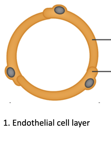
Drawn structure:
Endothelial cell layer
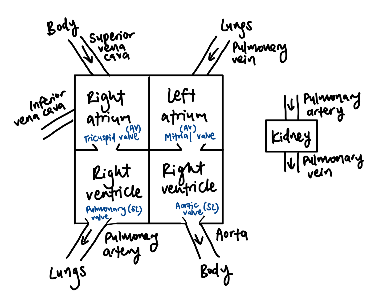
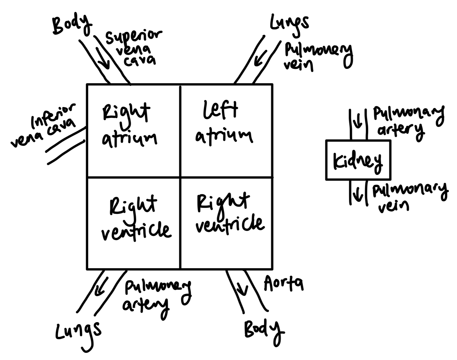
Draw the chambers and valves of the heart and the blood vessels connected to it. Describe the flow of blood through them.
Body —(vena cava)→ right atrium —(tricuspid/right AV valve)→ right ventricle —(pulmonary valve/right semilunar valve → pulmonary artery)→ lungs
Lungs —(pulmonary vein)→ left atrium —(mitrial/left AV valve)→ left ventricle —(aortic valve/left semilunar valve → aorta)→ body
Rules of blood travel (pressure changes for describing):
Atrium - When filled with blood, atrium contracts → forces blood into ventricle
Ventricle - Ventricle contracts → forces blood to exit the heart
Valve - When valves close, they prevent backflow of blood
Atrioventricular valve (mitral & tricuspid): Prevent backflow of blood back to the atrium
Semilunar valve (pulmonary & aortic): Prevent backflow of blood back to heart
Explain the cardiac cycle and pressure changes during it.
Cardiac cycle: Action of the heart from the ending of one heartbeat to the beginning of the next
When both atria & ventricles are relaxed (diastole), both atria fill with blood from veins (pressure rises).
When atria are filled, sinoatrial node sends impulses, causes atria to contract (systole) → blood is pushed into the ventricles (pressure rises).
AV valves are open, semilunar valves are closed.
When ventricles are filled, AV valves close to prevent backflow, ventricles contract (systole) → blood is pushed out through the semilunar valves → into pulmonary artery and aorta.
Explain how the heart circulates blood.
Overall, heart acts like a pump to circulate blood (Discovered by William Harvey)
How: Heartbeat is controlled by sinoatrial node within the heart (myogenic contraction) — heart muscle cells in the right atrium.
Sinoatrial node sends out an electrical impulse, stimulates contraction of the heart muscle tissue
Impulse causes atria to contract, stimulates atrioventricular (AV) node between atrium and ventricle
AV node sends signals via a nerve bundle (Bundle of His), stimulates nerve fibres (Purkinje fibres) in ventricular wall, causing ventricle to contract
Why: Sinoatrial node acts as a pacemaker — controls heart rate, and coordinates contraction of heart muscle
When interrupted, irregular contractions
Explain how the heart rate can be increased or decreased.
Consistently determined by myogenic contractions by SA node. But, can be changed by external impulses from:
Nerves:
Vagus nerve reduces heart rate
Cardiac sympathetic nerve increase heart rate
Hormones: Adrenal gland secretes epinephrine (adrenaline) hormone → heart, increases heart rate
Secreted in times of stress (e.g. exercise, heart failure, emotional stress/ excitement, pain)
Draw and explain the circulation systems in mammals versus fish. Explain the necessity of the former.
Mammals have double circulatory system — Blood passes through the heart twice per circuit.
Pulmonary circulation (separate circulation for lungs): Right pump sends deoxygenated blood from heart → to lungs (for oxygenation) → back to heart
Systemic circulation: Left pump sends newly oxygenated blood from heart → around body (to provide oxygen & nutrients to body tissues, becoming deoxygenated) → back to heart
Necessity of double pump:
Faster exchange of materials: Concentration gradient between blood & body cells is better maintained → Greater exchange rate of substances
Faster transport for oxygenated blood: Oxygenated blood is transported to tissues (e.g. muscles) that need it faster than single circulation systems
Fish have single pump circulation — Blood passes through the heartonce per circuit
Deoxygenated blood from body → atrium → ventricle → gills (to be oxygenated) → rest of body
Define occlusion of coronary arteries. Discuss its causes and consequences.
Occlusion of coronary arteries: Narrowing of arteries due to buildup of fats/ cholesterol/ other substances on artery walls.
Causes: SEGFORMS
Smoking
Exercise
Genetic predisposition
Forty-years old
Overweight
Male
Saturated fat-high diet
Consequences:
Tier 1: Atherosclerosis
Cholesterol builds up to form plaque → Artery walls lose elasticity
Lumen narrows, restricting blood flow
Blood clotting
If occurs in myocardial tissue, becomes…
Tier 2: Coronary heart disease
Coronary artery can become completely blocked