Chromosomal Abnormalities (Part I)
 Call Kai
 Learn
Learn Practice Test
 Spaced Repetition
 Match
Match Flashcards
 Knowt Play
1/76

There's no tags or description
Looks like no tags are added yet.
Name | Mastery | Learn | Test | Matching | Spaced | Call with Kai | Chat |
|---|
No analytics yet
Send a link to your students to track their progress
77 Terms
Meiosis
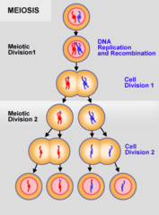
- A type of cell division that results in four daughter cells each with half the number of chromosomes of the parent cel
Diploid cells give rise to what in mitosis?
- Diploid (2N) give rise to haploid (1N) gametes
Meiosis is unique to which cells?
- Germ cells → spermatocytes and oocytes
Spermatocytes
- In testes, cells divide to create spermatocytes, which undergo meiosis (sexual div - identical # produced) to form 4 haploid spermatids.
Oocytes
- Female gametes, immature eggs
How is Meiosis Broken Down?
- Meiosis I
- Meiosis II
Meiosis I: Ploidy
- Diploid 2N → Haploid 1N
*Called reductive division
Where does the original Diploid (2N) cell come from before Meiosis I occurs?
- The zygote divides by mitosis to make all the body's diploid cells; some of those diploid cells in the testes or ovaries become germ cells and enter meiosis to make sperm or eggs.
How is ploidy determined?
- The amount of centromeres
Simplified Steps of Meiosis I
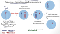
1. Diploid (2N, 2C) germ cell starts with 2 chromosomes (homologous).
2. Chromosomes within diploid cell are replicated during interphase. There are still 2 homologous chromosomes, except each chromosome now has a sister chromatid. Ploidy is now (2N 4C)
3. Meiosis I begins with crossover/recombination between maternal/paternal chromosomes
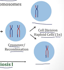
4. After recombination, cell divides → generates 2 haploid cells (1N, 2C)
Why are the cells 1N, 2C after cells divide in Meiosis 1?
- N: Each cell contains 1 centromere = 1N
- C: 2 copies of each chromosome as chromatids → 2C
Simplified Steps of Meiosis II
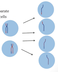
1. Starts with cells that just divided in Meiosis I, current ploidy is (1N, 2C)
2. Sister chromatids separate, cells divide → generates 4 haploid daughter cells (1N, 1C)
Meiosis I and II: Ploidy Summary
Meiosis I:
- Diploid cell (2N, 2C)
- Diploid cell after replication (2N, 4C)
- Cell divides, becomes haploid (1N, 2C) (2 Cells)
Meiosis II:
- Haploid cells from meiosis I separate sister chromatids and divide → 4x (1N, 1C) Cells
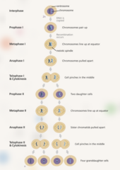
Steps and Ploidy of Spermatogenesis
1. Spermatogonium, Diploid (2N, 2C)
2. Mitosis, creating sister chromatids → 2x 1° Spermatocytes (2N, 4C)
3. Meiosis I separates homologous chromosomes → Each 1° generates 2x 2° Spermatocytes (1N, 2C)
4. Meiosis II → Sister chromatids separate → 4x Spermatids (1N, 1C)
5. Spermatids undergo spermatogenesis → Spermatozoa
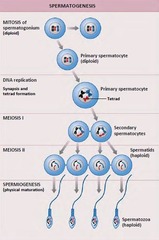
Steps and Ploidy of Oogenesis
1. 1° Oocytes (2N, 4C) are arrested in Meiosis I until puberty
2. At puberty, few 1° oocytes complete meiosis I → some form polar bodies, others form 2° Oocytes (1N, 2C)
3. In 2° Oocytes, meiosis II begins but is arrested in metaphase and is ovulated in this state
4. If ovulated 2° Oocyte is fertilized, meiosis II finishes → mature ovum (1N, 1C) + another polar body (1N, 1C)
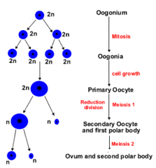
Aneuploidy
- Abnormal number of chromosomes.
Disomy, Monosomy, Trisomy
- Disomy: Two copies of each chromosome (Normal)
- Monosomy: One copy of each chromosome
- Trisomy: Three copies of each chromosome
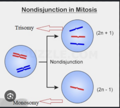
What causes Aneuploidy?
- Meiotic Nondisjunction: Failure of chromosome pairs to separate
When can Meiotic Nondisjunction occur?
- Can occur in either Meiosis I or II
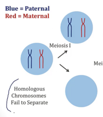
Meiosis I Nondisjunction
- Homologous chromosomes fail to separate
- Leaves one cell with homologous chromosomes, and the other with none
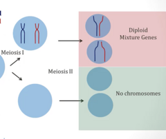
What happens during Meiosis II after a Meiosis I Nondisjunction?
- 2 diploid cells (only supposed to be 1N 1C)
- 2 cells with NO chromosomes
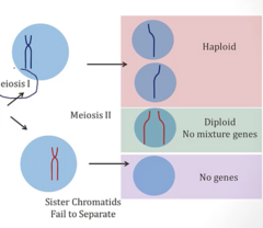
Meiosis II Nondisjunction
- Sister chromatids fail to separate
What does a Meiosis II Nondisjunction result in?
- Successful division of chromatids → 2x Haploid (1N, 1C)
- Unsuccessful division → 1x Diploid cell (2N, 2C) and 1x Cell with NO chromosomes
Where do the genes come from in the diploid cells created from Meiosis I/II Nondisjunction?
- Meiosis I: 2 Chromosomes, one from each parent
- Meiosis II: 2 Chromosomes, from SAME PARENT
When does Monosomy occur?
- Fertilization of a 1n (normal) with a 0n gamete → not typically viable
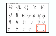
Turner Syndrome
- Females with only 1 X chromosome (due to monosomy)
When does Trisomy Occur?
- Fertilization of a 1n (normal) with 2n gametes (abnormal)
Trisomy 21
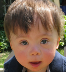
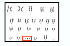
- 3 copies of chromosome 21 → Down syndrome
Trisomy 18
- 3 copies of 18 → Edward Syndrome
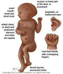
Edward Syndrome: Presentation
- Clenched fist showing overlapping fingers
- Rocker bottom feet
- Congenital heart defects
- abnormally shaped head, low set ears, mental retardation
- More common heart/GI defects than DS
Edward Syndrome: Prognosis
- Usually patients die within a few years of life
- 5-10% survive first year
Trisomy 13
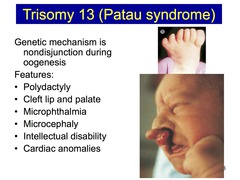
- Extra chromosome 13 → Patau Syndrome
Patau Syndrome: Presentation
- Severe Intellectual Disability (no separation of brain hemispheres)
- Extra fingers or toes (polydactyl)
- Cleft lip/palate
- Very small or absence of eyes
Common Cause of Trisomy
- Maternal Meiosis I Nondisjunction errors
What increases risk of trisomy?
- Advanced maternal age
Trisomy Genotype Example: Father with 21A/21B; Mother with 21C/21D
- 21ACD → Meiosis I NDJ (homologous chromosomes in mom failed to separate)
- 21ACC → Meiosis II NDJ (sister chromatids in mom failed to separate → 2 copies of same chromosome in fetus)
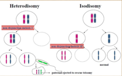
Uniparental Disomy
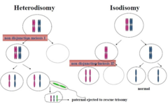
- Offspring receives 2 copies of a chromosome from 1 parent and no copies from the other parent.
Isodisomy
- Non-disjunction in meiosis II, 2 copies from father
Heterodisomy
- Non-Disjunction in Meiosis I, 2 copies from mother
Euploid
- An individual with the appropriate number of chromosomes but can lead to phenotype of recessive disease as they are inheriting both copies of chromosomes from ONE parent
*Child can inherit 2 copies of a recessive diseased gene from ONE parent*
What is the most common liveborn chromosome abnormality?
- Down Syndrome
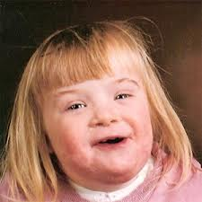
Down Syndrome: Presentation
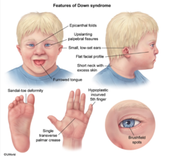
- Flat face/nose, low set ears, almond eyes, poor muscle tone, shorter height, palmar crease
- Heart/GI Malformations
- Intellectual disability, Early Alzheimer's
- ↑ Risk of leukemia
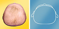
Is the posterior skull flat or rounded in Down syndrome?
- Flat
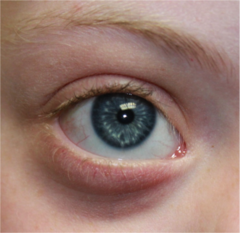
Brushfield Spots
- White spots in iris, found in Down syndrome
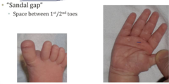
Down syndrome: Presentation found in hands and feet
- Short broad hands with large palmer crease
- Sandal gap on feet
50% of those with Down syndrome experience what?
- Congenital heart defects → most commonly endocardial cushion defects
5% of those with Down syndrome experience what?
- GI Abnormalities → duodenal atresia or stenosis
Why do those with Down syndrome often experience early alzheimer's?
- Amyloid precursor protein is found on chromosome 21 (have 3 copies)
- APP breaks down to form β-Amyloid → forms plaques → Alzheimers
Down Syndrome: Definitive Test
- Fetal Karyotype: Obtain sample of fetal cells, determining its number of chromosomes
Invasive and carries risk
Down syndrome: How is a fetal karyotype obtained?
- Chorionic Villus Sampling: Taking sample of placental tissue
- Amniocentesis: Inserting needle in amniotic cavity and taking sample of fluid which contains cells from the baby
Down syndrome: Karyogram
Down Syndrome: Noninvasive Screening
- Ultrasound
- Maternal Serum Testing
Down Syndrome: 1st Trimester Ultrasound Results
- Small, poorly-formed nasal bones
- Nuchal Translucency: Fluid under back of neck
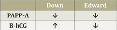
Down Syndrome: 1st Trimester Maternal Serum Results
- ↓↓ Plasma protein-A (PAPP-A)
- ↑↑ β-hCG
Down Syndrome: 2nd Trimester Maternal Serum Results
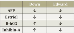
- ↓↓ α-Fetoprotein and Estriol (uE3)
- ↑↑ β-hCG and Inhibin A
Common Features of Trisomy Disorders
- All associated w/ advanced maternal age, due to NDJ
- Intellectual disability, physical deformities, heart defects
What is the 2nd most common trisomy in live births?
- Trisomy 18 → Edward Syndrome → SEVERE intellectual disability
Edward Syndrome: Occurrences in females vs males
- 3:1 Female to male ratio
Edward Syndrome: Screening
- So many physical and congenital abnormalities → easy to identify them in womb via ULTRASOUND
Edward Syndrome: 1st Trimester Maternal Serum Results
- ↓↓ PAPP-A
- ↓↓ β-hCG
Worse down syndrome → extra down
Edward Syndrome: 2nd Trimester Maternal Serum Results
- ↓↓ AFP
- ↓↓ Estriol
- ↓↓ β-hCG
- ↓↓ Inhibin-A
Worse down syndrome → extra down
How is Patau Syndrome detected?
- Fetal ultrasound > 90% of cases
Patau Syndrome: Prognosis
- Median survival is 7 days
- 91% die within first year of life
Patau Syndrome: 1st Trimester Maternal Serum Results
- ↓↓ PAPP-A
- ↓↓ β-hCG
Similar to edwards
Klinefelter and Turner Syndromes
- Klinefelter: Male with extra X (XXY)
- Turner: Female missing X (X0)
Klinefelter and Turner Syndromes: How can they be diagnosed?
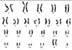
- Karyotype
Klinefelter XXY shown
When is Klinefelter less severe?
Mosaicism
- 46XY in some cells // 47XXY in others
What determines severity of Klinefelter?
- Ratio of estrogen:testosterone (↑ estrogen → more severe)
Klinefelter Syndrome: Presentation
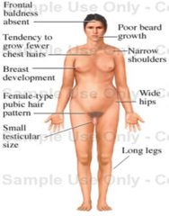
- Small/firm testes in males with atrophy in seminiferous tubules
- Low testosterone/delayed puberty
- Gynecomastia
- Female pubic hair pattern // Reduced facial and body hair
- Micropenis
- Long legs and arms
Klinefelter Syndrome: Hormone Levels
- ↑↑ FSH
- ↑ LH
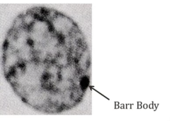
What can be found within the cells of pt's with Klinefelter's?
- Barr bodies in nucleus → inactivated X chromosome
Most common presentation of turner syndrome?
- 45XO
- Caused by sperm lacking an X Chromosome
Turner Syndrome: Presentation
- Short stature, broad chest
- Widely spaced nipples
- Webbed neck
- Swollen hands and feet
- Horse-shoe kidney (fused, sits lower in pelvis)
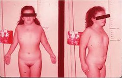
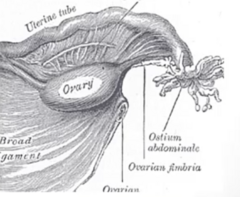
Turner Syndrome: Ovaries
- Non-functional → failure to menstruate → infertile
- "Streak Ovaries" Fibrous connective tissue in location of ovaries, very few follicles
Turner Syndrome: Hormones
- ↓↓ Inhibin B and Estrogen
- ↑↑ LH/FSH
Turner Syndrome: Cardiovascular Complication
- 30% Bicuspid aortic valve
- 5-19% Coarctation of aorta
- High BP
Turner Syndrome: Common associated diseases
- Osteoporosis
- TIID
- Thyroid Disease