Unit 3: Hip and Knee Joints + Ligaments
 Call Kai
 Learn
Learn Practice Test
 Spaced Repetition
 Match
Match Flashcards
 Knowt Play
1/67

There's no tags or description
Looks like no tags are added yet.
Name | Mastery | Learn | Test | Matching | Spaced | Call with Kai | Chat |
|---|
No analytics yet
Send a link to your students to track their progress
68 Terms
Hip Joint Type
Ball and Socket Joint (Synovial)
Hip Joint Characteristics
1. Stable - nearly the entire head of the femur is within the acetabulum
2. Weight bearing
3. Less mobile
Hip Joint is between
1. head of femur
2. acetabulum of os coxae
Hip Joint Movements
1. Flexion/Extension
2. Abduction/Adduction
3. Internal/External Rotation
4. Circumduction
What ligament supports head of femur?
Ligament of the head of the femur
Synovial Membrane attaches to : (3 things)
1. Femur
2. Acetabulum
3. Covers neck of the femur
Stabilizing Ligaments of the Hip Joint (3)
1. Iliofemoral Ligament
2. Pubofemoral Ligament
3. Ischiofemoral Ligament
What do the hip joint ligaments do when extended?
Become taut/tightened
Iliofemoral Ligament
Anterior to Hip Joint
Iliofemoral Ligament connects
Ilium between anterior inferior iliac spine and margin and base of acetabulum TO the intertrochanteric line of the femur
Pubofemoral Ligament
anterioinferior to hip joint
Pubofemoral Ligament connects
from iliopubic eminence + obturator membrane TO the fibrous joint capsule and the iliofemoral ligament
Ischiofemoral Ligament
posterior to hip joint
Ischiofemoral Ligament connects
from ischium (the posterioinferior part up until the acetabulum) TO the greater trochanter of the femur
Pelvic Girdle Joints (2)
1. Sacroiliac Joint
2. Pubic Symphysis
Sacroiliac Joint
Between sacrum and ilium of os coxae
Stabilizing Ligaments of Pelvic Girdle - Sacroiliac Joint
1. Anterior Sacroiliac Ligament
2. Posterior Sacroiliac Ligament
Stabilizing Ligaments of Pelvic Girdle - Pubic Symphysis
1. Superior Pubic Ligament
2. Inferior Pubic Ligament
Stabilizing Ligaments of Pelvic Girdle - Pelvic Wall
1. Sacrospinous Ligament
2. Sacrotuberous Ligament
Sacrospinous Ligament connects
from ischial spine TO the sacrum and coccyx
Sacrotuberous Ligament connects
from posterior superior iliac spine TO the coccyx and ischial tuberosity
How do structures get from abdomen + pelvis to the lower limb?
1. Obturator Canal
2. Greater Sciatic Foramen
3. Lesser Sciatic Foramen
4. Below Inguinal Ligament
Obturator Canal connects
abdominopelvic region and medial compartment of the thigh
Greater Sciatic Foramen formed by:
1. Greater sciatic notch
2. Sacrospinous ligament
3. Sacrotuberous ligament
4. Lateral border of the sacrum
Lesser Sciatic Foramen formed by
Lesser sciatic notch and sacrotuberous ligament
What passes through the Lesser Sciatic Foramen?
Tendon of Obturator Internus
Inguinal Ligament is formed by:
anterior superior iliac spine and pubic tubercle
Knee Joint is the Articulation Between (2 separate)
1. Femur and Tibia
2. Patella and Femur
What knee articulation = weight bearing?
Femur + Tibia
What kind of joint is the knee joint?
Hinge joint (flexion and extension)
Ligaments that reinforce the knee
Collateral Ligaments
2 Types of Collateral Ligaments
1. Tibial Collateral Ligament
2. Fibular Collateral Ligament
Cruciate Ligaments (2):
1. Anterior Cruciate Ligament (ACL)
2. Posterior Cruciate Ligament (PCL)
What do cruciate ligaments connect?
femur and tibia
When femoral condylar surfaces that articulate with tibia = flexed, what shape are they?
Curved/rounded
When femoral condylar surfaces that articulate with tibia = extended, what shape are they?
Flat
Menisci is made of
fibrocartilage
Shape of menisci
C-shaped
2 types of menisci
1. Medial Meniscus
2. Lateral Meniscus
Where do the menisci attach?
Intercondylar Eminence of the Tibia
Which meniscus is attached to joint capsule?
Medial = married
Which meniscus is more mobile?
Lateral = loose, not attached to the joint capsule
Where is medial meniscus located?
Between medial femoral and medial tibial condyles
Where is lateral meniscus located?
Between lateral femoral and lateral tibial condyles
What connects the medial and lateral menisci?
Transverse ligament of the knee
Function of the menisci
Help improve the fit between femoral and tibial condyles during movement
Cruciate ligaments are on (outside/inside) of synovial membrane
outside
What separates synovial membrane from patellar ligament?
Infrapatellar fat pad
4 Bursa of the Knee
1. Suprapatellar
2. Subcutaneous pre patellar
3. Deep infra patellar
4. Subcutaneous Infrapatellar
ACL function
Prevents anterior displacement of tibia relative to femur
PCL function
prevents posterior displacement of tibia relative to femur
Classic ACL vs. PCL injury
ACL - pivot/twist with planted foot
PCL - blow to anterior tibia/dashboard injury
what forms the roof of the ankle joint?
tibia
what forms the medial side of the ankle joint?
medial malleolus
what forms the lateral side of the ankle joint?
the lateral malleolus
what is at the bottom of the ankle joint? where does it connect?
talus bone
stabilizing ligaments of the ankle
1. Medial (Deltoid) Ligaments
2. Lateral Ligaments
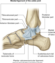
Medial (Deltoid Ligaments): (4 Parts)
4 parts:
1. Tibionavicular Part - Medial Malleolus of Tibia to Navicular
2. Tibiocalcaneal - Medial Malleolus of Tibia to Calcaneus
3. Posterior Tibiotalar Part - Posterior portion of Medial Malleolus of Tibia to Posterior Portion of Talus
4. Anterior Tibiotalar part - Anterior portion of Medial Malleolus of Tibia toAnterior Portion of Talus
Lateral Ligaments (3 Parts)
1. Anterior Talofibular Ligament - Anterior margin of lateral malleolus of fibula to the Talus
2. Posterior Talofibular Ligament - Medial side of lateral malleolus to posterior portion of Talus
3. Calcaneofibular Ligament - Lateral malleolus to the Calcaneus
Intertarsal Joints Function
Allow for:
- Inversion
- Eversion
-Supinating
-Pronating
Major Intertarsal Joints
1. Subtalar
2. Talocalcaneonavicular
3. Calcaneocuboid
Subtalar Joint
Between Talus and Calcaneus
- allows inversion/eversion of foot
stabilized by: lateral, medial, posterior talocalcaneal ligaments and interosseous talocalcaneal ligament
Talocalcaneonavicular
Synovial joint between:
- Talus and Calcaneus
- Talus and Spring Ligament
- Talus and Navicular
Allows for:
- Inversion/Eversion
-Supination/Pronation
Widest range of motion of tarsometatarsal Joint:
Metatarsal of hallux + Medial Cuneiform
Metatarsophalangeal Joints are reinforced by (2 ligaments)
1. Collateral Ligaments
2. Plantar Ligaments
Deep Transverse Metatarsal Ligaments
link heads of metatarsals together so they can act like a single unit
Fibular Retinacula
Binds tendons of Fibularis Brevis + Longus to lateral side of foot
Goes from Lateral Malleolus of Fibula to Calcaneus
Flexor Retinaculum
connective tissue above the Tom, Dick AN' Harry