A&P - Ch. 12
1/363
There's no tags or description
Looks like no tags are added yet.
Name | Mastery | Learn | Test | Matching | Spaced |
|---|
No study sessions yet.
364 Terms
Q: What is the starting structure of embryonic development?
A: The blastocyst, a hollow ball of cells.
Q: What does the blastocyst eventually do during early development?
A: It starts to fold on itself to form three germ layers: ectoderm, mesoderm, and endoderm.
Q: What does the endoderm and mesoderm give rise to?
A: Connective tissue and other internal structures (exact tissues vary by system; mesoderm is especially key for connective tissue).
Q: What does the ectoderm develop into?
A: The skin and the nervous system.
Q: What forms at the site where the ectoderm folds inward?
A: Neural folds and the neural groove.
Q: What does the folding of the ectoderm lead to?
A: Formation of the neural tube and neural crest.
Q: What does the neural tube become?
A: The central nervous system (CNS).
Q: What does the neural crest become?
A: The peripheral nervous system (PNS).
Q: What happens to the remaining ectoderm after folding?
A: It forms the epidermis.
Q: After the neural tube forms, what are the three primary brain vesicles that develop?
A: Prosencephalon (forebrain), Mesencephalon (midbrain), and Rhombencephalon (hindbrain).
Q: What structure forms below the rhombencephalon?
A: The future spinal cord.
Q: What does the Prosencephalon (forebrain) develop into?
A: The Telencephalon and the Diencephalon.
Q: What structures come from the Telencephalon?
A: The cerebrum and the eye cups (retina).
Q: What structures form from the Diencephalon?
A: The thalamus and hypothalamus.
Q: What does the Mesencephalon develop into?
A: The midbrain (remains as is).
Q: What does the Rhombencephalon develop into?
A: The Metencephalon and Myelencephalon.
Q: What structures form from the Metencephalon?
A: The pons and cerebellum.
Q: What structure forms from the Myelencephalon?
A: The medulla oblongata.
Q: What are the three primary brain vesicles that form from the neural tube?
Prosencephalon → Forebrain
Mesencephalon → Midbrain
Rhombencephalon → Hindbrain
Below these structures → Future spinal cord
Q: After the neural tube forms, what does it develop into structurally?
A: A longer structure with a central tube and three primary brain vesicles
the 3 brain vesicles in development:
Prosencephalon (______)
Mesencephalon (______)
Rhombencephalon (_____)
forebrain
midbrain
hindbrain
Q: What lies below the Rhombencephalon during development?
A: The future spinal cord.
Q: What secondary brain structures arise from the Prosencephalon?
Telencephalon → Cerebrum + Eye cups (optic vesicles)
Diencephalon → Thalamus + Hypothalamus
Q: What secondary brain structure forms from the Mesencephalon?
A: The midbrain (Mesencephalon does not subdivide further).
Q: What does the Rhombencephalon divide into?
Metencephalon → Pons and Cerebellum
Myelencephalon → Medulla oblongata
Q: What structure does the ventricular system originate from?
A: The central cavity of the neural tube, which expands and reshapes during brain vesicle formation.
Q: From which brain region do the lateral ventricles develop?
A: The Telencephalon, which comes from the Prosencephalon (forebrain).
Q: From which brain region does the third ventricle develop?
A: The Diencephalon, part of the Prosencephalon (forebrain).
Q: What is the function of the third ventricle?
:
Located in the midline between thalamic halves
Circulates CSF from lateral ventricles to the cerebral aqueduct
The _____ turns into the cerebrum
telencephalon
What does the diencephalon turn into?
Thalamus and hypothalamus
What does the metencephalon turn into ?
pons and cerebellum
What does the mylencephalon turn into?
medulla oblongata
Where does the lateral ventricles form?
Prosencephalon (it is initial)
Where does the third ventricles form?
in the diencephalon
where does the cerebral aqueduct form?
mid brain
where does the 4th ventricle form?
in the mylencephalon
_________ is the outermost germ layer (on the outside of the embryo)
Ectoderm
what does the Ectoderm turn into?
Epidermis (skin, hair, nails)
Central and Peripheral Nervous System (via the neural tube and neural crest)
Sensory organs (e.g., eyes, ears)
Tooth enamel
_______ is the Middle germ layer
Mesoderm
What is the Mesoderm turn into?
Muscles
Bones
Connective tissue
Cardiovascular system
Kidneys and reproductive organs
Dermis (deeper skin layer)
_______ is the Innermost germ layer
Endoderm
what does the Endoderm turn into?
Lining of the gut and respiratory tract
Organs such as the liver, pancreas, and lungs
______ are the Raised edges of the ectoderm that elevate and move toward the midline during neurulation
Neural Fold
What is the function of the neural fold?
They help form the neural tube by fusing together at the midline.
______ is a central depression or indentation between the neural folds
Neural Groove
What is the function of the neural groove?
Deepens as the folds elevate; eventually the folds meet and close over the groove to form the neural tube.
Q: What are the primary brain vesicles?
Prosencephalon (forebrain)
Mesencephalon (midbrain)
Rhombencephalon (hindbrain)
Q: What are the secondary brain vesicles?
Prosencephalon → Telencephalon + Diencephalon
Mesencephalon → stays as midbrain
Rhombencephalon → Metencephalon + Myelencephalon
Q: Which parts of the brain form inside the original neural tube cavity (within brain matter)?
The ventricles (lateral, third, cerebral aqueduct, fourth) form as the internal space, while surrounding tissue becomes brain matter (cortex, thalamus, etc.).
Q: Why do the ventricles appear "smaller" in the mature brain?
Because the surrounding brain tissue grows and expands around them, but the ventricle space itself remains as a CSF-filled cavity.
Q: What is spina bifida?
A: A neural tube defect in which the spinal column does not close properly during development.
Q: What are the three types of spina bifida?
Occulta
Meningocele
Myelomeningocele
Q: What is Spina Bifida Occulta?
Mildest form
Only vertebrae affected; spinal cord and nerves intact
Often no symptoms; sometimes a tuft of hair or dimple on back
No major deficits, often no treatment needed
Q: What is Meningocele?
A:
Meninges protrude through spinal defect, but spinal cord remains in place
Can cause mild deficits
Often treated with surgery
Q: What is Myelomeningocele?
Most severe form
Both meninges and spinal cord protrude through opening
Often results in motor, sensory, and bladder/bowel deficits
May require surgery, physical therapy, and lifelong support
Q: What nutrient helps prevent neural tube defects like spina bifida?
A: Vitamin B9 (folate or folic acid)
Q: Does folate deficiency guarantee neural tube defects?
A: No, folate deficiency increases risk, but doesn't always result in defects.
What does a zygote turn into?
Blastocyst (hollow ball of cells)
what does a Blastocyst turn into?
Embryo (which contains the ectoderm, mesoderm, and endoderm)
what does an Embryo turn into?
Fetus
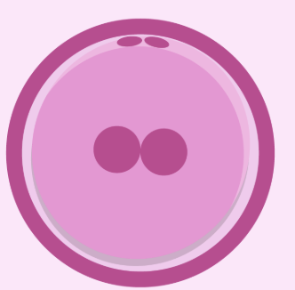
What is the following?
A zygote
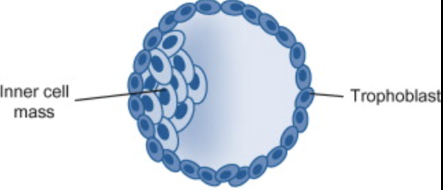
What is the following?
Blastocyst
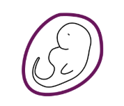
What is the following?
Embryo
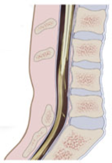
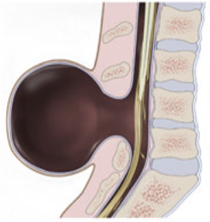
What type of spina bifida is this?
Spina bifida oculta (least severe)
What type of spina bifida is this?
miningocele
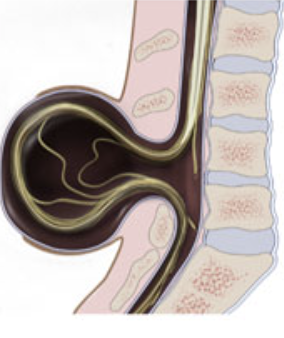
What type of spina bifida is this?
Myelomeningocele (most severe)
The ______ is a hollow, tube-like structure that forms from a layer of cells in the embryo.
neural tube
The neural tube eventuals becomes parts of the ______ nervous system which includes?
Central
Brain(upper part) and spinal cord (lower part)
Proper neural tube closure is critical. If it doesn’t close correctly, it can lead to neural tube defects (NTDs) such as?
Spina bifida – spinal cord doesn’t form properly.
Anencephaly – parts of the brain and skull are missing.
where is the central canal located?
in the spinal cord
Q: What is the basic structural pattern of the spinal cord in terms of white and gray matter?
A: The spinal cord has a central cavity surrounded by gray matter, with outer white matter.
Q: How is gray and white matter arranged in the brain stem?
A: The brain stem has additional regions of gray matter (brain nuclei) buried within the white matter.
Q: How is gray and white matter arranged in the cerebrum and cerebellum?
A: The cerebrum and cerebellum contain islands of gray matter (nuclei) within white matter, as well as an outer cortex of gray matter.
Q: What is gray matter composed of?
A: Neuron cell bodies and short nonmyelinated neurons.
Q: What is white matter composed of?
A: Mostly myelinated axons (with some nonmyelinated axons).
Q: What is the main function of cerebrospinal fluid (CSF) in the central nervous system?
A: CSF acts as a liquid cushion that provides buoyancy for the CNS.
Q: Besides acting as a cushion, what are two other roles of CSF?
A: It provides nutrients and circulates signaling factors.
Q: What are choroid plexuses and what is their function?
A: Choroid plexuses are layers of ependymal cells lining the ventricles that produce CSF from blood.
Q: What are meninges and their primary functions?
A: Meninges are connective tissue layers that protect nervous tissue, safeguard blood vessels, and contain cerebrospinal fluid (CSF).
Q: What is the dura mater and what does it enclose?
A: The dura mater is a two-layered sheet attached to bone that encloses the dural venous sinuses.
Q: What is the arachnoid mater and where is it located?
A: The arachnoid mater is a loose covering of the brain located beneath the dura mater, separated by the subdural space.
Q: What is contained in the subarachnoid space?
A: The subarachnoid space contains cerebrospinal fluid (CSF) and large blood vessels.
Q: What are arachnoid granulations and what is their function?
A: Arachnoid granulations are structures that protrude into the superior sagittal sinus to allow CSF to drain into the venous blood.
Q: What is the pia mater and how is it described?
A: The pia mater is a delicate membrane that closely wraps around the CNS like "shrink wrap."
Q: What structures form the blood-brain barrier?
A: Impermeable tight junctions between capillaries, supported by astrocytes and mesenchymal pericytes.
Q: What types of substances can pass through the blood-brain barrier?
A: Lipid-soluble substances such as fats, fatty acids, gases, alcohol, nicotine, and anesthetics.
Q: Which nutrients are allowed to cross the blood-brain barrier?
A: Glucose, some amino acids, and specific ions.
Q: What substances are blocked by the blood-brain barrier?
A: Metabolic waste, proteins, most toxins and drugs, potassium, and nonessential amino acids.
Q: What are the three functional areas of the cerebral cortex?
A: Motor areas, sensory areas, and association areas.
Q: What type of neurons are found in the cerebral cortex?
A: All neurons in the cerebral cortex are interneurons.
Q: How do the cerebral hemispheres relate to body function?
A: Each hemisphere is concerned with sensory and motor functions of the contralateral (opposite) side of the body.
Q: Are the two cerebral hemispheres structurally and functionally the same?
A: They are largely symmetrical in structure but not in function.
Q: What is lateralization in the context of the cerebral cortex?
A: Lateralization refers to the idea that functions are shared but not equally divided between hemispheres, resulting in cerebral dominance.
Q: Where is the primary motor cortex located?
A: In the precentral gyrus of the frontal lobe.
Q: What is the function of the primary motor cortex?
A: It controls voluntary movements of skeletal muscles.
Q: Where is the primary somatosensory cortex located?
A: In the postcentral gyrus of the parietal lobe.
Q: What is the function of the primary somatosensory cortex?
A: It receives and processes sensory information from the skin, skeletal muscles, and joints.
Q: What does "motor map in the precentral gyrus" mean?
A: It refers to a topographic organization of the body in the motor cortex, where different body parts are controlled by specific regions—known as the motor homunculus.
Q: What does "sensory map in the postcentral gyrus" mean?
A: It represents a map of sensory input across the body, with specific regions of the cortex receiving input from specific body parts—known as the sensory homunculus.