Ch 6: bones and Skeletal tissues
 Call Kai
 Learn
Learn Practice Test
 Spaced Repetition
 Match
Match Flashcards
 Knowt Play
1/39

There's no tags or description
Looks like no tags are added yet.
Name | Mastery | Learn | Test | Matching | Spaced | Call with Kai | Chat |
|---|
No analytics yet
Send a link to your students to track their progress
40 Terms
osteology
study of bones, bone is like a bank (it stores stuff)
ligament
hold bones together at the joint
tendon
attach muscle to bone
mineralization/calcification
accumulation and deposition of calcium and other mineral salts in body tissues
osseous tissue
bone tissue, a hard, specialized connective tissue that forms the rigid structural framework of the human body
Diaphysis
tubular shaft that forms long axis of bone
Consists of compact bone surrounding central medullary cavity that is filled with yellow marrow in adults

Epiphysis
ends of long bones that consist of compact bone externally and spongy bone internally
– Articular cartilage covers articular (joint) surfaces

epiphyseal plate
thin layer of hyaline cartilage located near the ends (metaphysis) of long bones in children and adolescents

Articular cartilage
Layer of hyaline cartilage that covers the joint surface where one bone meets another,
Allows joint to move more freely and relatively friction free

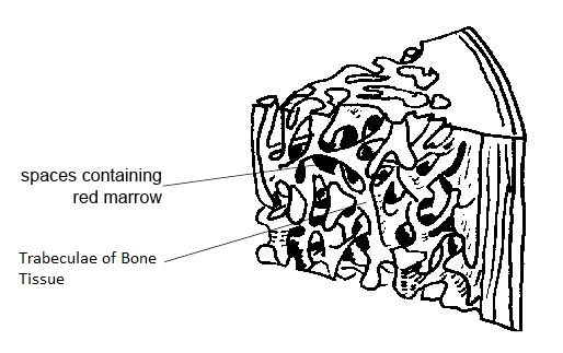
Trabeculae
needle-like or flat pieces of bone that make up spongy bone
Ossification (osteogenesis)
is the process of bone tissue formation
– Formation of bony skeleton begins in month 2 of development
– Postnatal bone growth occurs until early adulthood – Bone remodeling and repair are lifelong
Achondroplasia
the most common genetic cause of disproportionate short stature, or dwarfism
Orthopedics
a medical specialty focused on the diagnosis, treatment, prevention, and rehabilitation of conditions involving the musculoskeletal system
sella turcica
a saddle-shaped depression in the sphenoid bone at the base of the skull that holds and protects the pituitary gland
anabolic steroids
are synthetic versions of the male hormone testosterone that build muscle and treat specific medical conditions
Functions of skeleton
Support: Acts as a rigid framework that holds the body upright and gives it shape.
Movement: Works with skeletal muscles, tendons, and joints so the body can walk, run, and grasp items.
Protection: Shields fragile internal organs from external force, such as the skull protecting the brain or ribs guarding the heart and lungs.
Blood Cell Production: Houses red bone marrow inside certain bones to manufacture red blood cells, white blood cells, and platelets.
Storage: Keeps essential minerals like calcium and phosphorus—along with energy-storing fat in yellow marrow—ready for the body to use.
long bones
Longer than they are wide
▪Limb bones

flat bones
▪Thin, flat, slightly curved
▪Sternum, scapulae, ribs, most skull bones

Short bones
Cube-shaped bones (in wrist and ankle)
▪Sesamoid bones form within tendons (example:
patella) ▪Vary in size and number in different individuals

Irregular bones
▪Complicated shapes ▪Vertebrae and hip bones

compact bone
dense outer layer on every bone that
appears smooth and solid
spongy bone
made up of a honeycomb of small,
needle-like or flat pieces of bone called trabeculae
▪Open spaces between trabeculae are filled with red
or yellow bone marrow

Osteoblast (miotic)
Location: on the surfaces of bones where new bone tissue is actively formed or repaired
Function: Bone-forming cells that secrete unmineralized bone
matrix called osteoid

Osteoclast
Location: on the surfaces of bone tissue at sites of active bone remodeling and resorption
Function: Derived from same hematopoietic stem cells that
become macrophages
– Giant, multinucleate cells function in bone resorption
(breakdown of bone)
– When active, cells are located in depressions called resorption bays
– Cells have ruffled borders that serve to increase
surface area for enzyme degradation of bone
▪Also helps seal off area from surrounding matrix

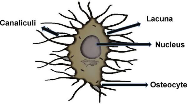
Osteocyte
Location: inside mature bone tissue, specifically residing in small cavities called lacunae located between the layers of the hardened bone matrix
Function: Mature bone cells in lacunae that no longer divide
– Maintain bone matrix and act as stress or strain sensors
▪Respond to mechanical stimuli such as increased force on bone or weightlessness
▪Communicate information to osteoblasts and osteoclasts (cells that destroy bone) so bone remodeling can occur
osteogenic cells
Location: Mitotically active stem cells in periosteum and
endosteum
Function: When stimulated, they differentiate into osteoblasts or
bone-lining cells, Some remain as osteoprogenitor stem cells

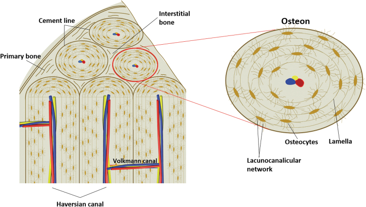
Osteon
is the structural unit of compact bone
– Consists of an elongated cylinder that runs parallel to
long axis of bone
▪Acts as tiny weight-bearing pillars
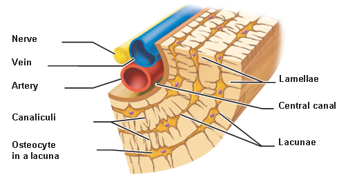
– An osteon cylinder consists of several rings of bone
matrix called lamellae
▪Lamellae contain collagen fibers that run in different
directions in adjacent rings
▪Withstands stress and resist twisting
▪Bone salts are found between collagen fibers
canaliculi
hairlike canals that connect lacunae to each other
and to central canal
When matrix hardens and cells are trapped, the canaliculi form
runs through core of osteon
▪Contains blood vessels and nerve fibers
Perforating (Volkmann’s) canals
canals lined with endosteum that occur at right angles to central canal
▪Connect blood vessels and nerves of periosteum, medullary cavity, and central canal

Lacunae
small cavities that contain osteocytes
Interstitial lamellae
Lamellae that are not part of osteon
▪Some fill gaps between forming osteons; others are
remnants of osteons cut by bone remodeling

Circumferential lamellae
Just deep to periosteum, but superficial to endosteum, these layers of lamellae extend around entire surface of diaphysis
▪Help long bone to resist twisting

red bone marrow
Location: Found primarily in the spongy (trabecular) bone of flat bones like the hip (ilium), sternum, ribs, skull, and vertebrae, as well as at the ends (epiphysis/metaphysis) of long bones in children. In adults, it is mainly restricted to the axial skeleton.
Function: Responsible for hematopoiesis—the production of red blood cells (carries oxygen), white blood cells (fights infection), and platelets (aids blood clotting). It also helps break down old red blood cells and store iron

Yellow bone marrow
Found mainly in the central medullary cavity (the hollow shaft or diaphysis) of long bones, such as the femur and humerus.
Function: Primarily stores adipose tissue (fat) to serve as a chemical energy reserve. If the body experiences severe, life-threatening blood loss, yellow marrow can convert back into red marrow to help produce missing blood cells.

Endochondral ossification
Endochondral ossification is the natural process where embryonic cartilage is gradually replaced by hard bone tissue to form most of the skeleton

intramembranous ossification
the direct process of making bone tissue from embryonic connective tissue sheets without using a cartilage model first

calcium homeostasis with excess blood calcium
High Blood Calcium (Hypercalcemia)
Detection: High blood calcium levels are detected by the parathyroid receptors.
Inhibition: The high calcium level acts as a negative feedback signal. It tells the parathyroid glands to stop releasing PTH.
Thyroid Action: The thyroid gland may also release calcitonin to help lower calcium by storing it back in the bones, though PTH control is much stronger.
Result: Calcium release stops, and blood levels drop back to the normal range.

calcium homeostasis with blood calcium deficiency
Low Blood Calcium (Hypocalcemia)
Detection: The parathyroid glands sense low calcium in the blood using special receptors.
Release: The glands release Parathyroid Hormone (PTH).
Bone Action: PTH activates osteoclasts to break down bone tissue, which releases stored calcium into the bloodstream.
Kidney Action: PTH tells the kidneys to reabsorb more calcium so less is lost in urine. It also helps create active vitamin D (calcitriol).
Intestine Action: Active vitamin D helps the gut absorb more calcium from food
Result: Blood calcium levels go back up to normal

osteoporosis
in older women and young women with low levels of body fat
group of diseases in which bone resorption exceeds deposit
small pores form and bone becomes brittle
• Matrix remains normal, but bone mass declines
– Spongy bone of spine and neck of femur most
susceptible
▪Vertebral and hip fractures common
process of ossification
the biological process of laying down new bone material by specialized cells called osteoblasts
1. Ossification centers are formed when mesenchymal
cells cluster and become osteoblasts
2. Osteoid is secreted, then calcified
3. Woven bone is formed when osteoid is laid down
around blood vessels, resulting in trabeculae
▪Outer layer of woven bone forms periosteum
4. Lamellar bone replaces woven bone, and red marrow
appears