PHSL Lecture 1
1/16
There's no tags or description
Looks like no tags are added yet.
Name | Mastery | Learn | Test | Matching | Spaced |
|---|
No study sessions yet.
17 Terms
What are the two circuits of the cardiovascular system and what side of the heart and valves are each associated with?
The systemic circuit (heart to body) which leaves from the left side of the heart through the aortic semilunar valve, and the pulmonary circuit (heart to lungs) which leaves through the right side of the heart through the pulmonary semilunar valve
What is the typical blood volume of a human
4-6L
What is the main functions (3) of the cardiovascular system?
Provide adequate supply of oxygen and nutrients
Remove any unwanted metabolic byproducts
Transporting substances and heat
Because diffusion of blood on its own is too slow
Where is the heart located and describe this structure
Within the chest cavity within the mediastinum, and is enclosed by a protective sac called the pericardium. The pericardium has two layers: fibrous pericardium and the inner serous pericardium, which includes a parietal layer and a visceral layer (adjacent to the heart)
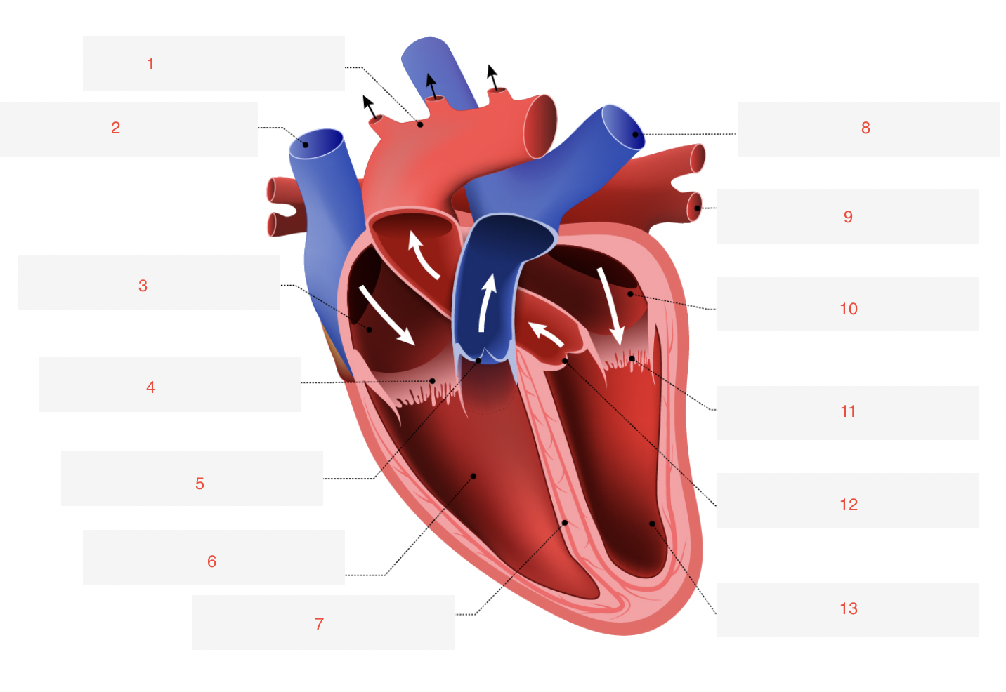
label the structures of the heart in the image
Aorta
Superior Vena Cava
Right Atrium
Tricuspid Valve
Pulmonary Valve
Right Ventricle
Septum
Pulmonary Artery
Pulmonary vein
Left Atrium
Mitral Valve
Aortic Valve
Left Ventricle
Which side of the heart is thicker (left or right) and why?
The left side of the heart is thicker because it needs to generate higher pressure to pump blood throughout the entire body, whereas the right side pumps blood only to the lungs so requires less pressure and has a thinner wall.
Does the ventricles or the atria have thicker walls and why?
The ventricles have thicker walls than the atria because they need to generate greater pressure to pump blood to the lungs and the rest of the body, unlike the atria, which primarily receive blood
what are the names of the two AV valves and the two semilunar valves and what sides of the heart are each associated with?
AV valves: mitral valve (left) and tricuspid valve (right)
Semilunar valve: aortic valve (LV-aorta) and pulmonary valve (RV-PA)
What are cardiac muscle cells called, and what structures help them contract efficiently?
Cardiac muscle cells are called cardiac myocytes are specialised for continuous rhythmic contraction. They contain key structures:
Sarcolemma: the plasma membrane of myocytes.
T-tubules: invaginations of the sarcolemma that carry electrical signals deep into the cell.
Sarcoplasmic reticulum (SR): stores calcium needed for contraction.
Mitochondria: abundant to meet the high energy demands of constant contraction
What are diastole and systole in the cardiac cycle?
Diastole is the relaxation phase when the heart fills with blood.
Systole is the contraction phase when the heart pumps blood out.
What are myofibrils and sarcomeres, and how do they relate to muscle contraction?
Myofibrils are long protein bundles in muscle cells made of repeating units called sarcomeres.
Sarcomeres are the basic contractile units containing actin and myosin filaments. Their shortening produces muscle contraction.
How are cardiac muscles connected together
Cardiac myocytes are connected end-to-end by intercalated discs, which contain gap junctions. These gap junctions enable electrical coupling between neighboring cells, allowing the heart muscle to contract as a synchronised unit
What are the physical and physiological factors that contribute to maintaining arterial blood pressure?
Physiological Factors: cardiac output (heart rate x stroke volume) and peripheral resistance
Physical Factors: Arterial blood volume and Arterial compliance
What is the mean arterial pressure equation and explain each component
MAP= cardiac output (CO) x total peripheral resistance (TPR)
CO = (flow) the volume of blood ejected by either ventricle in one minute
TPR: vessels in series and parallel determine total peripheral vascular resistance
What factors (equation) make up cardiac output and explain each component
CO = SV x HR
SV: volume of blood ejected from either ventricle per beat (L.beat^-1)
HR: beats per minute (beat.min^-1)
What is the average SV for an adult at rest and during intense excercise?
at rest: 5.4 Lxmin^-1
intense excercise: 28 Lxmin^-1
what is Poiseuille’s law?
Resistance = (8.n.l)/(pi.r^4)
where
n = viscosity of the fluid
l = length of the vessel
r = radius → largest determinant of resistance