Histology - Endocrine
1/89
There's no tags or description
Looks like no tags are added yet.
Name | Mastery | Learn | Test | Matching | Spaced |
|---|
No study sessions yet.
90 Terms
Adenohypophysis
What does #1, #4, and #5 form?

Pituitary
Identify the specimen.

Intermediate Lobe
Identify the structure labeled as 1.

Posterior Lobe
Identify the structure labeled as 2.

Pituitary Stalk
Identify the structure labeled as 3.
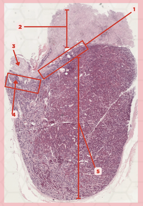
Pars Tuberalis
Identify the structure labeled as 4.

Anterior Lobe
Identify the structure labeled as 5.

Neural Ectoderm
What ectoderm is #2  derived from?

Oral Ectoderm
What ectoderm #1, #3, & #4 derived from?

Pituitary
Identify the specimen.
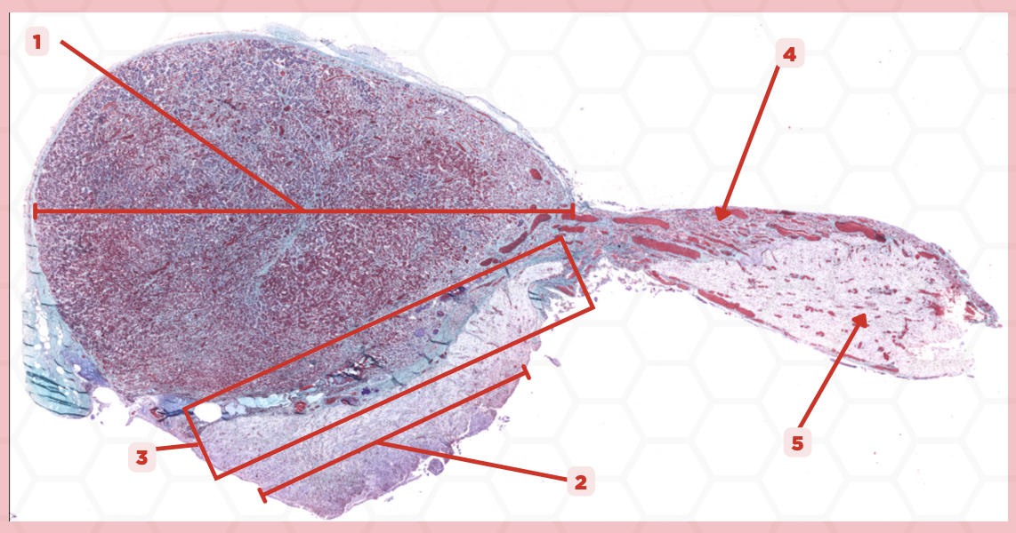
Anterior Lobe
Identify the structure labeled as 1.

Posterior Lobe
Identify the structure labeled as 2.

Intermediate Lobe
Identify the structure labeled as 3.

Pars Tuberalis
Identify the structure labeled as 4.

Pituitary Stalk
Identify the structure labeled as 5.

Pia Mater
#1 envelopes the pineal gland. From which structure is #1 derived from?

Pineal Body
Identify the specimen.

Capsule
Identify the structure labeled as 1.
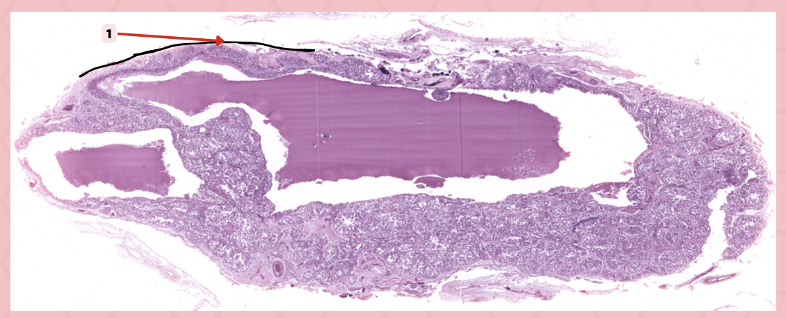
Fenestrated Capillaries
What type of capillaries are embedded inside #1?

Pineal Body
Identify the specimen.

Lobule
Identify the structure labeled as 1.
#5 Pinealocytes (Chief Cells)
Which among these structures are modified neurons that are responsible for synthesizing melatonin?

#1 Interstitial Cells
Which among these are supporting cells that are morphologically similar to astrocytes?

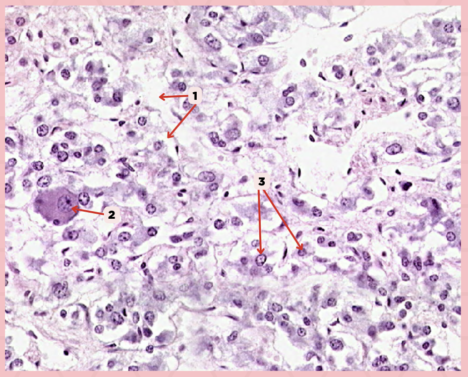
Interstitial Cells
Identify the structure labeled as 1.
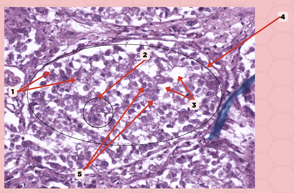
Clusters (Cords) of Cells
Identify the structure labeled as 2.

Capillaries
Identify the structure labeled as 3.

Lobule
Identify the structure labeled as 4.

Pinealocyte
Identify the structure labeled as 5.

True
True or False: These (#1) are extracellular/calcified bodies that increase with age.
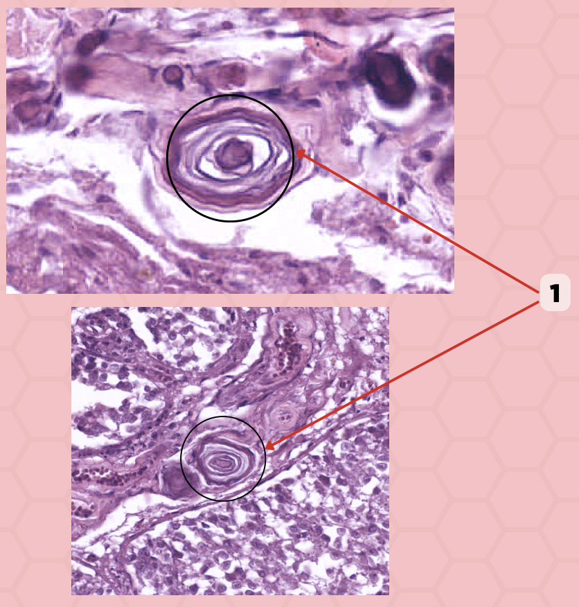
Corpora Arenacea
Identify the structure labeled as 1.

Dense Irregular Connective Tissue
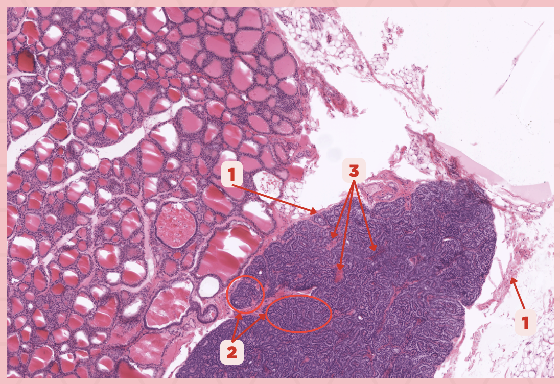
The capsule that covers the thyroid and parathyroid glands are made up of which type of connective tissue?

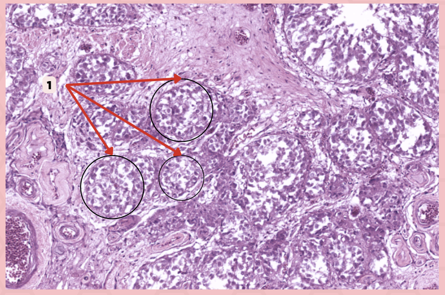
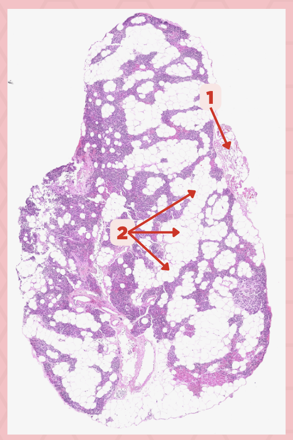
Thyroid-Parathyroid
Identify the specimen.
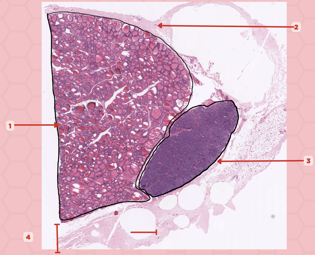
Thyroid Gland
Identify the structure labeled as 1.

Capsule
Identify the structure labeled as 2 and 4.

Parathyroid Gland
Identify the structure labeled as 3.

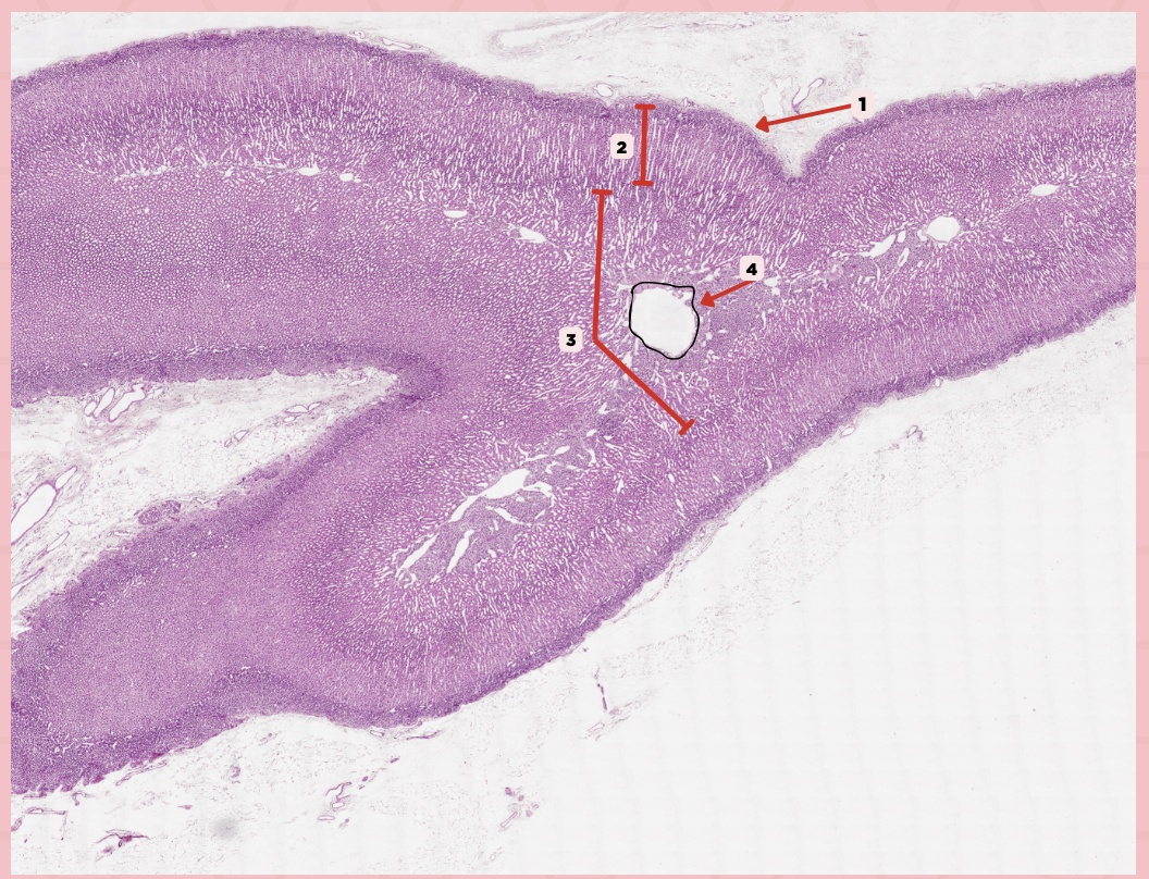
Thyroid
Identify the specimen.

Lobules
Identify the structure labeled as 1.

Follicles
Identify the structure labeled as 2.
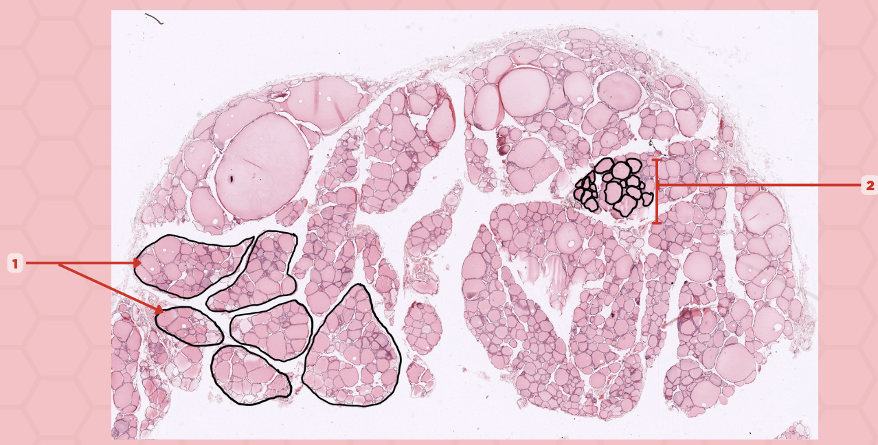
T3 and T4 Hormone
What type of hormone is being secreted by the pointer #1?
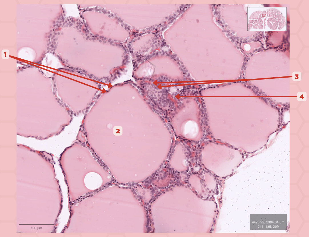
Follicular Cells
Identify the structure labeled as 1.

Colloid
Identify the structure labeled as 2.

Parafollicular Cells
Identify the structure labeled as 3.

Capillary
Identify the structure labeled as 4.

Lobules
The parathyroid gland is divided into _____, packed with epithelial cells that form cords and clusters.

False
TRUE or FALSE. The parathyroid gland is only enveloped by its own thin connective tissue capsule.

Parathyroid Capsule
Identify the structure labeled as 1.
Lobules
Identify the structure labeled as 2.

Septa
Identify the structure labeled as 3.

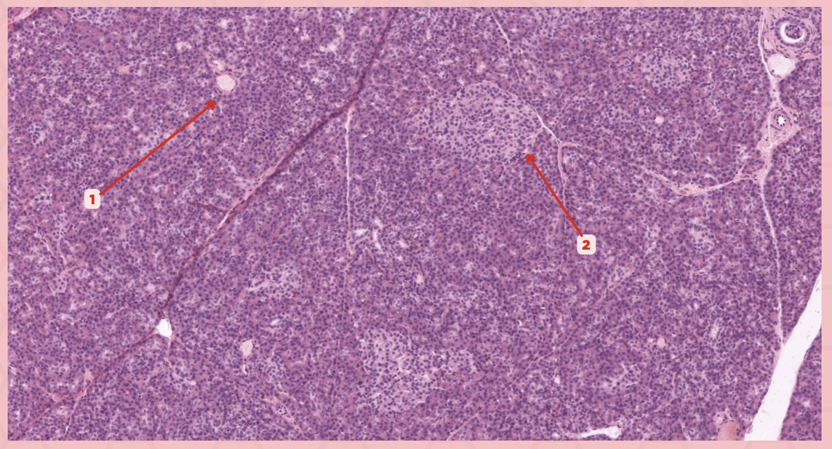
Parathyroid
Identify the specimen.

Chief Cells
Identify the structure labeled as 1. This comprise the majority of cells in the parathyroid gland.

Septa
Identify the structure labeled as 2.
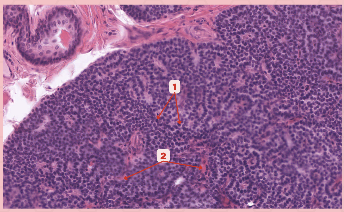
Parathyroid hormones
What hormones are synthesized and secreted by the cell in Pointer #1?

Oxyphil cells
What parenchymal cells in the parathyroid gland only appear shortly before puberty and increase in number with age?

Parathyroid
Identify the specimen.

Chief Cells
Identify the specimen labeled as 1.

Oxyphil Cells
Identify the specimen labeled as 2.

Adipocytes
Identify the specimen labeled as 3.

Capillaries
Identify the specimen labeled as 4.
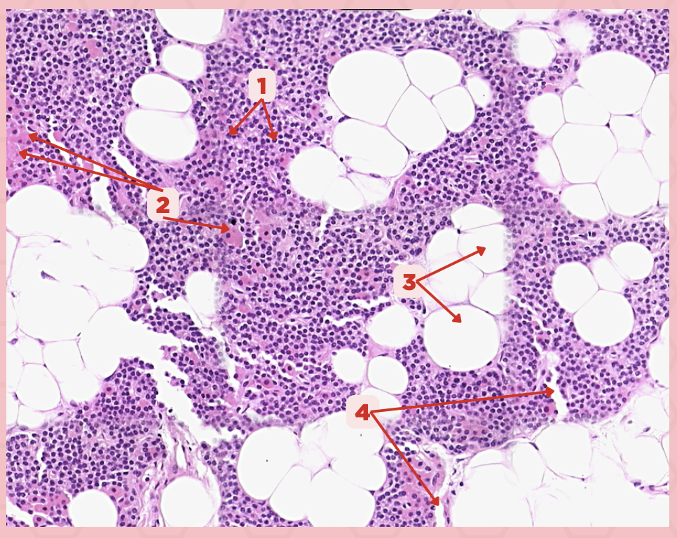
Newborns
Pointer #2 is hardly present in the parathyroid glands of _____.

Parathyroid
Identify the specimen.
Zone glomerulosa, Zone fasciculata, Zona reticularis
What are the zones present in pointer #2?
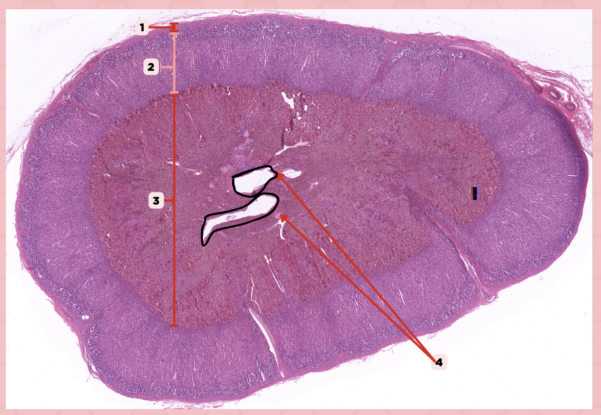
Catecholamines
What hormones are produced by pointer #3?

Adrenal
Identify the specimen.

Connective Tissue Capsule
Identify the structure labeled as 1.

Cortex
Identify the structure labeled as 2.

Medulla
Identify the structure labeled as 3.

Medullary Veins
Identify the structure labeled as 4.

Zone Glomerulosa
This layer seen in pointer #3 consists of rounded or pyramidal epithelial cells that arranges in irregular ovoid clusters that are separated by sinusoids
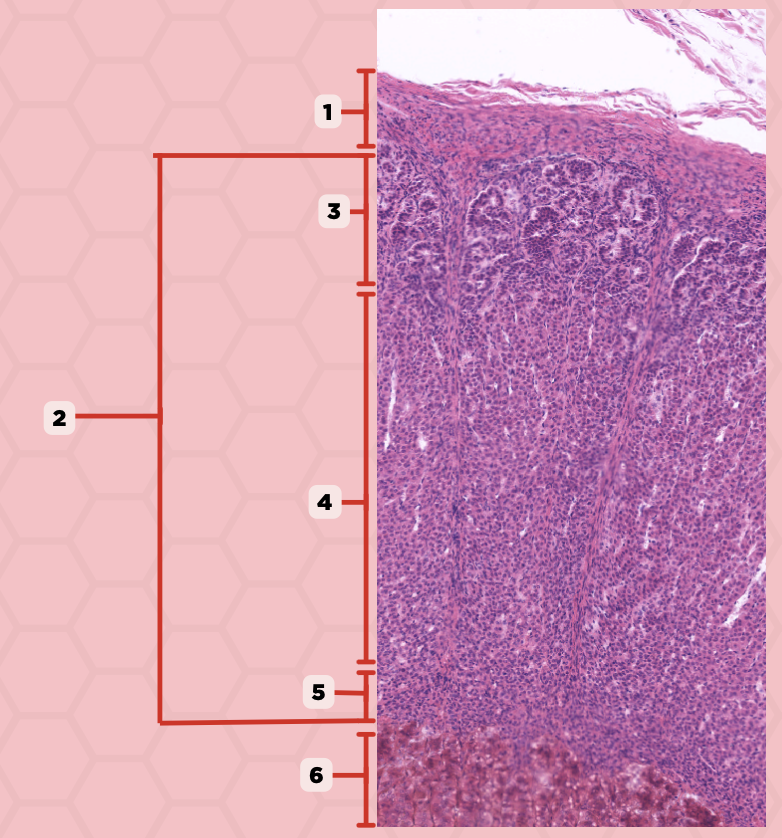
Mineralocorticoids, Glucocorticoids, Androgens
What are the 3 classes of Adrenocortical Hormones?

Zone Glomerulosa
Identify the structure labeled as 1.

Zona Fasciculata
Identify the structure labeled as 2.

Zona Reticularis
Identify the structure labeled as 3.

Androgens
What hormones are produced from the layer seen in pointer #5?

Zona Glomerulosa
Identify the structure labeled as 1.
Zona Fasciculata
Identify the structure labeled as 2.

Zona Reticularis
Identify the structure labeled as 3.

Adrenal Medulla
What region of the adrenal gland were the cells seen in pointer #2 and #3 are located?

Chromaffin cells (Pheochromocytes)
Which cells in the adrenal medulla are responsible for secreting catecholamines and exhibit a chromaffin reaction when treated with chromate?
Chromaffin Cells
Identify the structure labeled as 1.

Ganglion cells
Identify the structure labeled as 2.

Sinusoids
Identify the structure labeled as 3.

Renin-Angiotensin System
Which system primarily regulates the activity of parenchymal cells in the region seen in pointer #2 that produce aldosterone?
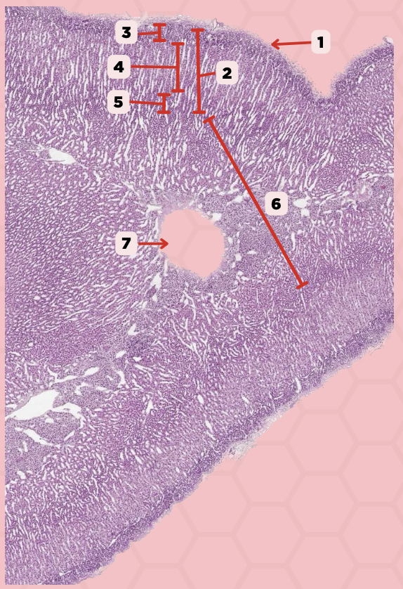
Beta-cells
Which cells occupy the central area of #1?

Pancreatic Acini
#3 delineates the pale- staining cells from the darker-staining cells. What do you call the darker-staining cells?

Pancreas
Identify the specimen.

Islets of Langerhans
Identify the structure labeled as 1.

Pancreatic Acini
Identify the structure labeled as 2.
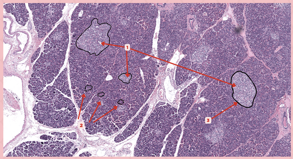
Reticular Tissue
Identify the structure labeled as 3.

Pancreatic Duct
Identify the structure labeled as 1.
Reticular Tissue
Identify the structure labeled as 2.
