RADPOS 1 - Finals (Anatomy)
1/84
There's no tags or description
Looks like no tags are added yet.
Name | Mastery | Learn | Test | Matching | Spaced |
|---|
No study sessions yet.
85 Terms
vertebral column or spine
The vertebral column or spine forms the central axis of the skeleton and is centered in the midsagittal plane of the posterior part of the trunk.
Enclose and protect the spinal cord
Act as support for the trunk
Support the skull superiorly
Provide attachment for deep muscles of the back and ribs laterally
The vertebral column or spine functions to:
ribs - sternum
sternum - shoulder girdle
vertebral column - hipbone
sacroiliac joints
(1) The upper limbs are indirectly supported through the ribs, which articulate with the sternum. (2) The sternum, in turn, articulates with the shoulder girdle. (3) The vertebral column also articulates with each hipbone at the (5) sacroiliac joints, supporting the trunk's weight and transmitting it through the hip joint to the lower limbs.
vertebrae
The vertebral column is made up of small segments called vertebrae.
Fibrocartilage
Fibrocartilage disks between vertebrae act as cushions.
Ligaments
Ligaments hold the vertebral column together.
jointed
curved
The vertebral column is jointed and curved, allowing flexibility and resilience.
33
The vertebral column consists of 33 small, irregular bones.
Cervical vertebrae (7)
Thoracic vertebrae (12)
Lumbar vertebrae (5)
Sacral vertebrae (5)
Coccygeal vertebrae (3-5)
The vertebral column is divided into five groups by region:
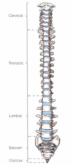
true or movable vertebrae
The 24 vertebrae in the cervical, thoracic, and lumbar regions remain separate throughout life and are termed true or movable vertebrae.
false or fixed vertebrae
The sacral and coccygeal vertebrae fuse to form the false or fixed vertebrae, with the sacral forming the sacrum and the coccygeal forming the coccyx.
Cervical curve
Thoracic curve
Lumbar curve
Pelvic (sacral) curve
Viewed from the side, the vertebral column exhibits four natural curves that arch anteriorly and posteriorly from the midcoronal plane.
Cervical curve
Convex anteriorly (lordotic); forms as the infant begins lifting the head (around 3–4 months) and sitting unsupported (by 8–9 months).
Thoracic curve
Concave anteriorly (kyphotic); present at birth.
Lumbar curve
Convex anteriorly (lordotic); develops when the child begins walking (around 1–1.5 years).
Pelvic curve
Concave anteriorly (kyphotic); present at birth.
cervical - thoracic
lumbar - pelvic
lumbosacral angle
The (1) cervical and thoracic curves merge smoothly, while the (2) lumbar and pelvic curves meet at an obtuse angle called the (3) lumbosacral angle, whose acuity varies among individuals. Females tend to have more pronounced lumbar and pelvic curves, creating a more acute lumbosacral angle.
Primary curves
Identify if Primary or Secondary Curves
Thoracic and pelvic; present at birth.
Secondary curves
Identify if Primary or Secondary Curves
Cervical and lumbar; develop after birth in response to posture and weight-bearing demands.
Kyphosis
Lordosis
Scoliosis
Abnormal Curvatures
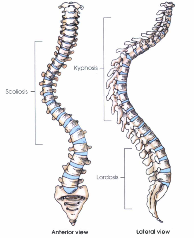
Kyphosis
Excessive posterior convexity (anterior concavity) usually in the thoracic region.
Lordosis
Excessive anterior convexity (posterior concavity) in the lumbar or cervical regions.
Scoliosis
Abnormal lateral curvature, often accompanied by vertebral rotation; may result from muscle imbalance or occupational posture.
mild lateral curvature
compensatory curves
From the frontal perspective, the vertebral column gradually widens from the second cervical vertebra down to the sacrum, then narrows sharply. A mild lateral curvature is common — typically rightward in right-handed individuals and leftward in left-handed ones — reflecting muscular influence. In scoliosis, compensatory curves develop to maintain head alignment over the feet.
anterior, weight-bearing body
posterior ringlike vertebral arch
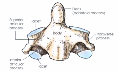
A typical vertebra consists of two main parts:
vertebral foramen
vertebral canal
A typical vertebra encloses the vertebral foramen, and in the articulated spine, the series of foramina form the vertebral canal that houses the spinal cord.
Vertebral Body
Cylindrical in shape and made primarily of cancellous bone, covered by compact bone.
The superior and inferior surfaces are flattened and coated with a thin plate of articular cartilage.
The anterior and lateral surfaces appear concave from the side view.
intervertebral disks
Between adjacent vertebral bodies lie intervertebral disks, which account for about one-fourth of the total spine length.
Nucleus pulposus
Annulus fibrosus
Each intervertebral disk consists of:
Nucleus pulposus
the central, soft, semi-gelatinous core.
Annulus fibrosus
the tough, fibrocartilaginous outer layer.
herniated nucleus pulposus (HNP)
Damage or rupture of the nucleus pulposus can compress spinal nerves, a condition known as herniated nucleus pulposus (HNP) or slipped disk, most common in the lumbar region.
2
The vertebral arch consists of two pedicles and two laminae, supporting:
Four articular processes (two superior, two inferior)
Two transverse processes
One spinous process
Pedicles
short, thick projections extending posteriorly from the vertebral body.
Their superior and inferior concavities form vertebral notches, which, when adjacent vertebrae articulate, create intervertebral foramina for nerve and vessel passage.
Laminae
broad, flat plates directed posteriorly and medially, forming the posterior portion of the vertebral arch.
spina bifida
A congenital defect where the laminae fail to unite posteriorly is spina bifida, which in severe cases allows spinal cord protrusion.
Transverse process
extend laterally and slightly posteriorly from where the laminae and pedicles meet.
Spinous process
projects posteriorly and inferiorly from the midline junction of the laminae.
Articular process
two superior and two inferior projections from the pedicle–lamina junction.
Each surface is covered with fibrocartilage and forms an articular facet.
Superior facets face posteriorly; inferior facets face anteriorly.
Facet orientation varies throughout the spine.
zygapophyseal joints
Articulations between transverse, spinous and articular processes form zygapophyseal joints (or interarticular facet joints), which guide and limit vertebral movement.
atlas
axis
C7
The cervical portion of the vertebral column consists of seven vertebrae (C1–C7). Among these, the first two (atlas and axis) and the seventh are atypical due to their distinct structural adaptations. The atlas and axis support the skull and allow head movement, while the seventh vertebra serves as a transitional structure between the cervical and thoracic regions.
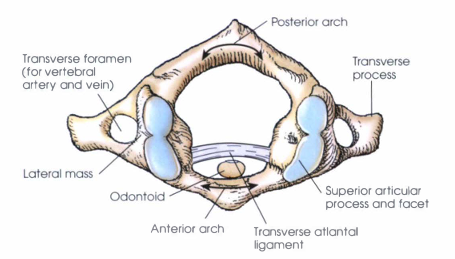
Atlas, C1
Ringlike structure without a body or a distinct spinous process.
Composed of an anterior arch, a posterior arch, two lateral masses, and two transverse processes.
The transverse atlantal ligament divides the ring into anterior and posterior portions.
The anterior portion holds the dens (odontoid process) of the axis.
The posterior portion transmits the upper spinal cord.
Each lateral mass contains a superior and inferior articular process.
Superior articular processes are large, concave, and horizontally oriented to articulate with the occipital condyles of the skull.
The transverse processes are long, extending laterally and slightly downward from the lateral masses.
Axis, C2
Characterized by the dens (odontoid process), a strong conical projection from the upper surface of the body that serves as the pivot for head rotation.
The dens fits into the anterior arch of the atlas, held in place by the transverse ligament.
Superior articular processes articulate with the inferior processes of the atlas.
Inferior articular processes are similarly oriented to those of the lower cervical vertebrae.
The laminae are broad and thick, while the spinous process is strong and positioned horizontally.
The unique structure of the axis and its joints with the atlas allows the head to rotate side to side (“no” motion).
Vertebra Prominens, C7
Distinguished by a long, prominent spinous process that projects nearly horizontally.
Easily palpable at the base of the neck, making it a key landmark for locating other vertebrae.
Provides transition from the cervical to thoracic spine.
Typical Vertebra, C3–C6
The body is small, transversely oval, and slightly elongated at the anteroinferior border, creating a mild overlapping effect in the articulated spine.
Each cervical vertebra contains three foramina: one vertebral foramen and a pair of transverse foramina.
The right and left transverse foramina transmit the vertebral artery and vein.
A deep concavity on the upper surface of each transverse process allows passage of spinal nerves.
Typical Vertebra, C3–C6
Transverse processes arise from both the vertebral body and arch and are short and wide.
Pedicles project laterally and posteriorly with equal superior and inferior notches.
Laminae are narrow and thin, completing the vertebral arch posteriorly.
Spinous processes are short, bifid (split into two points), and project posteriorly and slightly downward.
The palpable tip of each spinous process lies opposite the intervertebral space below the vertebra from which it extends.
Typical Vertebra, C3–C6
The superior and inferior articular processes are located posterior to the transverse processes at the pedicle-lamina junction.
These processes form short, thick articular pillars, which contain the fibrocartilaginous facets for articulation.
The zygapophyseal joints between C2 and C7 are positioned at right angles to the midsagittal plane and are best demonstrated in lateral radiographic projections.
Typical Vertebra, C3–C6
The cervical intervertebral foramina are directed anteriorly at a 45° angle to the midsagittal plane and 15° downward from the horizontal plane.
To visualize them radiographically:
Position the patient at a 45° oblique angle, or
Angle the central ray 15° longitudinally.
These foramina transmit spinal nerves and vessels exiting the spinal cord.
Thoracic Vertebrae
The thoracic vertebrae, twelve in total, form the midsection of the vertebral column and articulate with the ribs. Their size and shape transition between the smaller cervical vertebrae above and the larger lumbar vertebrae below.
Thoracic Vertebrae
The vertebral bodies increase in size from the first to the twelfth vertebra.
The upper thoracic vertebrae resemble cervical bodies, while the lower ones resemble lumbar bodies.
Typical thoracic vertebrae (T3–T9) have triangular bodies that are deeper posteriorly than anteriorly, with posterior surfaces concave from side to side.
Thoracic Vertebrae
Each thoracic vertebra has costal facets (or demifacets) on the posterolateral margins for rib articulation.
T1: One whole superior facet for the first rib, and an inferior demifacet for the second rib.
T2–T8: Two demifacets, one superior and one inferior, for articulation with adjacent ribs.
T9: Only a superior demifacet.
T10–T12: Each has a single whole facet on the superior margin for articulation with its corresponding rib (no inferior facets).
Thoracic Vertebrae
Transverse processes project obliquely, laterally, and posteriorly.
Except for T11 and T12, each has a small concave facet on the anterior surface at its extremity to receive the tubercle of a rib.
Laminae are broad and thick, overlapping those of the vertebra below.
Spinous processes are long. From T5 to T9, they project sharply downward and overlap, becoming less vertical above and below this region.
The palpated tip of a spinous process at this level lies opposite the interspace below the vertebra from which it arises.
Thoracic Vertebrae
The zygapophyseal (facet) joints of the thoracic vertebrae, except those of T12, are angled anteriorly at approximately 15–20 degrees, forming a 70–75 degree angle to the midsagittal plane.
Radiographic visualization requires the patient to be rotated 70–75 degrees from the anatomic position or 15–20 degrees from the lateral position.
Thoracic Vertebrae
The intervertebral foramina of the thoracic spine lie perpendicular to the midsagittal plane.
They are best demonstrated radiographically with the patient in a true lateral position.
During imaging, the arms should be elevated to lift the ribs and prevent them from obscuring the foramina.
Lumbar Vertebrae
The lumbar region of the spine consists of five vertebrae (L1–L5) that form the lower back. These vertebrae are the largest in the vertebral column, designed to support the weight of the upper body and withstand significant pressure during movement and posture maintenance.
Lumbar Vertebrae
The bodies are large, bean-shaped, and increase in size from L1 to L5.
They are deeper anteriorly than posteriorly, giving a slightly wedge-shaped appearance.
The superior and inferior surfaces are flat or slightly concave.
The posterior surface is flattened front to back, while the anterior and lateral surfaces are concave vertically.
Lumbar Vertebrae
Transverse processes are smaller than those of thoracic vertebrae.
The upper three pairs extend laterally.
The lower two pairs incline slightly upward.
Pedicles are short, strong, and direct posteriorly.
Laminae are thick and broad, lying behind the pedicles and transverse processes.
Spinous processes are large, thick, blunt, and project almost horizontally.
The palpable tip aligns with the intervertebral space below its vertebra.
Mamillary process
Accessory process
Pars interarticularis
Unique Features of Lumbar Vertebrae
Mamillary process
Unique Features of Lumbar Vertebrae
rounded projection on the posterior side of each superior articular process.
Accessory process
Unique Features of Lumbar Vertebrae
small projection at the posterior base of each transverse process.
Pars interarticularis
Unique Features of Lumbar Vertebrae
the segment between the superior and inferior articular processes within the lamina.
L5
The fifth lumbar vertebra has a highly wedge-shaped body, deeper in front for articulation with the sacrum.
Its intervertebral disk is also wedge-shaped to match the curvature of the lumbosacral junction.
The spinous process is shorter and smaller, while the transverse processes are thicker compared to those of upper lumbar vertebrae.
Lumbar Vertebrae
Zygapophyseal joints are inclined posteriorly from the coronal plane, forming an angle (open posteriorly) of 30–50 degrees to the midsagittal plane.
These joints are best visualized in oblique radiographic projections.
Intervertebral foramina are generally at right angles to the midsagittal plane, except for the fifth, which angles slightly forward.
The upper four pairs are seen best in a true lateral projection.
The fifth pair requires slight body rotation for clear visualization.
Spondylolysis
a bony defect or fracture in the pars interarticularis of a vertebra.
spondylolisthesis
If bilateral, it can progress to spondylolisthesis, a condition where one vertebra slips forward over another, most often the fifth lumbar over the sacrum.
Scottie dog
neck
In radiographs, the oblique “Scottie dog” appearance represents the posterior elements of a lumbar vertebra; the “neck” corresponds to the pars interarticularis, where defects appear.
Sacrum
The sacrum is a large, triangular-shaped bone formed by the fusion of five sacral vertebrae. It sits between the iliac bones of the pelvis and forms the posterior part of the pelvic cavity. The sacrum supports the vertebral column above and transmits weight to the pelvis.
Sacrum
The sacrum’s broad base faces superiorly and anteriorly, while its apex points inferiorly and posteriorly.
It is wedged between the iliac bones at the sacroiliac joints.
The bone’s size, curvature, and angulation vary among individuals.
In males: longer, narrower, more evenly curved, and more vertical.
In females: shorter, wider, more sharply curved (especially in the lower half), and lies in a more oblique plane, producing a greater lumbosacral angle.
Sacrum
The upper surface, or base, of the first sacral segment resembles that of a lumbar vertebra.
The base articulates with the fifth lumbar vertebra to form the lumbosacral junction.
Concavities on the upper pedicles of the first sacral segment align with those on the lower pedicles of L5, forming the last pair of intervertebral foramina.
The superior articular processes of the first sacral segment articulate with the inferior articular processes of L5, forming the last pair of zygapophyseal joints.
sacral promontory
a prominent ridge at the superior anterior margin of the sacral base, marking the anterior boundary of the pelvic inlet.
sacral canal
a continuation of the vertebral canal located posterior to the sacral bodies, through which sacral nerves pass.
Anterior (pelvic) sacral foramina
Posterior sacral foramina
The canal’s anterior and posterior walls are pierced by four pairs of sacral foramina, which transmit nerves and blood vessels:
Anterior (pelvic) sacral foramina on the concave anterior surface.
Posterior sacral foramina on the convex dorsal surface.
ala
Each side of the sacral base has a large, wing-shaped ala (Latin for “wing”).
auricular surface
The auricular surface, located on the superior anterior part of the ala, articulates with the iliac bone of the pelvis, forming the sacroiliac joint.
apex of the sacrum
The apex of the sacrum, located inferiorly, bears an oval articular facet that joins the coccyx.
sacral cornua
coccygeal cornua
The sacral cornua, two small projections from the last sacral segment, extend downward to articulate with the coccygeal cornua of the first coccygeal segment.
Coccyx
The coccyx, often referred to as the tailbone, is the terminal segment of the vertebral column. It consists of three to five rudimentary vertebrae, most commonly four, that typically fuse into a single bone during adulthood.
Coccyx
The coccyx decreases in size from its broad base to its narrow apex.
It articulates superiorly with the sacrum at the sacrococcygeal joint.
From this articulation, it curves inferiorly and anteriorly, often deviating slightly from the body’s midline.
The coccygeal cornua, small projections extending upward from the posterolateral aspect of the first coccygeal segment, articulate with the sacral cornua of the sacrum.
Intervertebral Joints
Located between adjacent vertebral bodies.
Classified as cartilaginous symphysis joints.
Permit only slight movement individually, but provide significant overall mobility when combined.
The intervertebral disks act as cushions, allowing limited motion and absorbing shock.
Zygapophyseal Joints
Found between the articular processes of vertebral arches.
Classified as synovial gliding joints.
Allow free movement and contribute to vertebral flexibility, enabling motions such as flexion, extension, lateral flexion, and rotation.
Atlanto-occipital joints
Atlantoaxial joint
Costovertebral joints
Costotransverse joints
Specialized Articulations
Atlanto-occipital joints
Synovial ellipsoidal joints between the atlas (C1) and occipital bone; permit flexion and extension (nodding “yes”).
Atlantoaxial joint
Comprises both a synovial pivot and gliding articulation between the atlas (C1) and axis (C2); allows rotation (nodding “no”).
Costovertebral joints
Synovial gliding joints where the heads of the ribs articulate with the bodies of the thoracic vertebrae.
Costotransverse joints
Synovial gliding joints formed between the rib tubercles and the transverse processes of the thoracic vertebrae.