Lecture 3 - protein function
 Call Kai
 Learn
Learn Practice Test
 Spaced Repetition
 Match
Match Flashcards
 Knowt Play
1/27

There's no tags or description
Looks like no tags are added yet.
Name | Mastery | Learn | Test | Matching | Spaced | Call with Kai | Chat |
|---|
No analytics yet
Send a link to your students to track their progress
28 Terms
Aquaporin
protein that facilitates the travel of water through the cell membrane
homotetramer → 4 of the same subunits (hydrophilic residues inside, hydrophobic residues outside)
water passes though the hydrophilic residues
Globins
Family of proteins that sense and carry oxygen
Myoglobin
Single molecule of heme bonded to a globin
delivers oxygen to muscles
single polypeptide (153 AAs)
8 alpha helices connected by bends, together they bind the heme group
Heme
molecule that helps carry oxygen. because oxygen is poorly soluble and can’t be carried throughout long distances, evolution drove heme to carry oxygen
Its protoporphyrin ring binds Fe²⁺, and Fe²⁺ binds oxygen
nitrogens stabilizes iron
Fe²⁺ is not stable in free heme, so proteins that contain heme stabilize oxygen by tucking the heme into their 3D structure
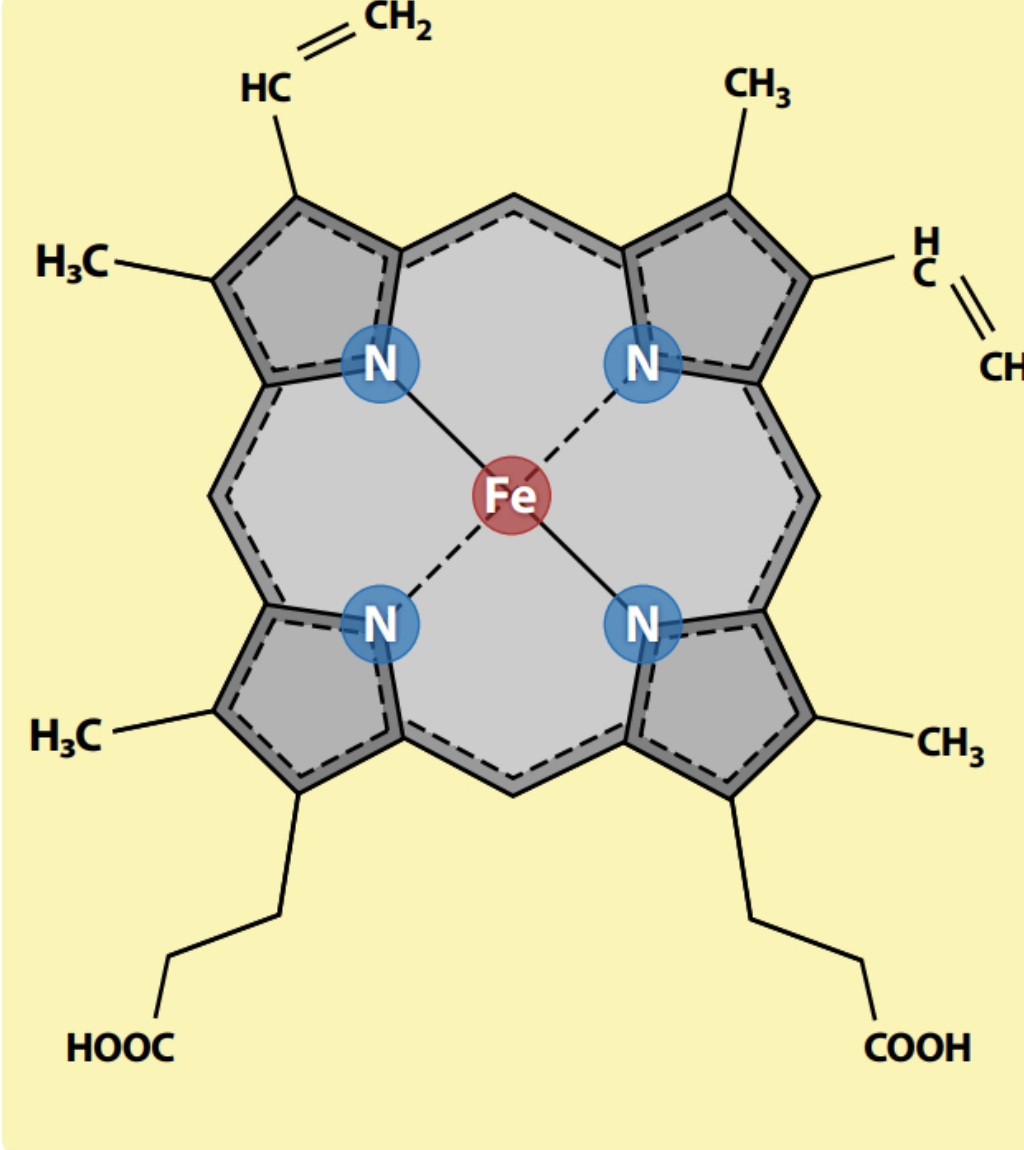
Histidine residues and oxygen binding to heme
Histidines on the heme facilitate oxygen binding
proximal His: Stabilizes the Fe²⁺ center
distal His: stabilizes O2 binding
(stabilizing is done via ionic bonding)
Proximal and distal His are highly conserved
Hemoglobin
oxygen-carrying protein in red blood cells
4 subunits of myoglobin-like proteins
4 hemes
2 conformations: R state (relaxed) and T state (tense)
allosteric
Explain what this means: oxygen binding to hemoglobin is cooperative
hemoglobin becomes better at grabbing oxygen as it grabs more oxygen → this is because it has 4 oxygen binding spots (cooperative binding)
this matters because if it’s in the lungs, with a high affinity for oxygen → it quickly loads up oxygen. however, in the tissues, with low oxygen affinity, hemoglobin releases O2 quickly as it releases O2.
this is an example of allosteric activity
R state vs T state
T state: tense state
oxygen has LOW affinity for binding to heme, heme is tucked
R state: relaxed state
oxygen has HIGH affinity for binding to heme, heme is outward facing
Allosteric protein
A protein where the binding of one site effects the binding properties of another site on the same protein
Ligand
a molecule or ion that binds to another molecule (usually a protein) at a specific binding site
ex) Oxygen binds to hemoglobin, so oxygen is the ligand
Homotropic vs heterotrophic
Homotropic: When the ligand that binds is the SAME molecule as the one whose binding is being affected
ex) O2 affects the binding of O2 (same molecule)
Heterotrophic: When the ligand that binds is a DIFFERENT molecule than the one whose binding is being affected
ex) H+ affects O2 binding (different molecules)
Class example: actin/myosin muscle contraction
actin/myosin muscle contraction is an example of protein/ligand interaction
Muscles contain actin and myosin filaments
Process:
A nerve impulse triggers the release of calcium ions
Calcium ions bind onto actin filaments → this causes the actin filament to turn, exposing the myosin binding sites
The head of each myosin is bound to an ADP and a phosphate
Myosin head releases the phosphate and binds to actin filaments through the myosin binding sites
The filaments then glide past each other → as myosin and actin move, they release the ADP
ATP then binds to myosin heads, severing the bond between actin and myosin
ADP and phosphate is then restored on the myosin heads → repeat process
Phosphorylation
Adds a phosphoryl group to reactive hydroxyls in proteins (Ser, Thr, Tyr)
Process:
Signal comes in
Protein kinase grabs a phosphoryl group off of ATP
Kinase transfers phosphorly group onto a reactive hydroxyl from Ser, Thr, or Tyr → enzyme is now phosphorylated → turns enzyme from inactive to active
Enzyme does its job → protein phosphatase removes phosphate group from enzyme
Phosphate is released as Pi (inorganic phosphate)
Enzyme goes from active → active (switch off)
Kinase: phosphorylates proteins
Phosphatase: removes phosphate groups
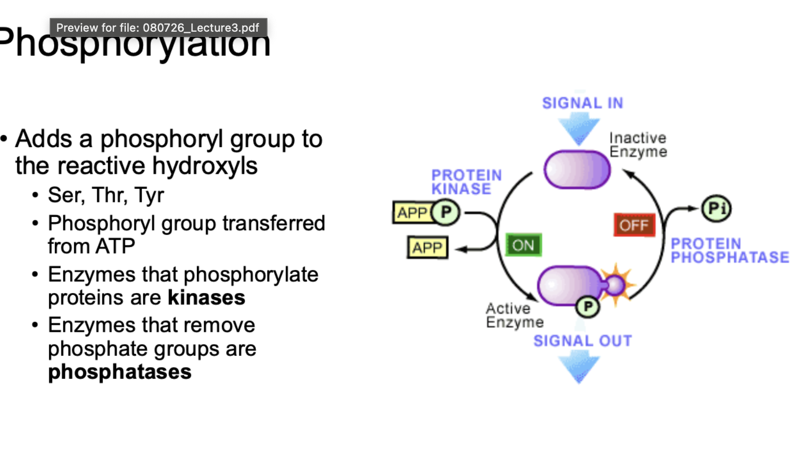
Kinases
enzymes that phosphorylate proteins
Phosphatases
enzymes that remove phosphate groups
What is signal transduction via phosphorylation cascade?
Signal from outside the cell gets “relayed” into the cell by a chain of kinases turning each other on (like dominoes) until it triggers a final response
extracellular ligand binds a receptor (a kinase)
Triggers kinase cascade, where each active kinase phosphorylates (activates) the next
ends in a biological response
Ubiquitination
Protein waste program
Ubiquitin - peptide
Ubiquitin’s terminal Glycine (COOH group) and the substrate’s Lysine (NH2 group) come together
Bond forms between the carboxyl carbon of Glycine and amino group of Lysine → releases water → result is an isopeptide bond linking Ubiquitin to the substrate
The more ubiquitous attached to a protein, the more it signals for the protein to get trashed
Ubiquitin
Peptide that’s added to a Lysine residue through an isopeptide bond with Ubiquitin’s C-terminus
Ubiquitin and proteasome “shredder”
Ubiquitin tags protein
protein is fed into the proteasome “shredder”
Part 1: E1-E2-E3 cascade
E1(enzyme) : activates ubiquitin
E2 (enzyme): takes from E1
E3 (ligase): takes from E2, catalyzes attaching ubiquitin onto protein
Part 2: proteasome
Ubiquitin tagged protein is recognized by the receptor on the proteasome’s 19S cap
DUBs (enzymes that remove ubiquitin tags) pop off, recycling ubiquitin molecules so the cell can reuse them
The protein is fed into the 20S core → Beta subunits inside the 20S core cut the protein → protein gets chopped into oligopeptides, which are released
Silent mutation
No change in amino acid
Nonsense mutation
introduces STOP codon
Missense mutation (conservative)
Change in amino acid, but the new amino acid has similar biophysical properties to the original amino acid
Missense mutation (non-conservative)
Change in amino acid, but the new amino acid has different biophysical properties to the original amino acid
Loss of function mutation
protein loses its normal activity
Gain of function mutation
protein does more than normal, does something new, or is active when it shouldn't be
Which amino acid is best for mutation, and why?
Alanine
simple/unreactive side chain (CH3)
Mutation nomenclature
Format: [Original amino acid][position number of that original amino acid][new amino acid]
ex) G12V = Glycine-12 mutated to Valine (or Gly12Val)
Mimetic mutations
Replacing AAs with AAs that have biological reactivity to mimic a function on that protein
ex) Phosphomimetics
ex) Aspartate looks and acts like phospho-serine → by replacing Serine with Aspartate, you fake an amino acid that is already phosphorylated