Overview of the Sensory Inputs and Motor Outputs of the Brain (Week 1, Mod 8)
1/21
There's no tags or description
Looks like no tags are added yet.
Name | Mastery | Learn | Test | Matching | Spaced |
|---|
No study sessions yet.
22 Terms
What are the 3 basic functions that neurons perform in the nervous system?
1) Sensory input: Neurons that collect information about the external environment and internal conditions of the body
2) Integration: Neurons that receive the sensory input, analyze it, and make decisions about the appropriate response to make
3) Motor output: Neurons that carry out the instructions that result from integration by impulses sent to various effector organs that include muscles and glands
Define “sensation”…
Sensation: A physical feeling or perception for something that happens to or comes into contact with the body
Where, anatomically, in the vertebrae is a neuronal signal received?
Enters through the DORSAL portion of the vertebrae (into the dorsal horn of the gray matter), through the dorsal root of the spinal nerve
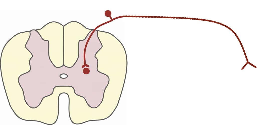
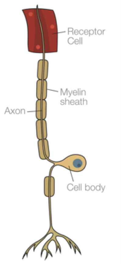
Describe the morphology of the sensory nerve… is it multipolar, bipolar, or unipolar?
Unipolar
What is the difference between how a spinal nerve receives stimuli and how a CRANIAL nerve receives stimuli?
Spinal nerve: Carries a signal between the body and the spine
Cranial nerve: Carries a signal between the body and the BRAIN; interacts directly with the brainstem
What are the two different ways we can classify a receptor?
1) Anatomically - how they look, ex: encapsulated vs. non-encapsulated
2) Location - based on location in the body; location will = function
What are the 3 kinds of receptors based on their location in the body?
1) Exteroreceptors: Near the surface of the body. Sensitive to change in external environment
2) Proprioceptors: Sensitive to movement of muscles, tendons and joints
3) Interoceptors: Located within the viscera. Sensitive to change in internal environment
What are the 2 different classifications of the SENSORY portion of the PNS?
Somatic afferent: Dendritic zone is ON or NEAR the surface of the body
Visceral afferent: Dendritic zone is in the WALL of different VISCERA of the body
What are the 2 types of fibers associated with the somatic afferent PNS?
General somatic afferent (GSA):
Touch, temperature, proprioception and noxious stimuli
Cranial nerve V for the head
Spinal nerves for the rest of the body
Special somatic afferent (SSA):
Vision: Cranial nerve II
Sound: Cranial nerve VIII
What are the two types of nerve fibers associated with the visceral afferent PNS?
General visceral afferent (GVA):
Organ content, distention, chemicals
Cranial nerves VII, IX and X to visceral structures in the head
Cranial nerve X and spinal nerves to the viscera and blood vessels of the rest of the body
Special visceral afferent (SVA):
Taste: Cranial nerves VII, IX, and X
Olfaction: Cranial nerve I
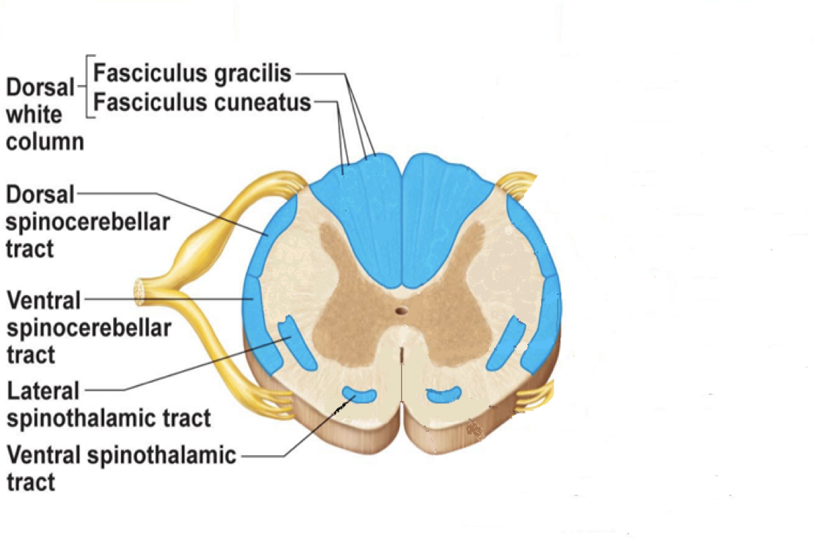
What are the 4 different sensory spinal tracts of white matter that nerve axons travel through to communicate with the CNS, and what kind of information does each receive?
Fasciculus gracilis:
Proprioceptive information from pelvic limbs
Fasciculus cuneatus:
Proprioceptive information from thoracic limbs
Dorsal and ventral Spinocerebellar tracts :
Information to cerebellum for coordination
Spinothalamic tracts:
Information to thalamus about pain, itch, touch and temperature
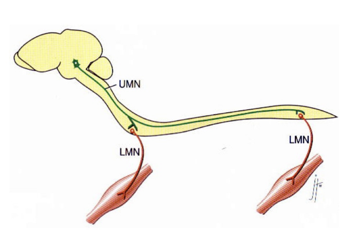
What is a “lower motor neuron”? Are there upper motor neurons?
Lower motor neuron is simply a motor neuron that controls the muscle movements from everything BELOW the neck
There are upper motor neurons… these are nerves that originate in the motor region of the cerebral cortex / brainstem and stretch ALL THE WAY DOWN THE CNS
Carry motor information DOWN to the lower motor neurons
Prof explained it as this: the thought / desire to move your hand comes from the UPPER MN, sending the signal to your LOWER MN to actually act on the movement
Upper neuron activated before lower neuron
Where are the lower motor neurons (LMNs) located in the individual vertebrae?
In the VENTRAL horn of the gray matter of the spinal cord… neuron signal LEAVES through this pathway
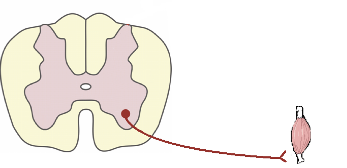
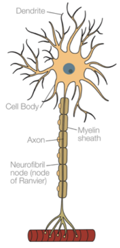
What is the morphology of a lower motor neuron?
Has a LONG axon and a HUGE cell body, with multiple dendrites involved
What are the 2 different classifications of the MOTOR portion of the PNS? What is it classified by?
Is based on the location of the axon terminals in the body
General somatic efferent (GSE):
Axon terminals in striated muscle throughout the body
Cranial nerves: all of them except from I, II and VIII
Ventral nerve roots and spinal nerves for the rest of the body
General Visceral efferent (GVE):
Axon terminals in involuntary smooth muscle of viscera
Cranial nerves: III, VII, IX, X and XI
Spinal nerves for the rest of the body
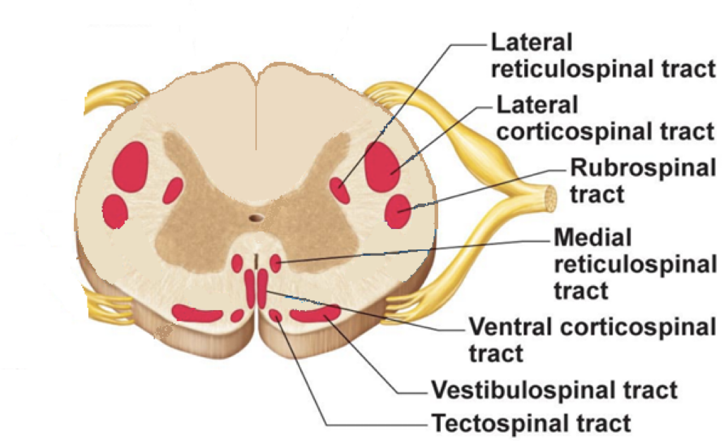
What are the 5 different MOTOR spinal tracts of white matter that nerve axons travel through to communicate with the CNS, and what kind of information does each receive?
Corticospinal tracts (pyramidal system):
Motor information directly from cerebral cortex
Poorly developed in domestic animals
For FINE MOVEMENTS
Rubro(red)spinal tracts (extrapyramidal system):
Motor information from red nucleus in the brainstem
Better developed in domestic animals
Reticulospinal tracts (extrapyramidal system):
Motor information from reticular formation in the brainstem
Vestibulospinal tracts (extrapyramidal system):
Motor information from vestibular nuclei in the brainstem
Tectospinal tracts (extrapyramidal system):
Motor information from the tectal region in the brainstem
What are the 7 tests that can be performed to assess proprioception in the dog?
1) Proprioceptive placing (knuckling response)
2) Hopping reaction
3) Hemiwalking
4) Wheelbarrowing
5) Extensor postural thrusting
6) Hip sway
7) Visual and tactile placing
(watch lecture for videos, not in the presentation)
Which pathway are we assessing by testing proprioception?
Afferent pathway + upper motor neurons + lower motor neurons
Afferent pathway senses that paw is in the wrong position; sends signal up to the brain
Upper motor neurons then send unconscious signal to CORRECT this
Lower motor neurons receive signal and physically correct it as a reflex
What are 4 tests we can use to assess MOTOR function?
1) Evaluate gait
2) Evaluate muscle tone and bulk
3) Patellar reflex
4) Withdrawal (flexor) reflex
We want to be able to tell whether or not its the UPPER motor neurons or the LOWER motor neurons that are effected… in the cases of muscle tone, spinal reflexes, and muscle atrophy, what should we expect to see if we have a UMN lesion or an LMC lesion?
UMN lesion:
Muscle tone - normal / increased
Spinal reflexes - normal or exaggerated
Muscle atrophy - Little and late (from disuse)
LMN lesion:
Muscle tone - DECREASED
Spinal reflexes - decreased or absent
Muscle atrophy - Early and SEVERE (neurogenic)
How do we assess the SEVERITY of damage to sensory function? What are the parameters?
Normal →
Loss of proprioception →
Loss of superficial pain perception →
Loss of DEEP pain perception
How do we assess the severity of damage to MOTOR function? What are the parameters?
Normal →
Back pain →
Weakness →
TOTAL paralysis