EXAM 2 - Spinal Cord and PNS
1/106
There's no tags or description
Looks like no tags are added yet.
Name | Mastery | Learn | Test | Matching | Spaced | Call with Kai |
|---|
No analytics yet
Send a link to your students to track their progress
107 Terms
Central Nervous System (CNS)
brain and spinal cord
PNS
cranial nerves, spinal nerves and ANS
PERIPHERAL NERVES
⦿ Bundles of axons outside the CNS
⦿ Most are myelinated
⦿ Myelinating cells are schwann cells/neurolemnocytes
Connective tissue coverings in peripheral nerves
Nerves arranged in bundles (fascicles)
3 covering: (outer to inner)
1. epineurium
2. perinerium
3. endoneurium
motor endplate
synapse of neuron to muscle
Ganglion
bundle of nerve cell bodies in the peripheral nervous system
plexus
network of nerves; ventral rami of sp n
(brachial plexus)
31 pairs of spinal nerves
8 cervical
12 thoracic
5 lumbar
5 sacral
1 coccygeal
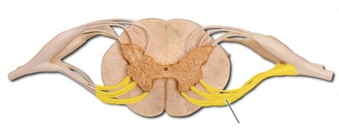
2 parts of the nerve root
1. dorsal (sensory)
2. ventral (motor)
clinical application for dorsal nerve roots
viruses can lay dormant here - chicken pox, shingles, herpes viruses
clinical application of ventral nerve roots
polio virus, ALS both attack here
destroy motor neurons that innervate skeletal muscle
each Nerve then divides into
1. dorsal ramus
2. ventral ramus
characteristics of dorsal rami
small, mixed (sensory and motor), NEVER form plexuses
Characteristics of ventral rami
large & thick, mixed (sensory and motor), form plexuses
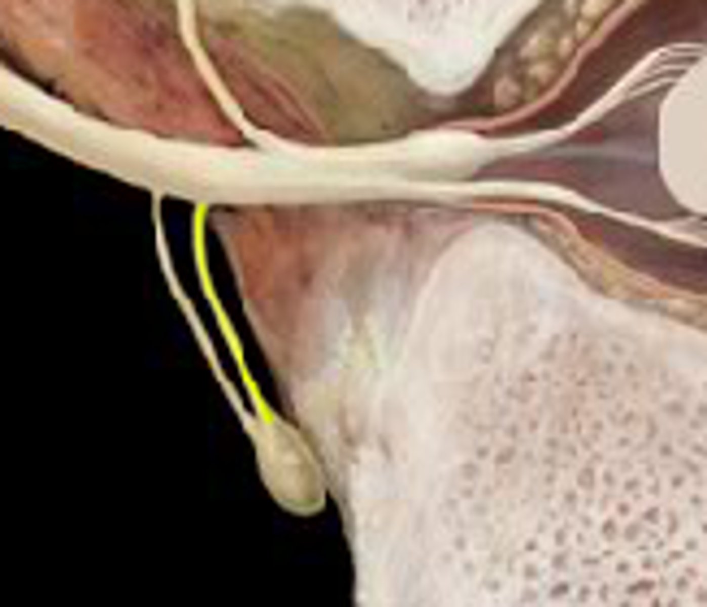
where are dorsal rootlets
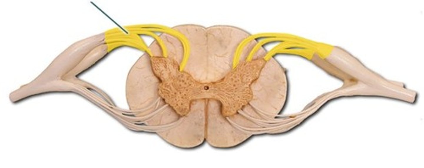
where are ventral rootlets
where are dorsal roots

where are ventral roots

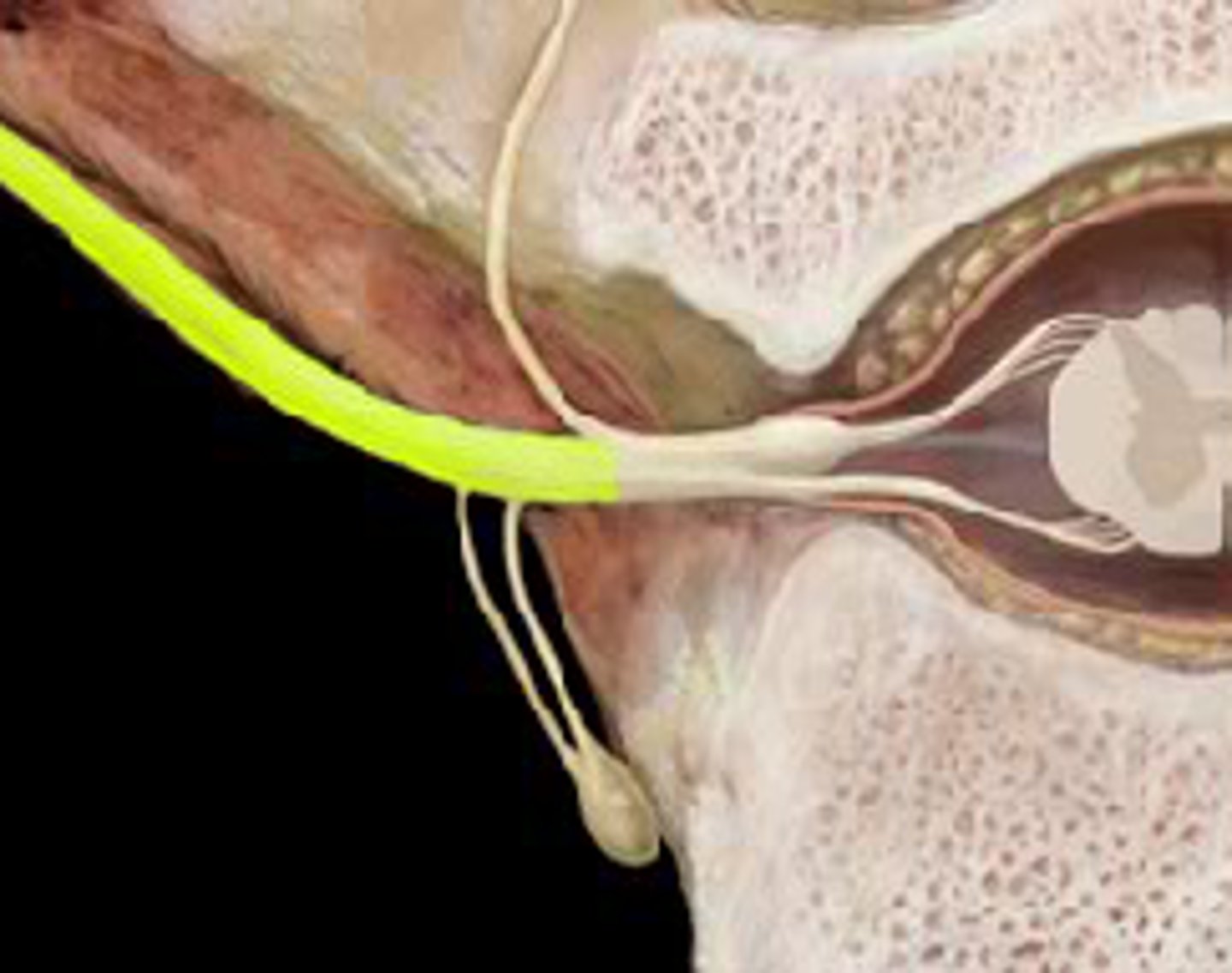
dorsal root ganglion location
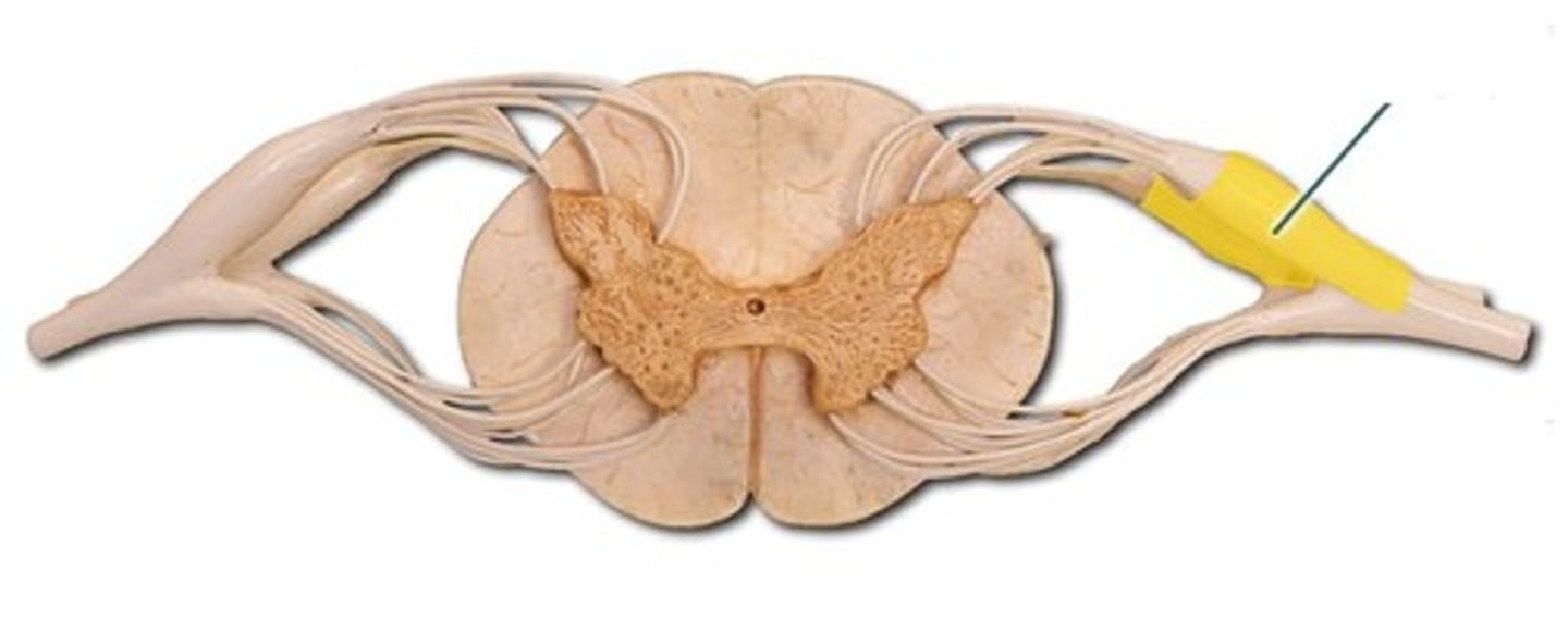
what makes a spinal nerve
where the ventral and dorsal roots combine into the spinal nerve
Dorsal ramus
the division of posterior spinal nerves that transmit motor impulses to the posterior trunk muscles and relay sensory impulses from the skin of the back
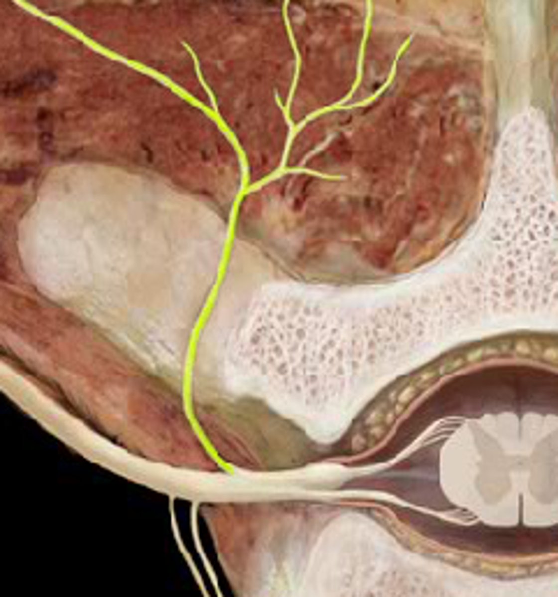
ventral ramus
the anterior division of spinal nerves that communicate with the muscle and skin of the anterior and lateral trunk
gray ramus
unmyelinated nerves, return from sympathetic ganglion to rejoin spinal nerve (postganglionic fibers)
more medial
sympathetic ganglion
Ganglion (bundle of nerve cell bodies) that receives preganglionic sympathetic fibers. part of sympathetic nervous system that has fight or flight response
white ramus
Carries visceral motor fibers to sympathetic ganglion of autonomic nervous system (preganglionic fibers)
more lateral
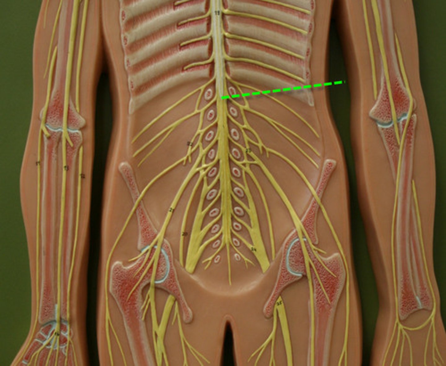
Spinal cord and age
Length varies with age
◼ Foramen magnum to:
⭘ Adult: between L1 and L2
⭘ Child : L3
⭘ Fetus: Full length
General cord features
the spinal cord is CNS and continuous with brain stem
length of spinal cord
length varies with age
adult: foramen magnum to between L1 and L2
child: foramen magnum to L3
fetus: foramen magnum to full length
*shortens as you develop
Cervical & lumbosacral enlargements locations
locations:
C4-T1
T11-L1
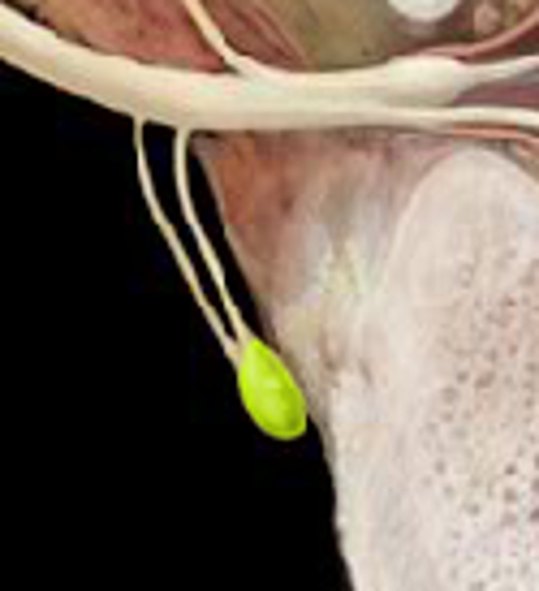
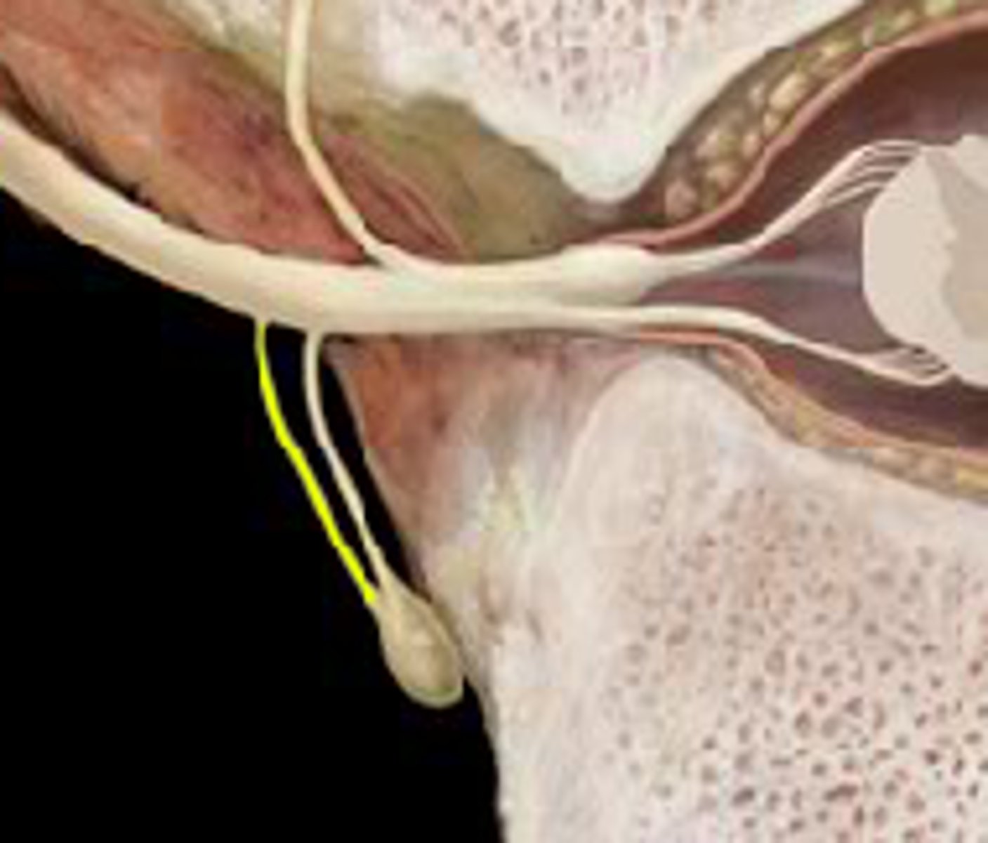
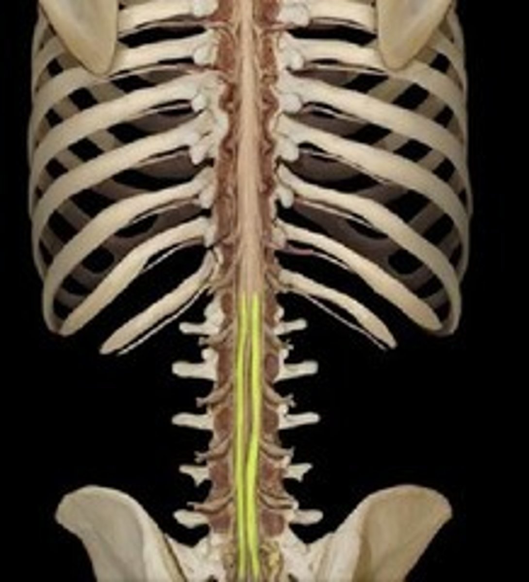
Conus medullaris (medullary cone)
L1 - end of spinal cord
Filum terminale (terminal filament)
pia mater
anchors spinal cord to coccyx
silver/shiny
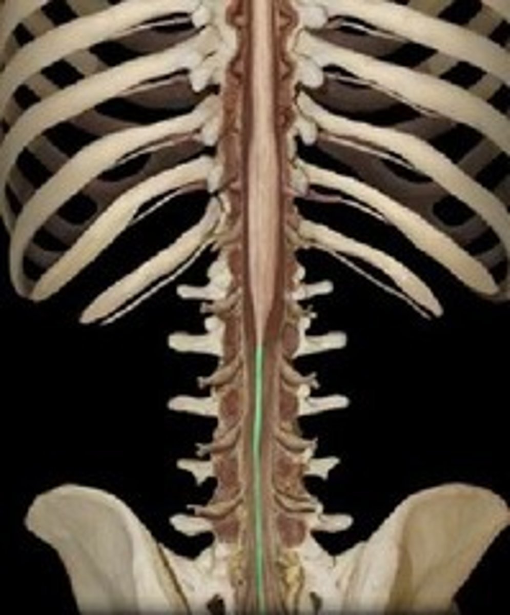
Cauda equina
collection/bunch of spinal nerves below the end of the spinal cord
"horse tail"
meninges of the spinal cord
dura mater, arachnoid mater, pia mater
Dura mater
outermost
◼ Ends at S2 - forms dural sac at that point
◼ Epidural space - continues with the epineurium of spinal n - between the dura and the bone of vert (coats the dura mater)
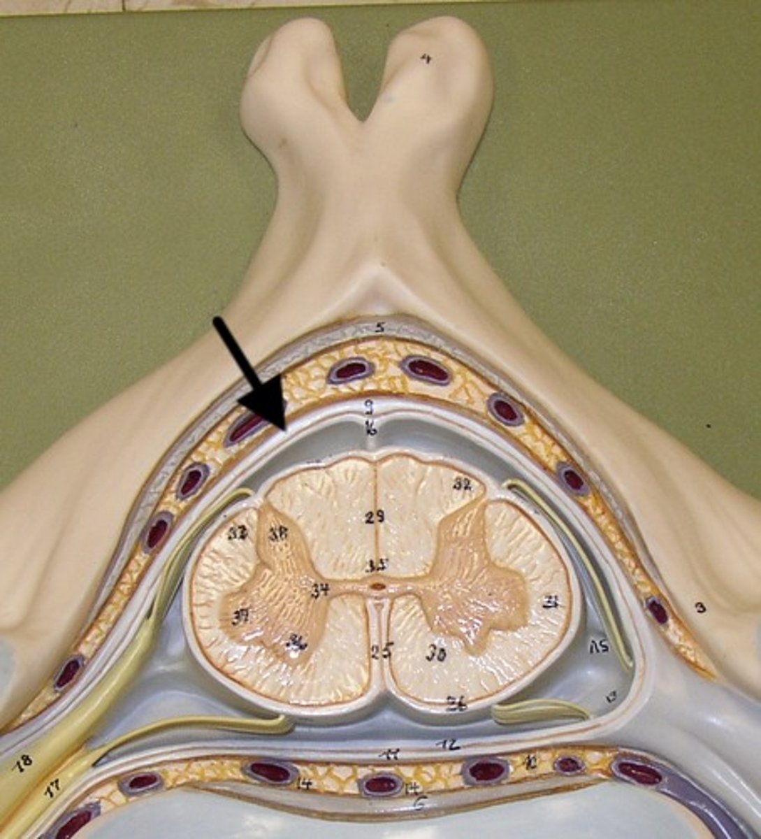
Arachnoid mater
◼ Delicate & avascular; impermeable; fibrous & elastic
CT; ends S2 (dural sac)
◼ Subarachnoid space - CSF flows
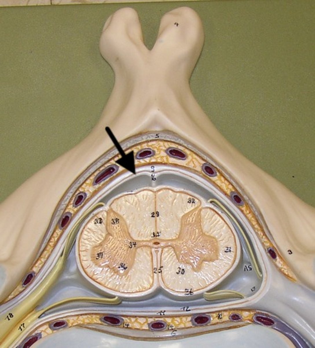
Pia mater
makes terminal filament
Ligamentum denticulum AKA denticulate ligaments
- looks like teeth
- serves as an anchor for the spinal cord but delicate
- fibrous bands that help separate anterior and posterior roots
Cord grows slower than column
◼ Upper cord segments adjacent to same # verts
◼ Spinal nerves L2-S4 - form cauda equina
Spinal nerves exit vertebrae via?
intervertebral foramen
Where do spinal nerves exits along the vertebrae?
◼ C1: between the occipital bone and the atlas
◼ C2-C7: above the same # vert
◼ C8: between C7 and T1 vert
◼ T1-S5: exit below same # vert
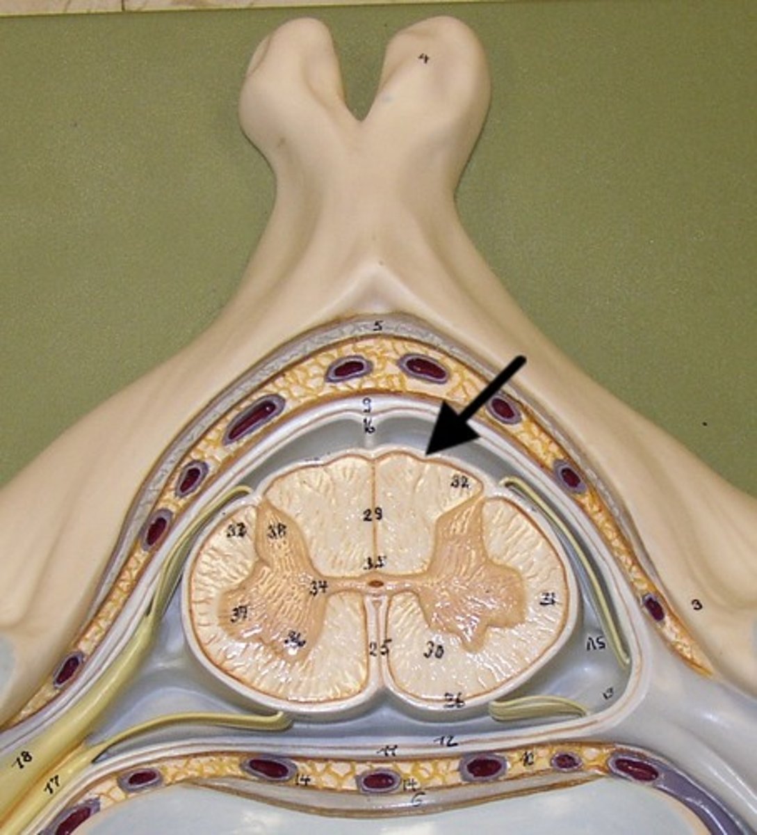
Dorsal median fissure
Defines left and right gracilis tubercles
splits dorsal white matter in half
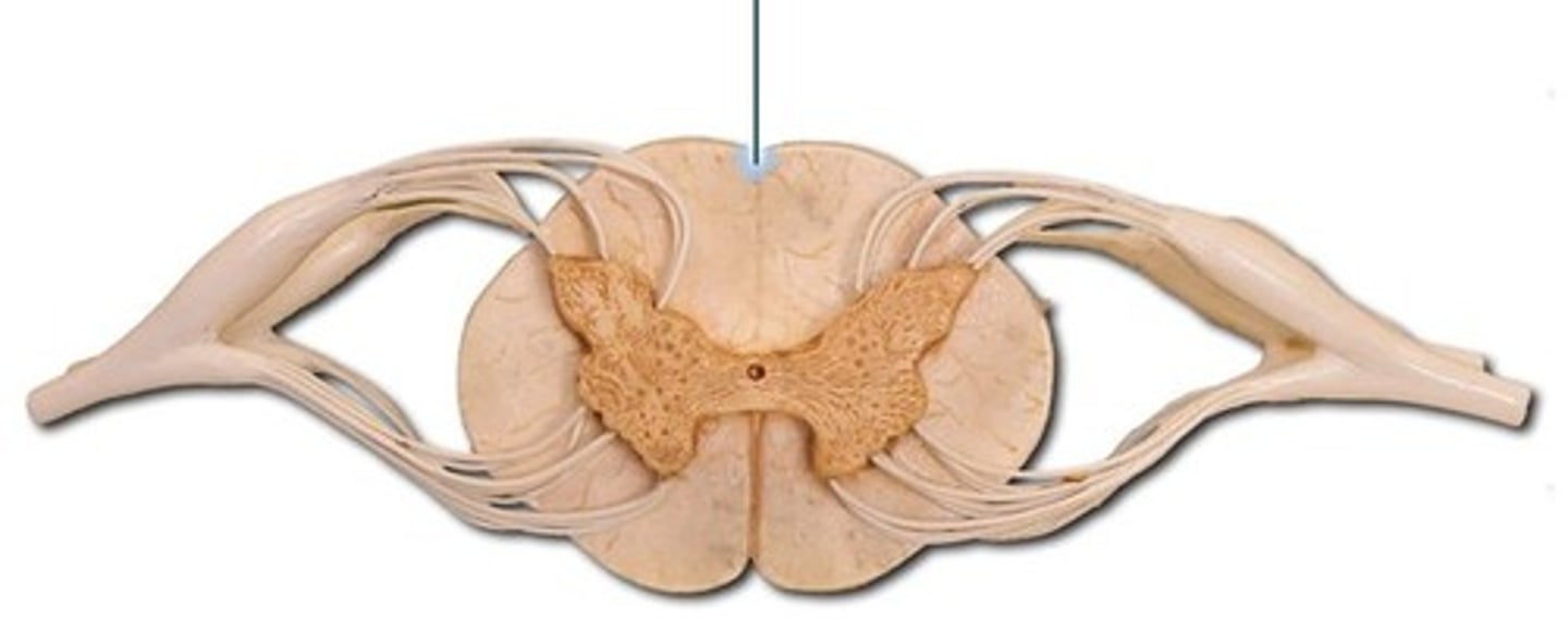
Ventral median sulcus
splits ventral white matter in half
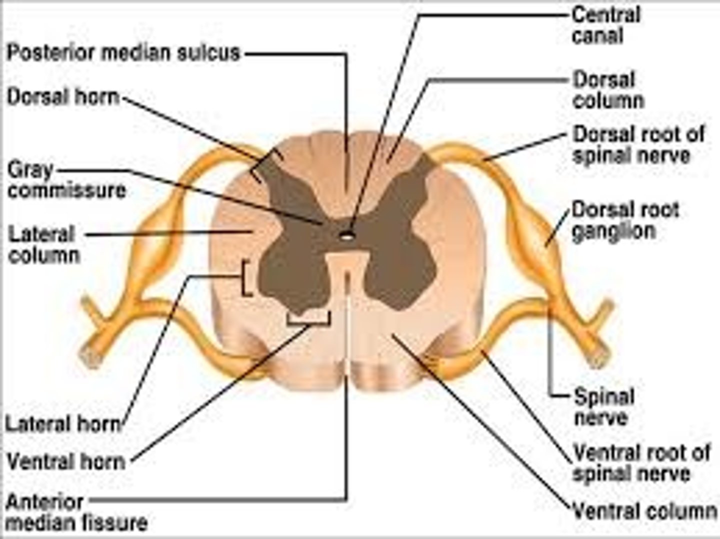
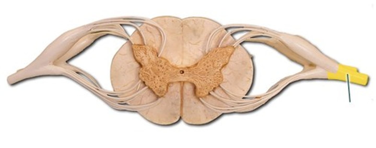
gray matter of spinal cord
INNER: Butterfly shaped
Surrounds the central canal of spinal cord
Contains unmyelinated mostly cell bodies neuron cell bodies (also have minimlal axons)

white matter of spinal cord
OUTER part of sp. cord
myelinated mostly axons

central canal
A tiny channel found within the spinal cord and inferior medulla oblongata for CSF to flow in
Where does the white/gray matter change happen
at the level of the medulla
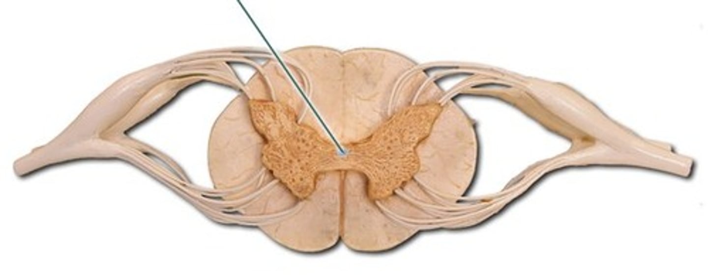
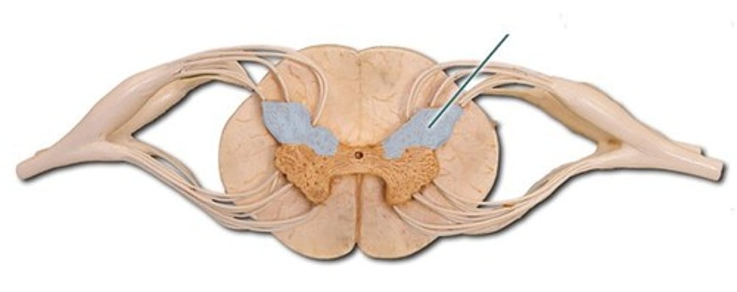
Dorsal horn
termination sensory fibers
◼ integration for relay; reflex
Ventral horn
cell bodies lower motor fibers
◼ medial & lateral columns
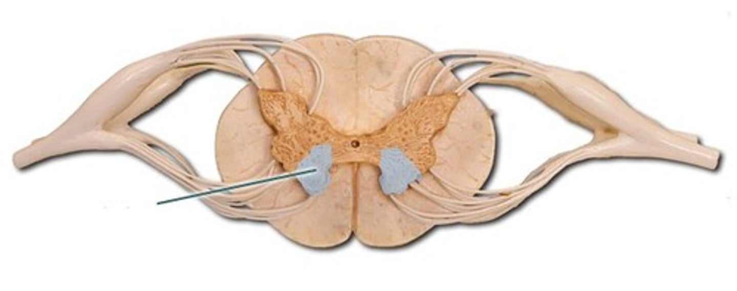
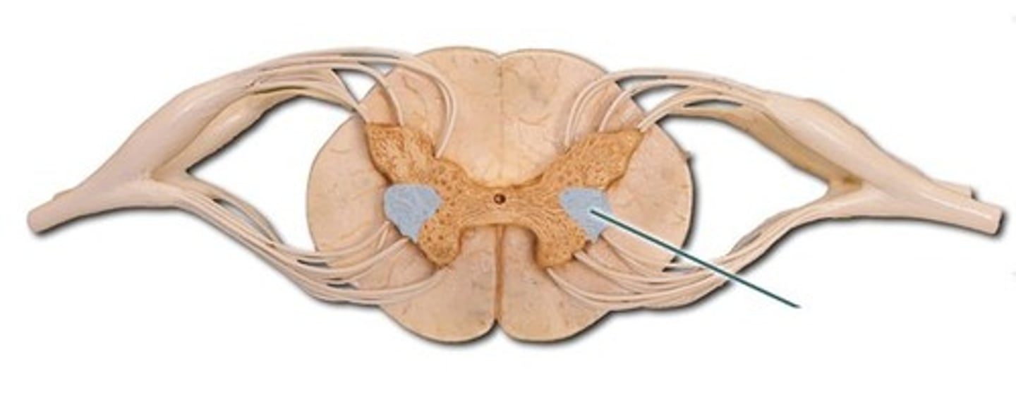
Lateral horns
preganglionic sympathetic cell bodies
◼ Cord levels T1-L3 only
WHITE MATTER COLUMNS
⦿ Dorsal (posterior) ⦿ Ventral (anterior) ⦿ Lateral
TRACTS OR FASCICULI
⦿ Definition
groups of neuronal fibers in CNS with similar function
named for origin and termination
EXAMPLES:
- peduncles
- columns
- funiculus
Commissures
connect 2 hemispheres
TRACTS (WHITE MATTER)
⦿ Ascending
sensory
TRACTS (WHITE MATTER)
⦿ Descending
motor
***Usually a 2 neuron pathway (upper & lower)
Lateral Spinothalamic tract
sensory tract for pain and temperature sensation
ascending spinal cord tract to the thalamus
clinical app - chronic pain
dorsal column tract
2 parts:
fasciculus gracile
fasciculus cuneatus
- functions for conscious perception of position (position of limb) and 2 point discrimination
clinical application: lesion = inability to recognize form by touch (astereognosia)
Lateral Corticospinal tract
descending motor output tract...cortex to spinal cord
- functions: largest human tract; fine motor movements (writing)
clinical app: LCST hemiplegia = lesion in tract causes weakness/loss of voluntary control of muscles ipsilaterally
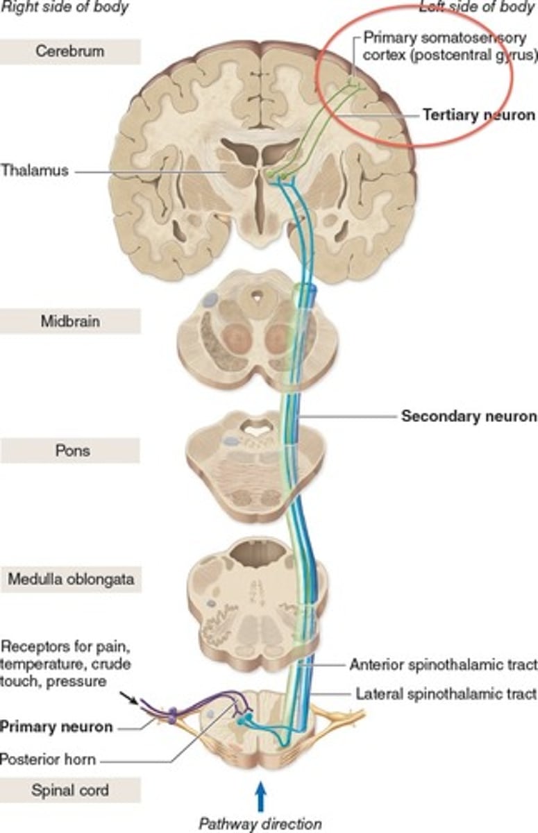
SPINOTHALAMIC PATHWAY
One of the smaller tracts of the anterolateral system.
- passes up the anterior and lateral columns of the spinal cord
carries signals for pain, pressure, temp, light touch, tickle, and itch
first order neurons end in posterior horn
synapse with second order neurons which decussate to other side of spinal cord and form ascending spinothalamic tract
third order neurons continue from thalamus to cerebral cortex
sends signals to the contralateral cerebral hemisphere
GRACILE FASCICULUS PATHWAY
-carries signals from the midthoracic and lower parts of the body (dorsal column ascending)
-Below T6, it composes the entire posterior column (at T6 joins cuneate fasciculus)
-First order nerve fibers end in the gracile nucleus on the ipsilateral side and synapse with second order neurons in the medulla. Second order neuron decussate in medulla forming medial lemniscus. Synapse with third order neuron at thalamus where third order carries signals to contralateral cerebral hemisphere
carries signals for vibration, visceral pain, deep and discriminative touch and proprioception (non-visual sense of position and movement of body) from lower limbs and lower trunk
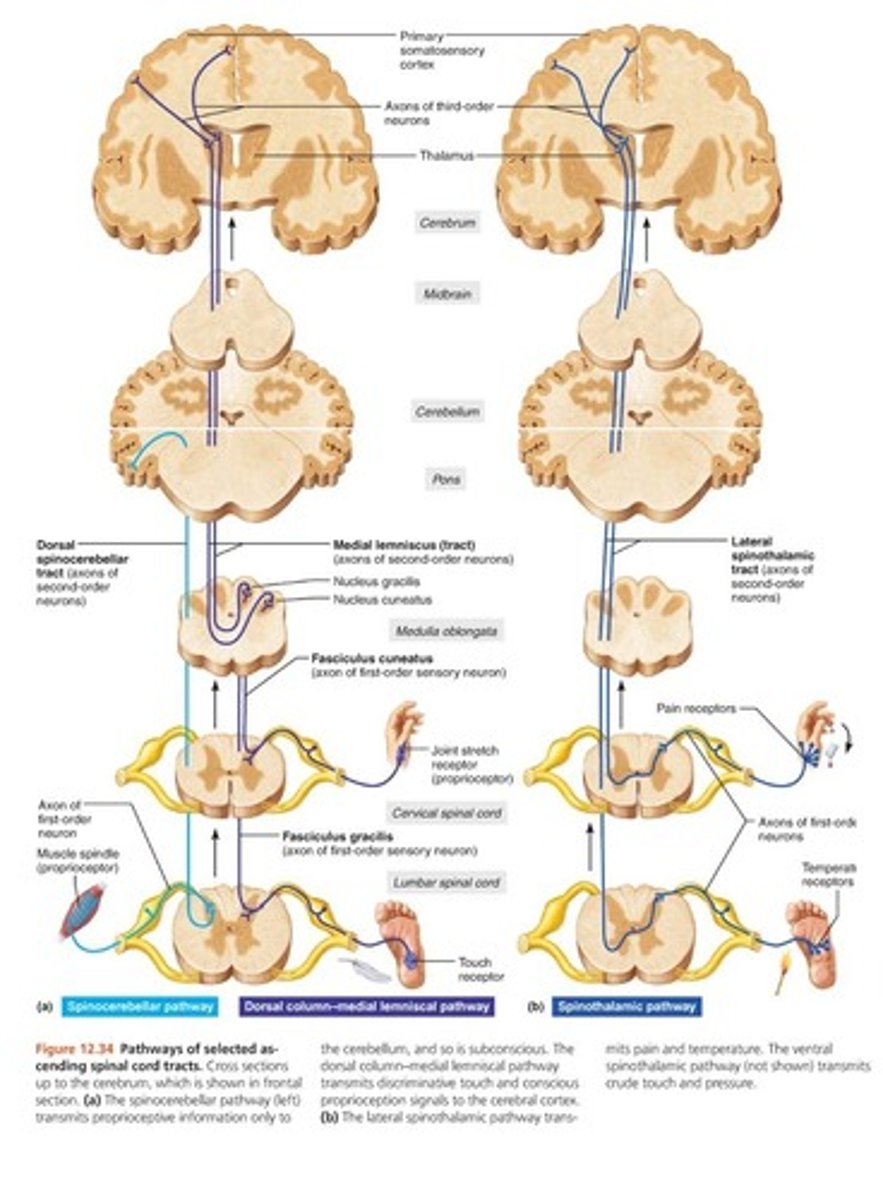
CUNEATE FASCICULUS PATHWAY
Joins the Gracile Fasiculus at T6
occupies lateral portion of the posterior column
carries the same type of sensory signals
originates from the level of T6 and up (upper limb and chest)
First order nerve fibers end in the cuneate nucleus on the ipsilateral side of the medulla oblongata and synapse with second order neurons in the medulla. Second order neuron decussate in medulla forming medial lemniscus. Synapse with third order neuron at thalamus where third order carries signals to contralateral cerebral hemisphere
medial lemniscus is formed from what?
from the second order neurons of the gracile and the cuneate systems that decussate in the medulla
CORTICOSPINAL TRACTS PATHWAY
From cerebral cortex to spinal cord for precise finely coordinated limb movements
pyramids (anterior ridges) in medulla formed from fibers in this system
decussate in the lower medulla
LATERAL corticospinal tract - contralateral side of the spinal cord (the brain signals go to opposite side of spinal cord at medulla)
ANTERIOR corticospinal tract - on ipsilateral side of the spinal cord
two neuron pathway (upper and lower motor neurons)
Paresis
damage to spinal cord causing partial paralysis, weakness
Paraplegia
damage to spinal cord causing paralysis from the waist down typically
lower extremity paralysis
Quadriplegia
damage to spinal cord causing paralysis of all four extremities
Can lead to:
⭘ respiratory paralysis(ventilation), loss of sensation or motor
control
⭘ disorders of bladder, bowel and sexual function
Damage to spinal cord from strokes or
other brain injuries
hemiplegia
a R sided stroke - leads to L sided paralysis/paresis
hemiplegia
weakness or paralysis of one side of the body
not always permanent
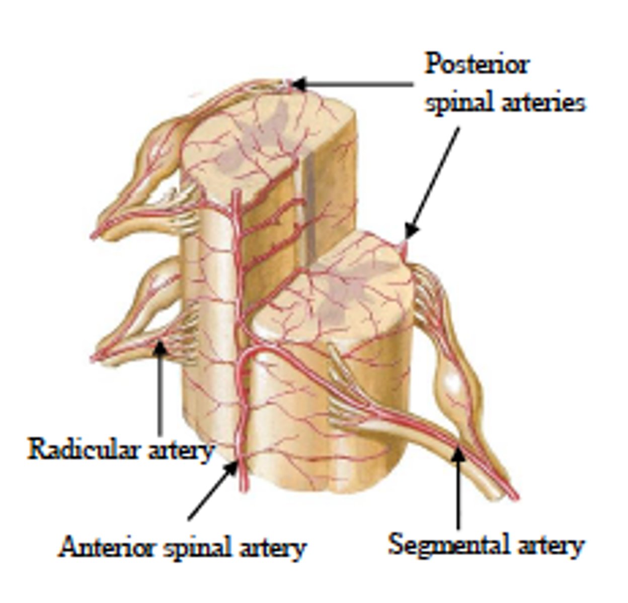
VASCULAR ANATOMY OF CORD
⦿ Arterial
◼ 3 longitudinal arteries supply cord
⭘ 1. anterior (1) spinal artery
⭘ 2. Posterior (2) spinal arteries
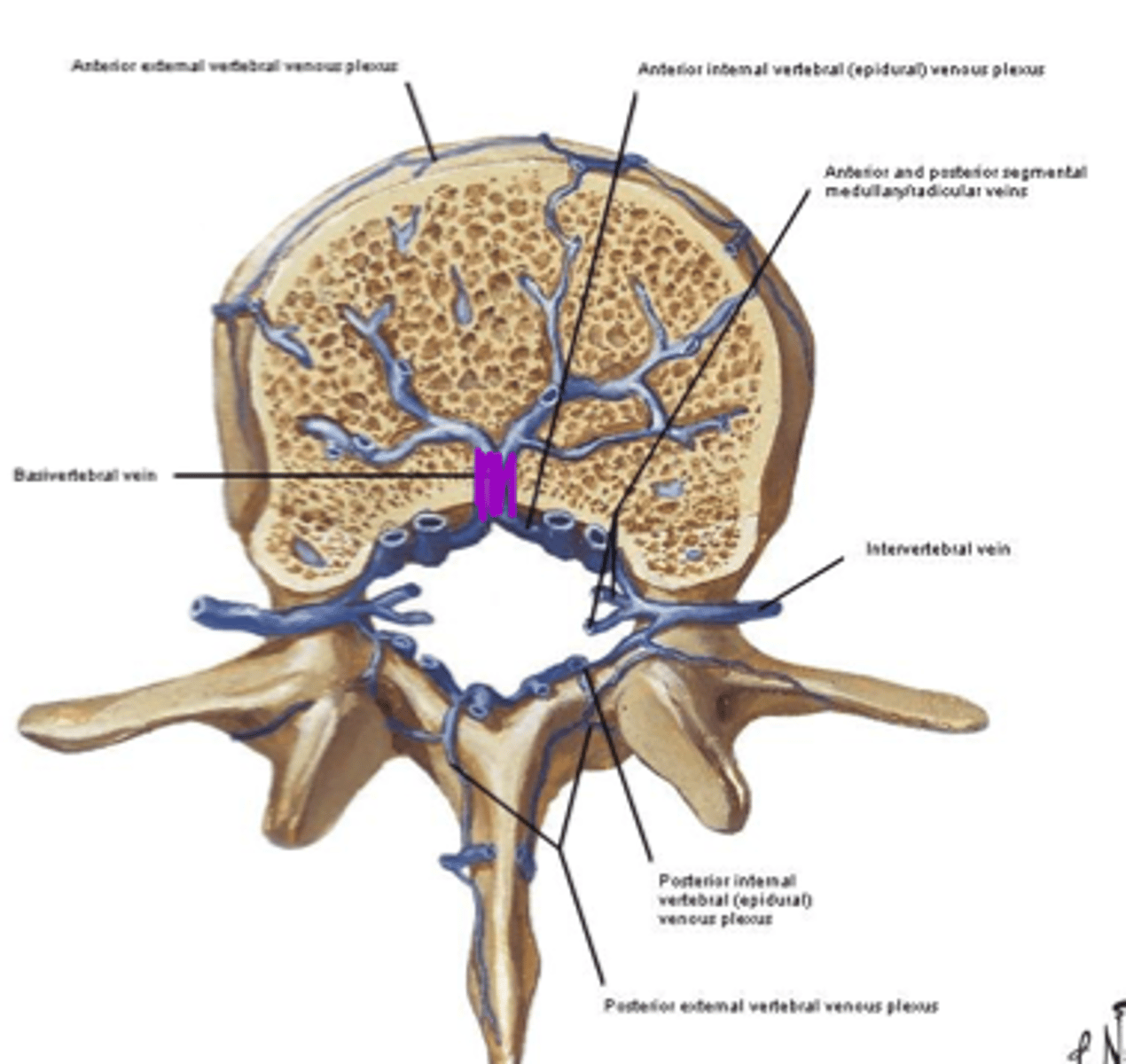
VASCULAR ANATOMY OF CORD
⦿ Venous
◼ Internal & external venous plexuses
◼ Basivertebral & intervertebral veins
◼ 6 longitudianl veins drain the cord
⭘ 1. Anterior (3) spinal veins
⭘ 2. Posterior (3) spinal veins
REGIONAL DIFFERENCES WHITE VS GRAY
⦿ Observe differences in distribution of gray & white matter
⦿ Pay special attention to general progression
from cervical to sacral
⦿ Note: Amount of gray matter increases as you travel cervical to sacral (because you add lateral gray matter horns)
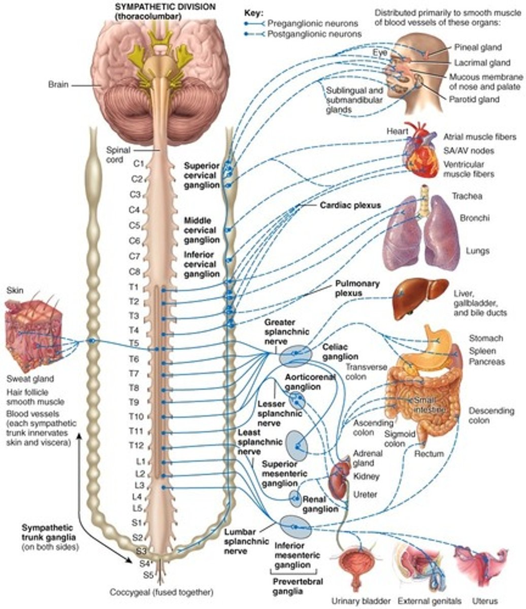
AUTONOMIC NERVOUS SYSTEM ( ANS)
⦿ 2 Parts of ANS
1. sympathetic
2. parasympathetic
CNS neuron
1 neuron to somatic effector (target)
ANS neuron
2 neuron pathway to target effector
- preganglionic neuron
- postganglionic neuron
two types of ANS pathways
visceral sensory afferents
Motor efferents
Visceral Sensory Afferent Pathway
◼ Somatic (body wall) poorly understood
◼ Visceral (gut) better understood clinically
⭘ Use DORSAL ROOT (sensory=dorsal)
Motor Efferents Pathway
◼ Message for action at target organ
⭘ Use VENTRAL ROOTS (motor=ventral)
BASIC COMPARISON DIVISIONS - SNS & PSNS
⦿ Location of Pre-ganglionic cell bodies (CNS
connections)
⦿ Location of fibers
⦿ Pre & post-ganglionic fiber length
⦿ Ratio of pre to post ganglionic fibers
⦿ Neurotransmitters & Fate of NT
⦿ General function(s)
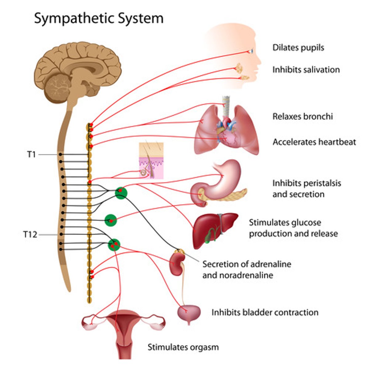
LOCATION OF PRE- GANGLIONIC CELL BODIES (SNS)
⦿ SNS
◼ Thoracolumbar division
◼ Spans - T1-L2 (lateral gray column)
LOCATION OF PRE- GANGLIONIC CELL BODIES (PSNS)
⦿ PSNS: Craniosacral division
⭘ Brainstem
-Contains - axons of CN III, VII, IX, X (nuclei in brainstem=cell bodies)
⭘ Sacral
-Spans - S2-S4 (lateral gray column)
LOCATION OF FIBERS (SNS) (postganglionic fibers)
⦿ SNS
◼ Chain of ganglia on each side of vertebral column
-paravertebral
◼ # ganglia per chain = 31
-also prevertebal ganglion and splanchic nerve
LOCATION OF FIBERS (PSNS) (postganglionic fibers)
⦿ PSNS
◼ Ganglia run uninterrupted from CNS to terminal
ganglia close to organ they innervate
◼ ***No ganglia chain (cannot see it with being so close to target)
-near or in walls of viscera
FIBER LENGTH (SNS)
⦿ SNS
◼ Pre-gang = SHORT
◼ Post-gang = LONG
FIBER LENGTH (PSNS)
⦿ PSNS
◼ Pre-gang = LONG
◼ Post-gang = SHORT
RATIO OF PRE TO POST (SNS and PSNS)
⦿ SNS
◼ 1 pre- to many post-
◼ Ratio 1:25
⦿ PSNS
◼ 1 pre- to few post-
◼ Ratio 1:5
NEUROTRANSMITTER SNS
⦿ SNS
◼ Pre-gang = Ach
◼ Post-gang = NE - Norepinephrine
NEUROTRANSMITTER PSNS
⦿ PSNS
◼ Pre-gang = Ach
◼ Post-gang = Ach
CHOLINERGIC FIBERS - secrete
ACh
CHOLINERGIC FIBERS - seen in
SNS and PSNS
CHOLINERGIC FIBERS - types of receptors
◼ 1. nicotinic
⭘ Fxn: always stimulatory
◼ 2. muscarinic
⭘ Fxn: inhibitory or stimulatory
Where are muscarinic receptors stimulatory or inhibitory
they are in PSNS:
stimulatory in smooth muscle
inhibitory in cardiac muscle
ADRENERGIC FIBERS - Secrete
Norepinephrine (NE)
ADRENERGIC FIBERS - found
in SNS
ADRENERGIC FIBERS - types of receptors
◼ (1) alpha (α): mostly stimulatory
⭘ 2 types:
◼ (2) beta (β): mostly inhibitory
⭘ 2 types:
2 types of alpha adrenergic receptors
A1 - constrict blood vessels
A2 act on platelets - stimulate blood clotting
2 types of Beta Adrenergic Receptors
B1 - stimulatory on the heart
B2 - inhibit respiratory structures
FATE OF NEUROTRANSMITTER in SNS
⦿ SNS
◼ NE is retrieved - active transport which takes energy and time
◼ Length of effect - prolonged effect
FATE OF NEUROTRANSMITTER in PSNS
⦿ PSNS
◼ Ach destroyed - quickly by enzyme acetylecholinesterase
◼ Length of effect - short lived
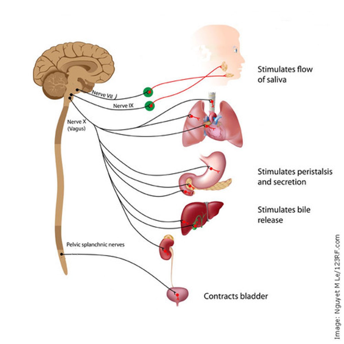
SUMMARY OF PARASYMPATHETIC EFFERENT DISTRIBUTION (CN III, CN VII, CH IX, CN X, S2-S4
Know that PSNS has craniosacral division
⦿ CN III: paravertebral chain- to orbital area
⦿ CN VII: para- to lacrimal area; submandibular &
sublingual gland
⦿ CN IX: para- to parotid gland
⦿ CN X: Para- to heart, lungs, upper gut
⦿ S2-S4: para- to distal gut
SYMPATHETIC MOTOR DISTRIBUTION
-thoracolumbar division
⦿ Throughout body (blood vessels)
⦿ L/R paravertebral chains
◼ Rami communicantes:
⭘ White -carry preganglionic fibers
⭘ Gray - carry post ganglionic fibers
⦿ Prevertebral ganglia & splanchnic nerves
Prevertebral ganglia & splanchnic nerves
◼ Def - paired nerves carrying ANS fibers to the visceral organs
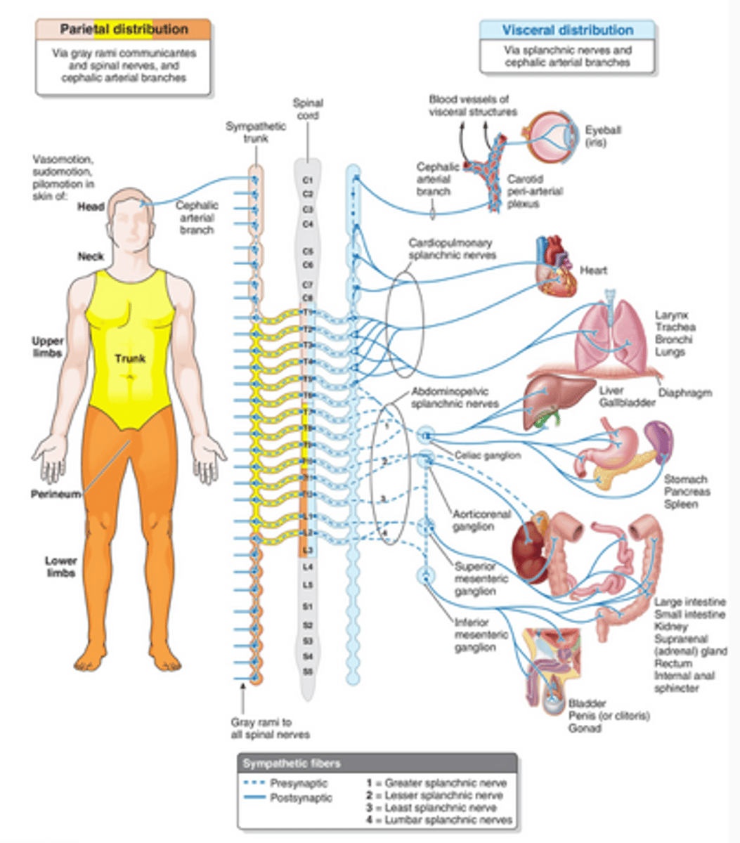
***Thoracolumbar Division SYMPATHETIC PATHWAYS
⦿ Once a sympathetic preganglionic fiber has entered
the paravertebral chain, it can do one of 4 things to
synapse with a postganglionic fiber:
1. End at level it entered
2. Run up the chain
3. Run down the chain
4. Pass out to a prevertebral ganglion via a splanchnic nerve that goes directly to organ