Physiology Exam 3
1/31
There's no tags or description
Looks like no tags are added yet.
Name | Mastery | Learn | Test | Matching | Spaced | Call with Kai |
|---|
No analytics yet
Send a link to your students to track their progress
32 Terms
The function of the _____ circuit is to pump blood from the heart, to the lungs, and back to the heart.
Pulmonary or pulmonary circuit
which ion is not used in autorhythmic of cardiac muscle?
a. calcium
b. sodium
c. potassium
d. chloride
e. none of the above
d. chloride
Identify the structure in the heart that is leaky to sodium?
SA node
Funny channel
Sinoatrial node
In the SA (sinoatrial) node, there are three types of channels: voltage gated sodium, potassium, and _____ channels, which allow the influx of calcium.
T type
L type
funny
Identify the heart structure that directly depolarizes the ventricular myocardia.
a. pulmonary semilunar valve
b. bundle of his
c. interventricular septum
d. tricuspid valve
e. Purkinje fibers
f. bicuspid valve
g. aortic semilunar valve
h. Sa node
e. Purkinje fibers
Identify the atrioventricular valve that separates the left atrium from the left ventricle.
Bicuspid valve
mitral
mitral valve
Identify the correct sequence of electrical conduction in the heart.
AV node
SA node
interatrial pathway
internodal pathway
Purkinje fibers
bundle of HIS
right and left bundle branches
ventricular myocardia
Order
a. 2-4-1-3-6-7-8-5
b. 1-4-3-2-7-6-5-8
c. 2-3-4-1-6-7-5-8
d. 1-3-4-2-8-7-5-6
c. 2-3-4-1-6-7-5-8
The conduction pathways in the heart (ex. internodal, intratrial, bundle of HIS, etc.) are not really composed of nerves; instead they are modified ______ cells that rapidly conduct the cardiac action potential. (Hint: only the proper technical term)
Pacemaker
myocardia
myocardial
heart
cardiac
Identify the heart structure located between the right ventricle and the left ventricle.
a. SA node
b. Bundle of HIS
c. interventricular septum
d. bicuspid valve
e. Purkinje fibers
f. AV node
g. pulmonary semilunar valve
h. Tricuspid valve
j. aortic semilunar valve
c. interventricular septum
Identify the heart structure located between the left atrium and left ventricle.
a. SA node
b. Bundle of HIS
c. interventricular septum
d. bicuspid valve
e. Purkinje fibers
f. AV node
g. pulmonary semilunar valve
h. Tricuspid valve
j. aortic semilunar valve
d. bicuspid valve
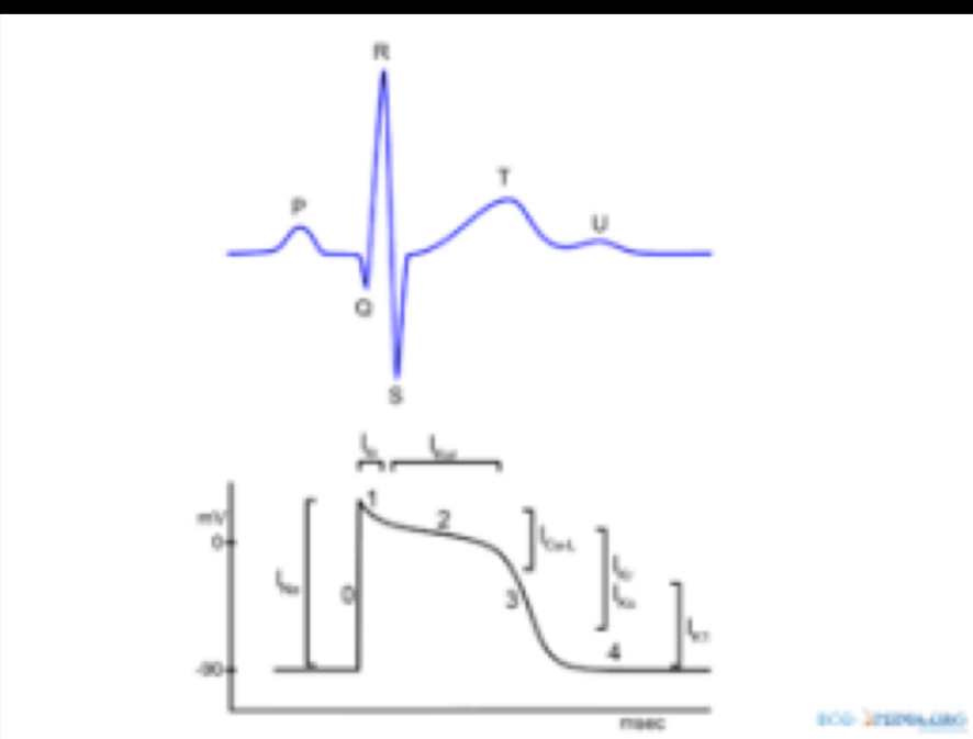
One of these images is a pacemaker potential in the SA node.
True or false
False
This type of action potential occurs within the _______________ .
Ventricle
The SA and AV nodes of the heart are dual reciprocally innervated by the Vagus nerve (cranial nerve X; which slows the heart rate) and the ________________________ (which increases heart rate).
Sympathetic cardiac nerve
In an normal EKG (ECG):
There are no visible, distinguishable waves and there is no distinguishable T-P interval
The P-wave is absent, there is a large QRS complex, and the T-wave is large and there is a distinguishable T-P interval
The P-wave is present, there is a large QRS complex, and the T-wave is present and there is a distinguishable T-P interval
The P-wave is present, there is a small QRS complex, and the T-wave is present and there is a distinguishable T-P interval
The P-wave is present, there is a large QRS complex, and the T-wave is present and there is a very large T-P interval
None of these answers are correct
The P-wave is present, there is a large QRS complex, and the T-wave is present and there is a very short T-P interval
3 . The P-wave is present, there is a large QRS complex, and the T-wave is present and there is a distinguishable T-P interval
In an normal EKG (ECG):
There are no visible, distinguishable waves and there is no distinguishable T-P interval
The P-wave is absent, there is a large QRS complex, and the T-wave is large and there is a distinguishable T-P interval
The P-wave is present, there is a large QRS complex, and the T-wave is present and there is a distinguishable T-P interval
The P-wave is present, there is a small QRS complex, and the T-wave is present and there is a distinguishable T-P interval
The P-wave is present, there is a large QRS complex, and the T-wave is present and there is a very large T-P interval
None of these answers are correct
The P-wave is present, there is a large QRS complex, and the T-wave is present and there is a very short T-P interval
The P-wave is present, there is a large QRS complex, and the T-wave is present and there is a distinguishable T-P interval

Identify the segment of the ECG that corresponds to ventricular contraction (both tonal and ejection phases).
P wave
ST segment
PR interval
QT interval
T wave
QT interval
Identify the region on the ECG that corresponds to ventricular repolarization.
PQ Segment
QT Interval
Q
P
ST Segment
QRS Complex
T
S
R
PR Interval
PQ Segment
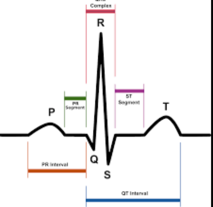
Identify the region of the ECG that corresponds to ventricular depolarization
T
P
SA node
QT Interval
ST Segment
PR Interval
QRS Complex
Q
PQ segment
QRS Complex
Identify the region on the ECG that corresponds to atrial depolarization.
AV node
T
QRS Complex
S
P
Q
R
PQ Segment
ST Segment
SA node
PR Interval
P
In a heart paced by the AV node:
The P-wave is present, there is a large QRS complex, and the T-wave is present and there is a very short T-P interval
The P-wave is present, there is a large QRS complex, and the T-wave is present and there is a very large T-P interval
There are no visible, distinguishable waves and there is no distinguishable T-P interval
The P-wave is absent, there is a large QRS complex, and the T-wave is large and there is a distinguishable T-P interval
None of these are correct
The P-wave is present, there is a large QRS complex, and the T-wave is present and there is a distinguishable T-P interval
The P-wave is present, there is a small QRS complex, and the T-wave is present and there is a distinguishable T-P interval
The P-wave is absent, there is a large QRS complex, and the T-wave is large and there is a distinguishable T-P interval
In tachycardia:
The P-wave is present, there is a large QRS complex, and the T-wave is present and there is a very large T-P interval
None of these are correct
The P-wave is absent, there is a large QRS complex, and the T-wave is large and there is a distinguishable T-P interval
There are no visible, distinguishable waves and there is no distinguishable T-P interval
The P-wave is present, there is a large QRS complex, and the T-wave is present and there is a distinguishable T-P interval
The P-wave is present, there is a small QRS complex, and the T-wave is present and there is a distinguishable T-P interval
The P-wave is present, there is a large QRS complex, and the T-wave is present and there is a very short T-P interval
All heart attacks lead to acute cardiac arrest (i.e. within minutes, without medical intervention).
True
False
False