skeletal system: metabolic and neoplastic diseases
 Call Kai
 Learn
Learn Practice Test
 Spaced Repetition
 Match
Match Flashcards
 Knowt Play
1/29

There's no tags or description
Looks like no tags are added yet.
Name | Mastery | Learn | Test | Matching | Spaced | Call with Kai | Chat |
|---|
No analytics yet
Send a link to your students to track their progress
30 Terms
metabolic disease
a disease that disrupts normal functions of the body
osteoporosis (sub)
The structural integrity of the trabecular bone pattern is destroyed. Porous and brittle bone
It is identified as a bone mass less than 648 mg/cm2.
Major cause of hip, spine, and wrist fractures in women over 50.
osteopenia
A radiographically visible decrease in bone density
Occurs as a result of osteoporosis or osteomalacia.
Identified as a bone mass between 648 and 833 mg/cm2.
primary osteoporosis
Postmenopausal is the most common form of this type of osteo
caused by a lack of estrogen which weakens the bone matrix
Senile
secondary osteoporosis
Most commonly associated with an existing disease process:
Endocrine diseases
Neoplastic diseases
Malnutrition
Chronic renal failure
Hereditary diseases
-Or as a result of drug therapy.
osteomalacia (sub)
Caused by lack of calcium in tissues and failure of bone tissue to calcify.
Results from inadequate intake or absorption of vitamin D.
rickets
Termed used if osteomalacia occurs before growth plate closure. happens in kids
osteomalacia xray
Appears similar to osteoporosis.
Bowing and greenstick fractures

Paget’s Disease (sub and des)
Fairly common in the older population.
cause is unknown
Usually begins in the 5th decade of life.
Can affect one or more bones.
Most common in the pelvis, spine, skull, and long bones.
two stages of paget’s disease
Osteolytic Stage: Bone undergoes continuous destruction.
Osteoblastic Stage: bone replaced by abnormally soft and poorly mineralized material. It is thicker than the normal bone, and adds weight and more stress to the bone which causes deformities and fractures.
paget’s disease xray
cotton wool appearance

acromegaly (add)
An endocrine disorder caused by a disturbance in the function of the pituitary gland.
More common in females than males.
Caused by excessive secretion of Growth hormone in adults.
This produces a thickening and coarsening of bone.
Individuals have:
A prominent forehead and jaw
Widened teeth
Abnormally large, spade-like hands (thich hands)
acromegaly xray
An enlarged sella turcica (pituitary is in the sella turcica)
Changes in the skull
benign tumors
localized tumor of well differentiated cells that don’t invade surrounding tissues or metastasize
neoplastic
new abnormal tissue growth
skeletal benign neoplastic diseases
Osteochondroma (Additive) – Radiography and MRI
Osteoma (Additive) – Radiography and MRI
Giant cell tumors (Subtractive) – Radiography and MRI
skeletal malignant neoplastic diseases
Osteosarcoma (Additive) – Radiography and MRI
Ewing Sarcoma (Both) – Radiography and MRI
Chondrosarcoma (Subtractive) – Radiography and MRI
Osteochondroma (additive)
Most common benign bone tumor.
3x more common in men than women.
Arises from the metaphysis.
Most commonly, involves the lower femur or upper tibia.
can have extoses
extoses
Excessive benign bone growth.
May appear as singular or multiple lesions.
Normally diagnosed in childhood or adolescence.
osteochondroma xray

osteoma (additive)
Most commonly located in the skull.
Composed of very dense, well-restricted, normal bone tissue.
Usually projects into the orbits or paranasal sinuses.
Slow growing and of little significance, unless they cause:
Obstruction
Impinge on the brain or eye
Interfere with the oral cavity
osteoma xray

Giant Cell Tumors (GCT) (subtractive)
Also known as osteoclastomas.
Characterized by the presence of numerous, multinucleated osteoclastic cells.
it is subtractive because it degrades the bone
Affect women more than men between the ages of 20 and 30.
GCTs can be either benign or malignant.
Affects the epiphyses of long bones, especially the lower femur, the upper tibia, and the lower radius.
giant cell tumor xray
soap bubble

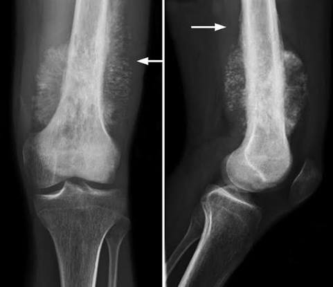
Osteosarcoma (additive)
Also referred to as osteogenic sarcoma.
Most common primary malignancy of the skeleton.
The cause is linked genetically to deletion of genetic material on chromosome 13.
Most frequently found in the metaphysis of long bones.
Approximately 50% affect the knee.
75% occur in patients younger than 20 years old.
spicules (small new bone cells) lifts the periosteum from the bone
Highly aggressive with a poor prognosis.
osteosarcoma xray
sunburst or sunray
Ewing sarcoma (sub and add)
Occur usually between the ages of 5 and 15 years.
More common in boys than girls.
Often involves the entire shaft of a long bone. Specifically comes from the medullary canal
Effects the extremities and the pelvis.
ewing sarcoma xray

Chondrosarcoma (subtractive)
Arises from cartilage.
Common locations are the pelvis, shoulder, and ribs.
Men are 3x more likely than women to develop.
lesions have the ability to seed. the traveling of the cancerous cells
can destroy the bone
chondrosarcoma xray
Irregular or circular radiolucencies with areas of calcification.
