lab exam 2
 Call Kai
 Learn
Learn Practice Test
 Spaced Repetition
 Match
Match Flashcards
 Knowt Play
1/33

There's no tags or description
Looks like no tags are added yet.
Name | Mastery | Learn | Test | Matching | Spaced | Call with Kai | Chat |
|---|
No analytics yet
Send a link to your students to track their progress
34 Terms
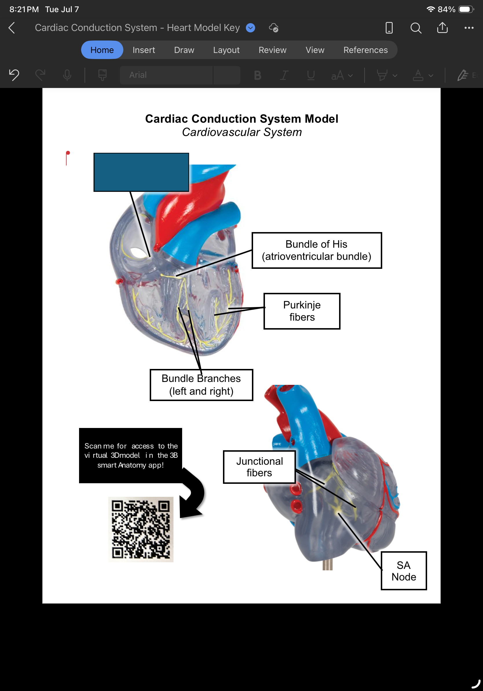
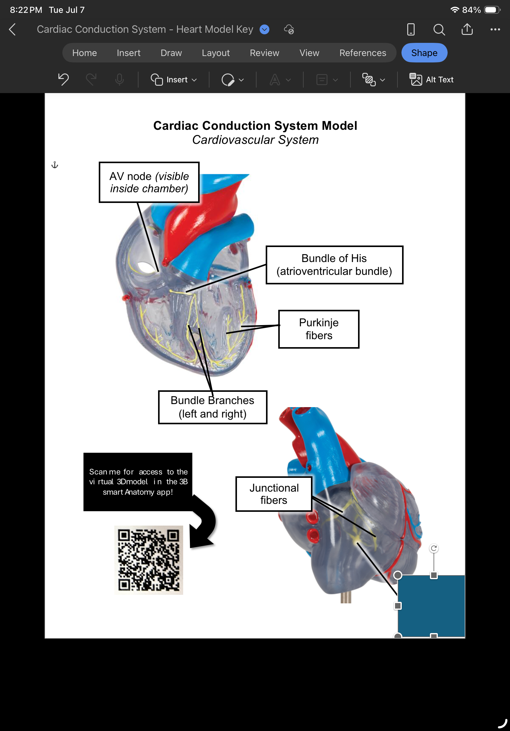
AV node
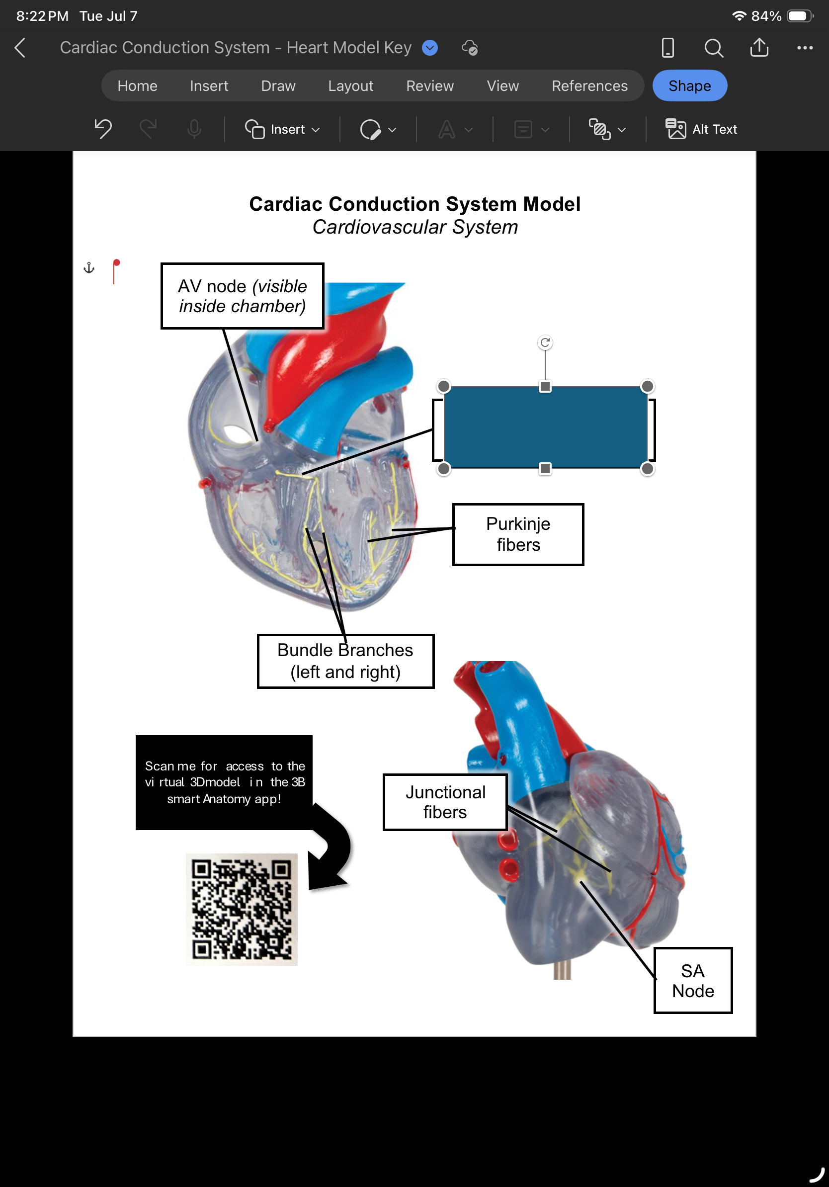
Bundle of His
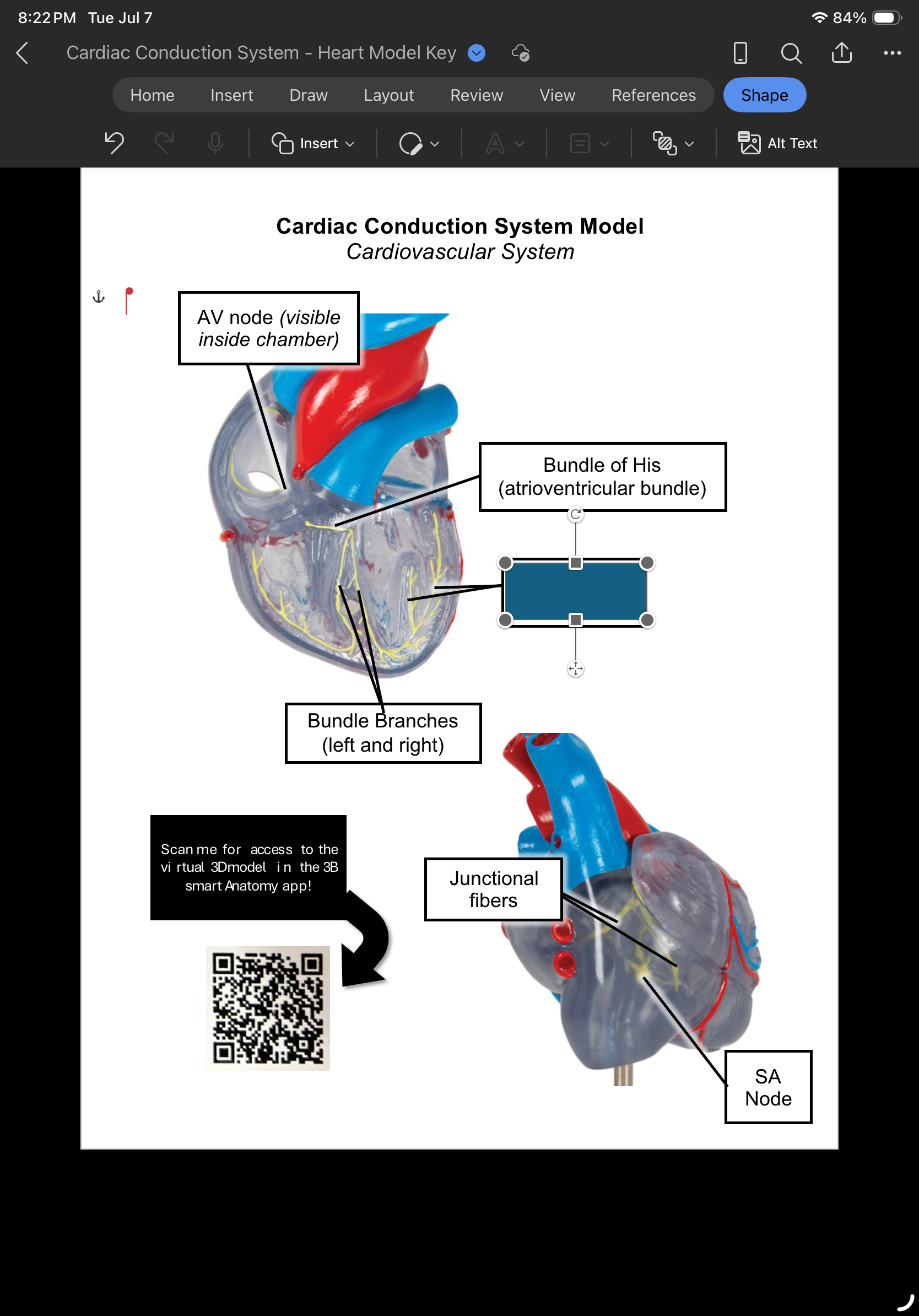
Purkinje fibers
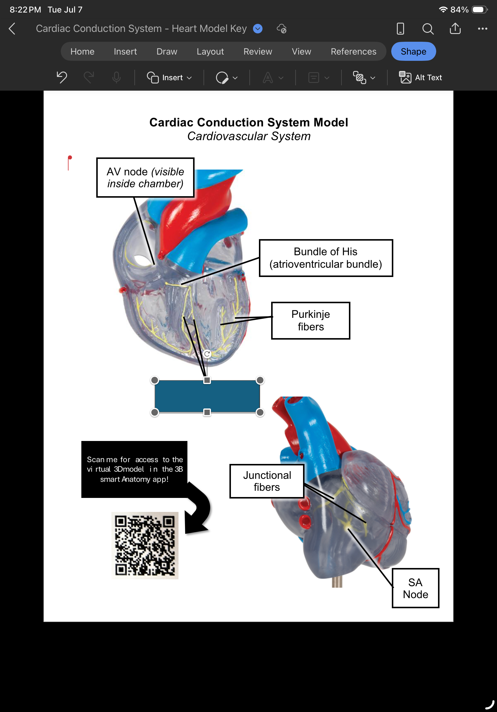
Bundle branches
List the cardiac conduction system starting with the SA node.
Sa node, AV node, AV bundle, bundle branches, & purkinje fibers.
Sa node
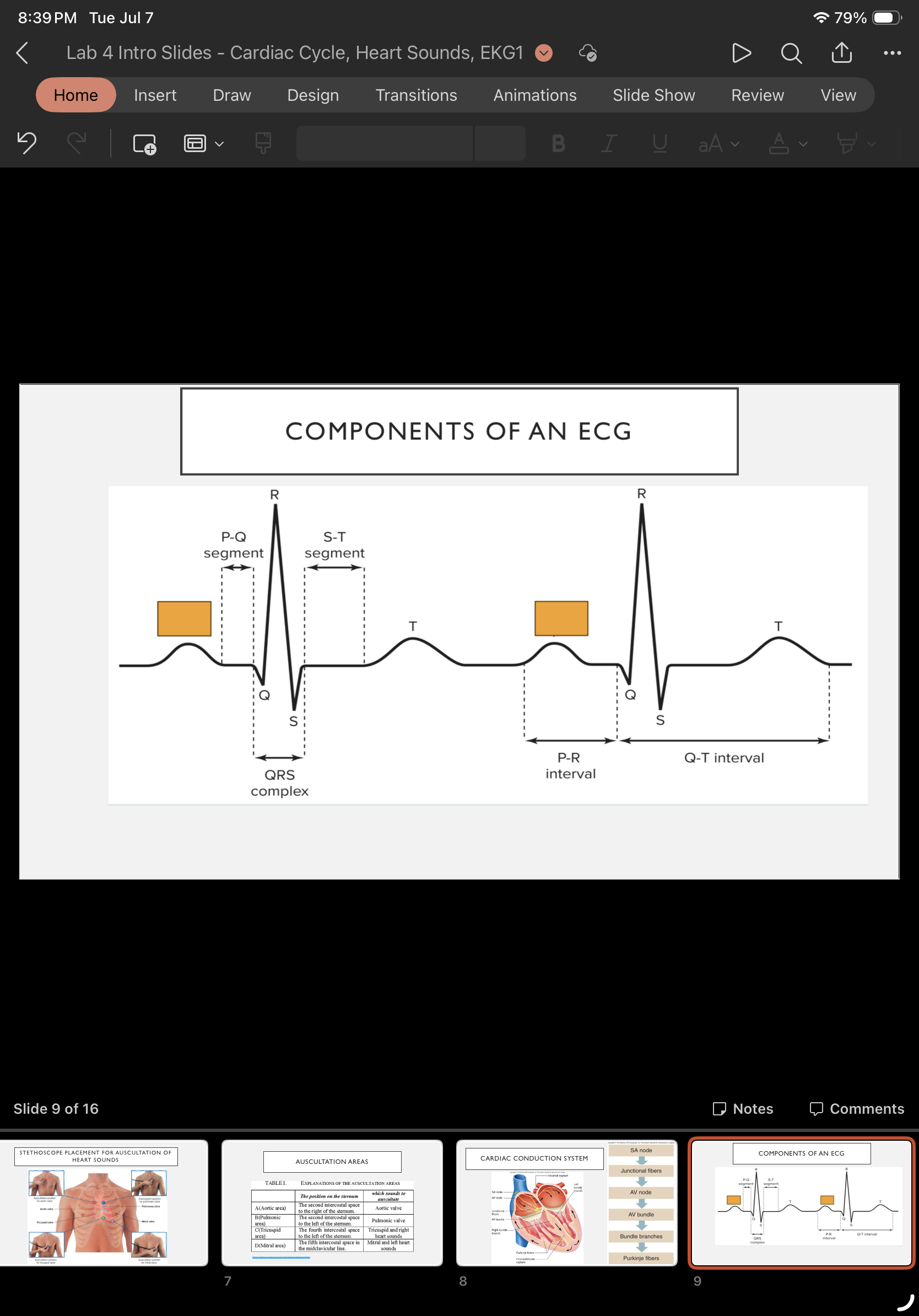
What wave is this & what does it mean?
P wave: depolarization of SA node and atria
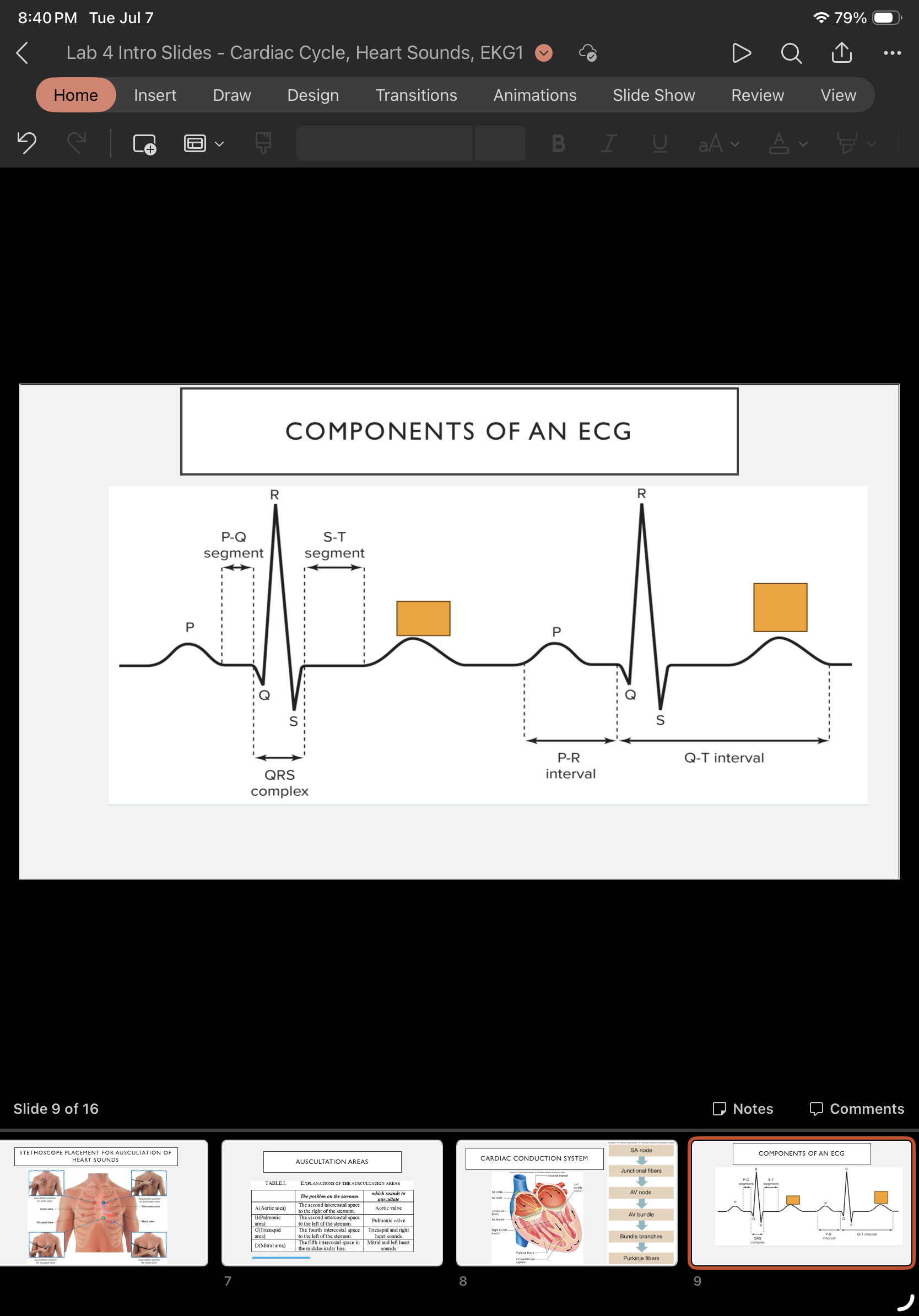
What wave is this & what does it mean?
T wave: ventricular repolarization
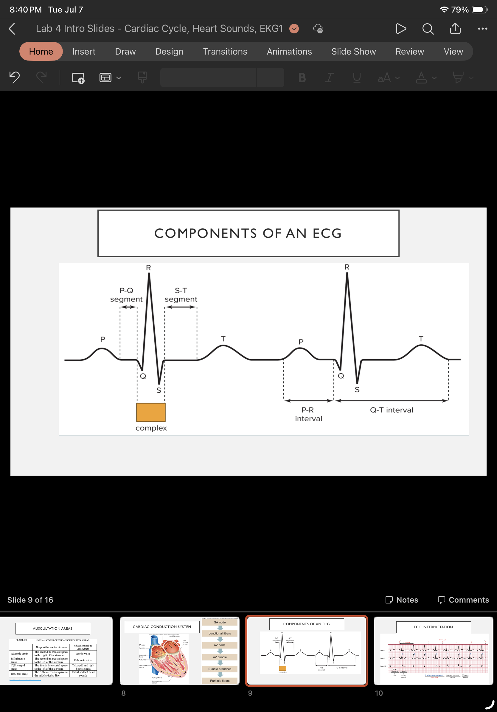
What complex is this & what does it mean?
QRS complex: ventricular depolarization and atrial repolarization
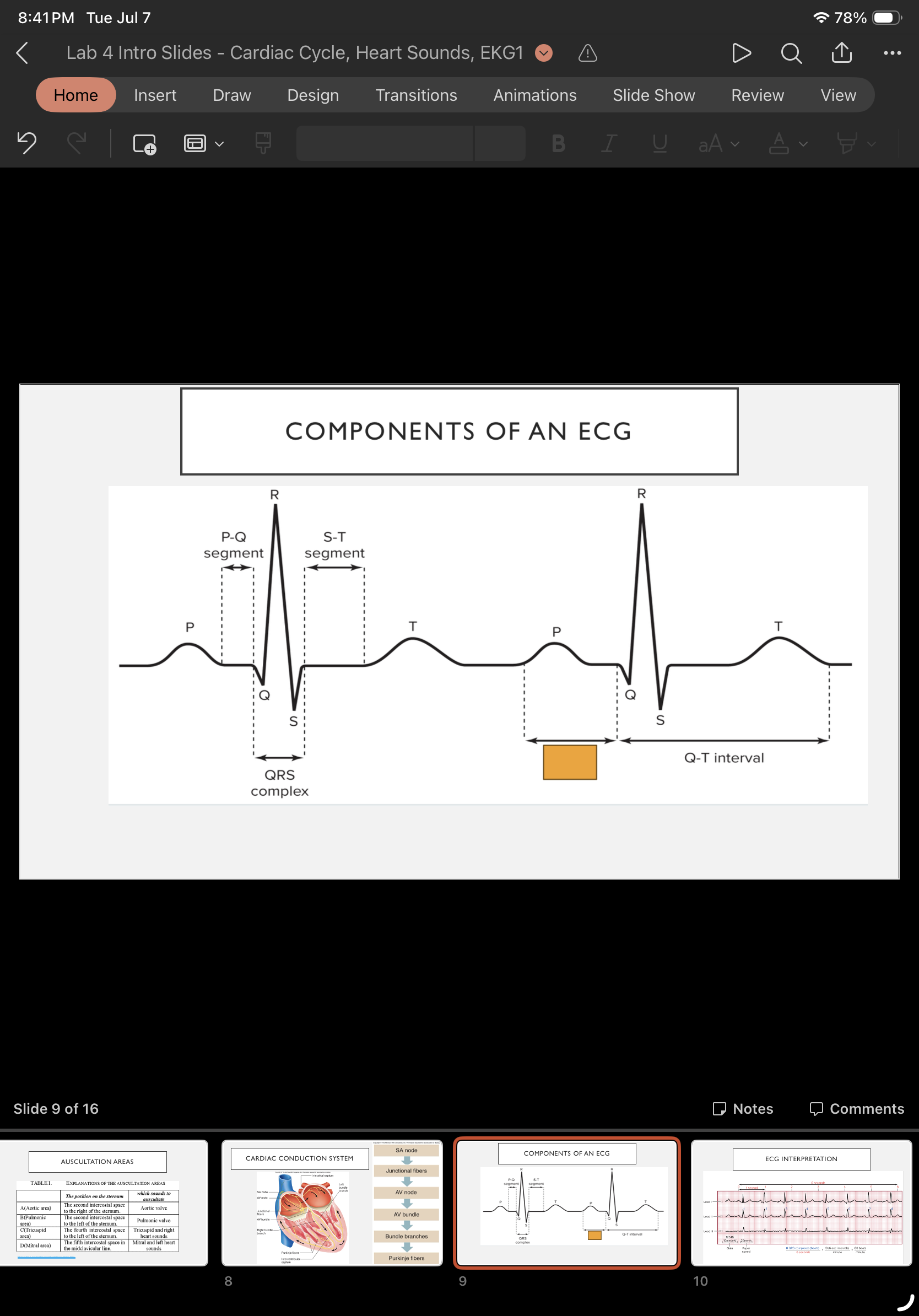
what interval is this & what does it mean?
P-R interval: beginning of atrial excitation to beginning of ventricular excitation
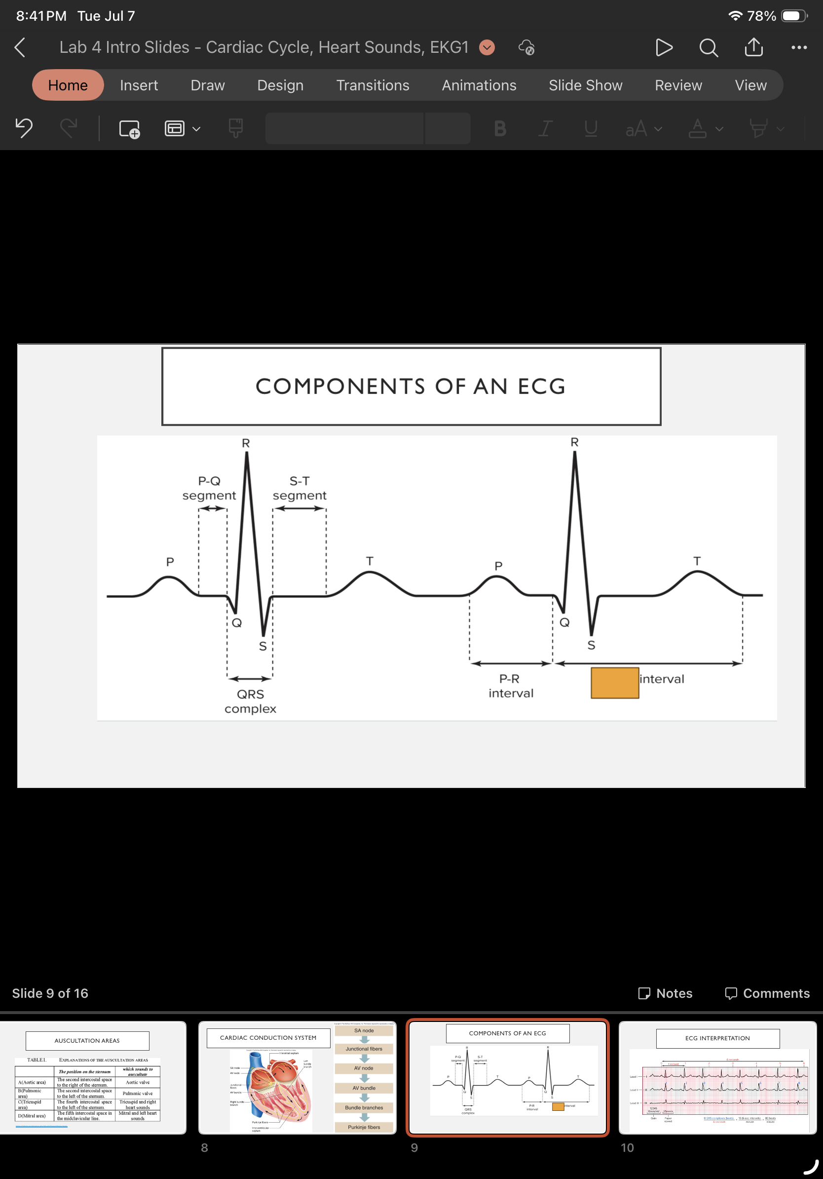
what interval is this & what does it mean?
Q-T interval: beginning of ventricular depolarization through ventricular repolarization
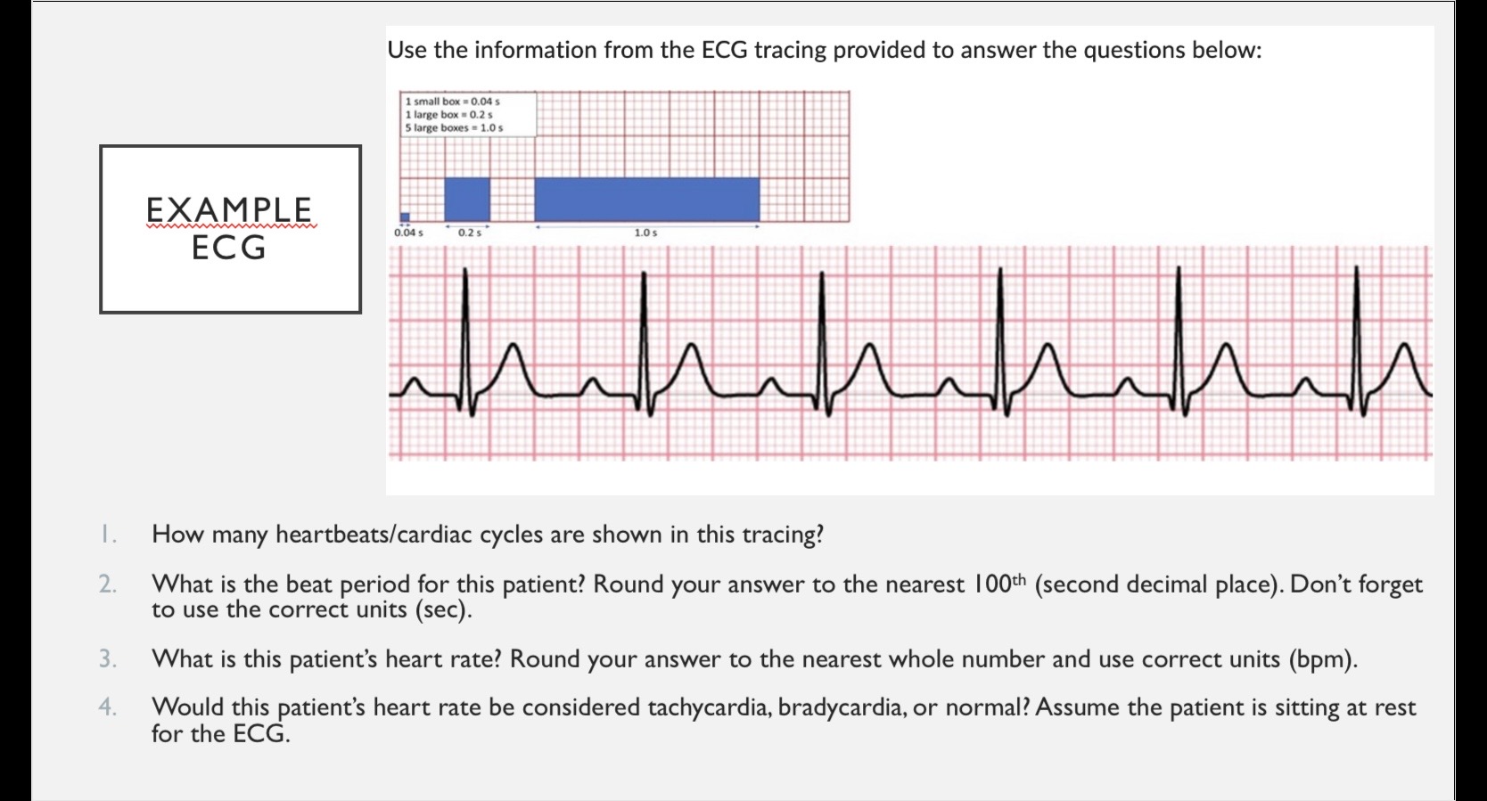
6 beats
0.8
75 beats per minute
Normal
What causes the first heart sound. “LUB”
the closing of the atrioventricular valves
What causes the second heart sound. “Dub”
Closing of the semilunar valves.
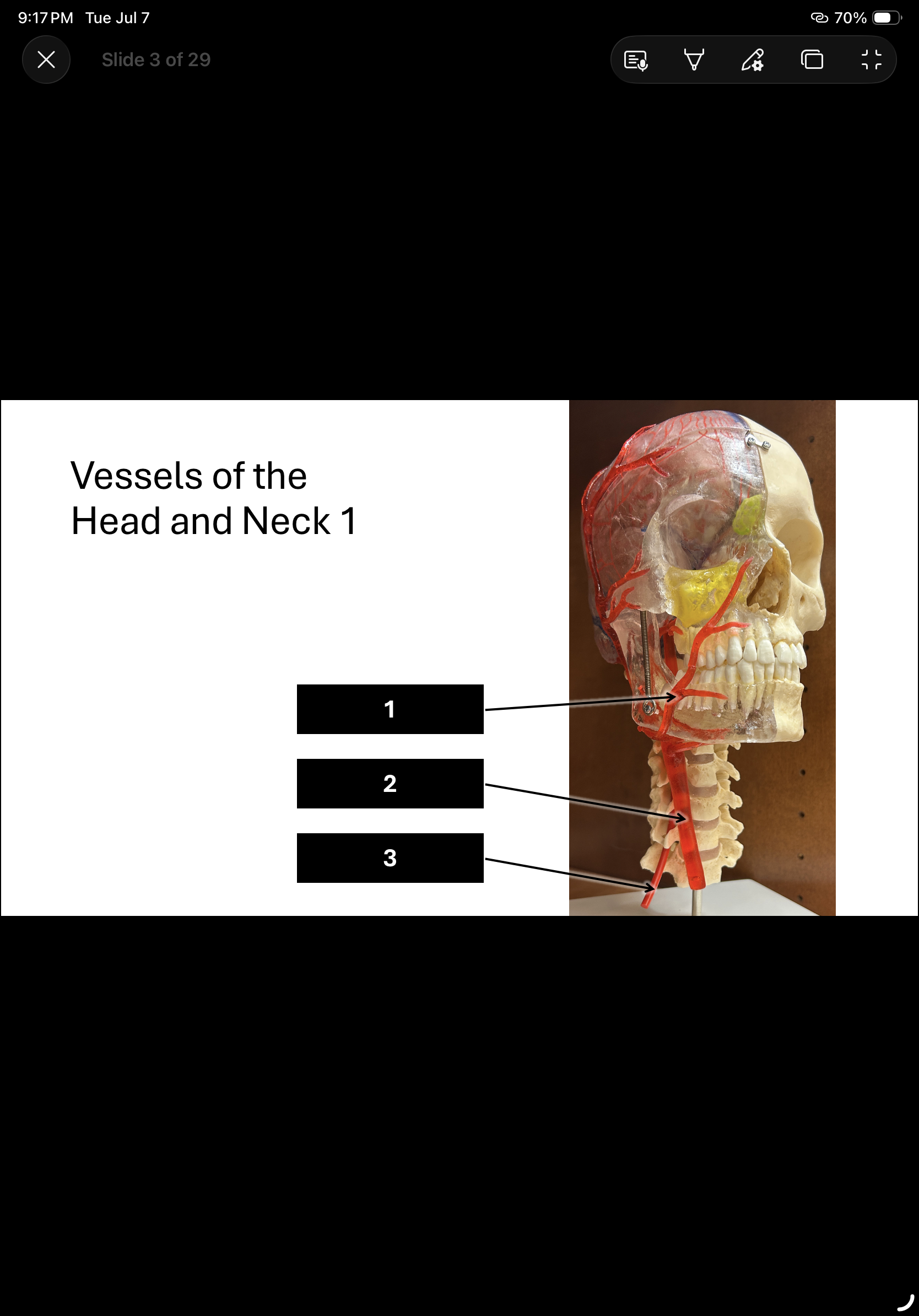
What is 1 & 2?
Facial artery 2. Common carotid.
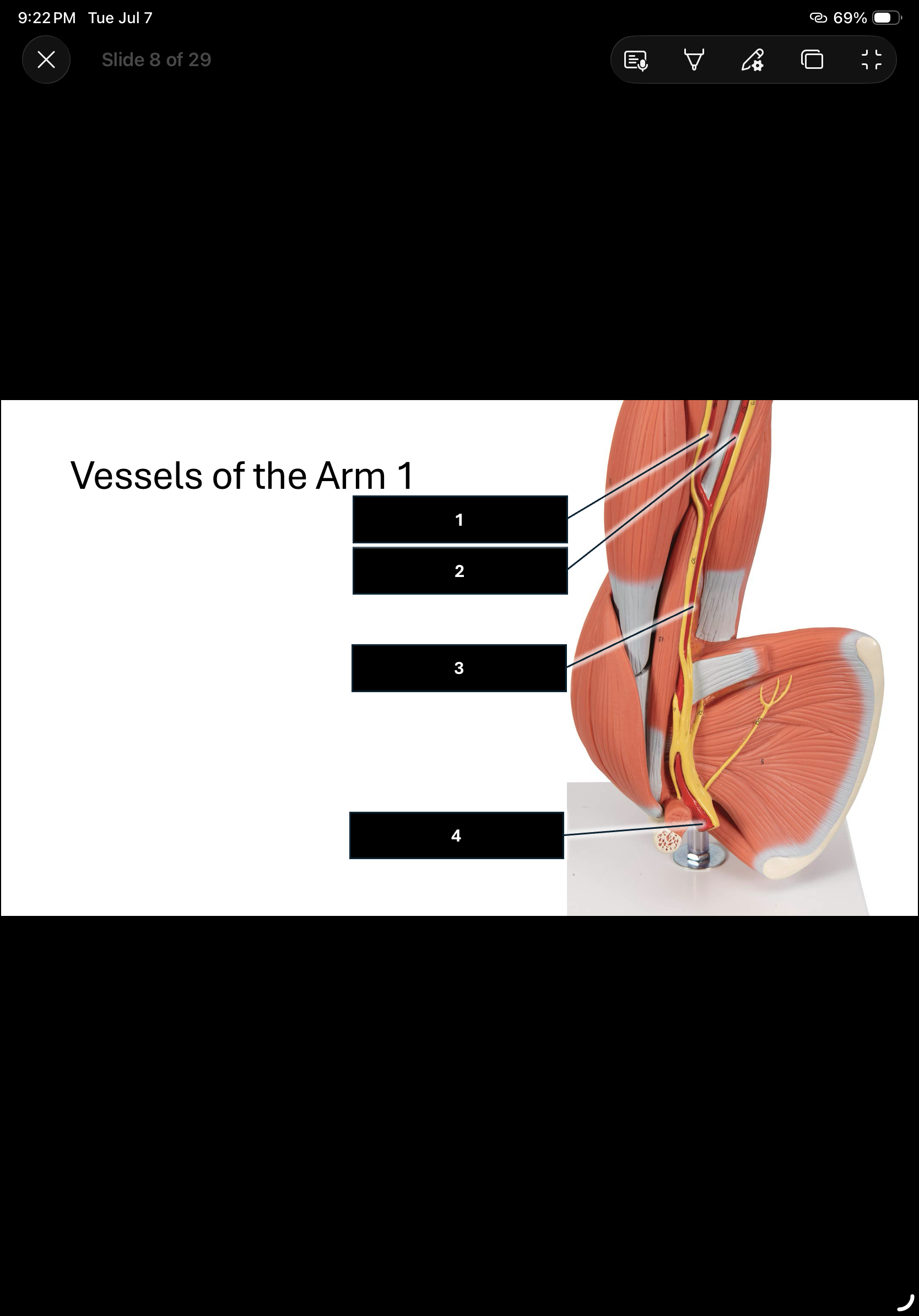
In order.
Radial artery
Ulnar artery
Brachial artery
Axillary artery
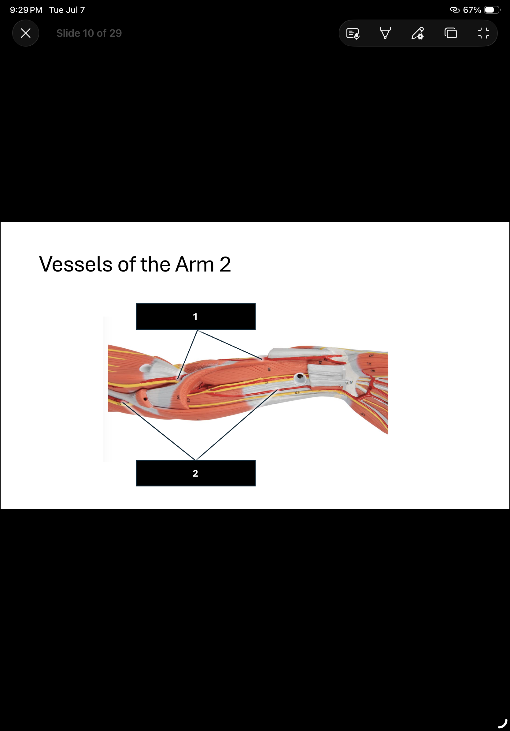
In order.
Radial artery (thumb side)
Ulnar artery (pinky side)
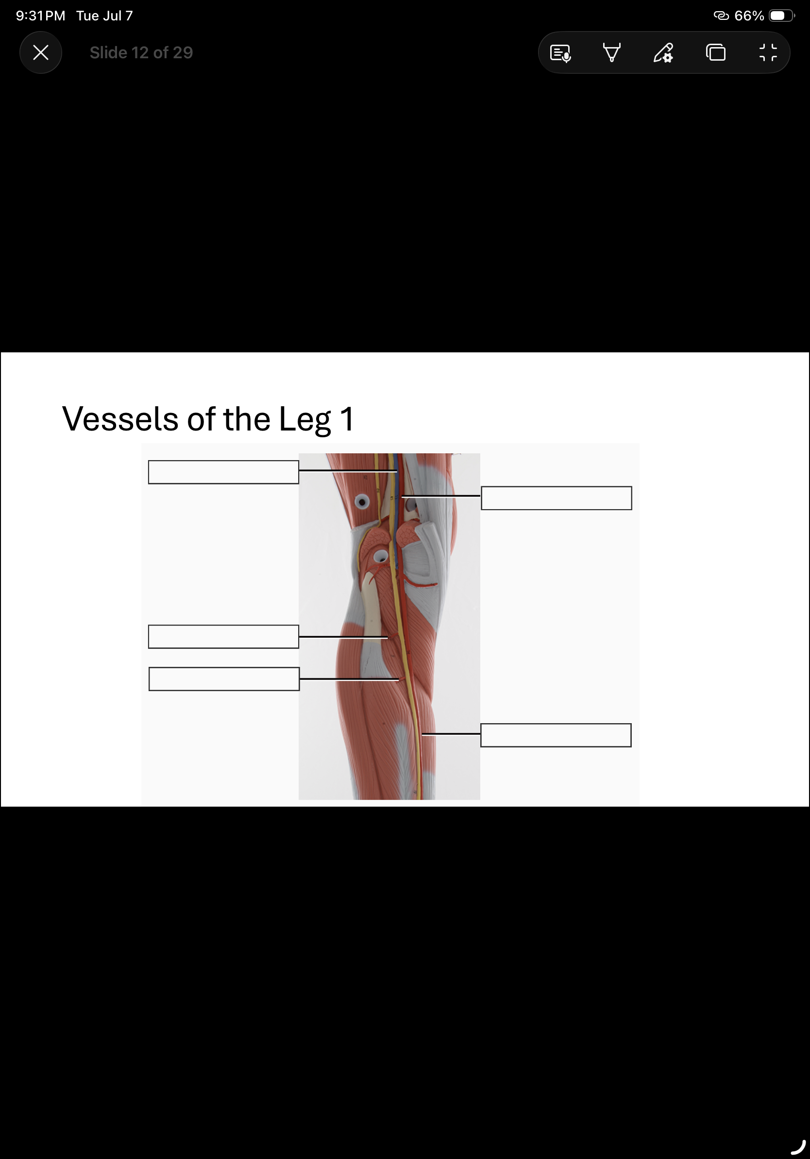
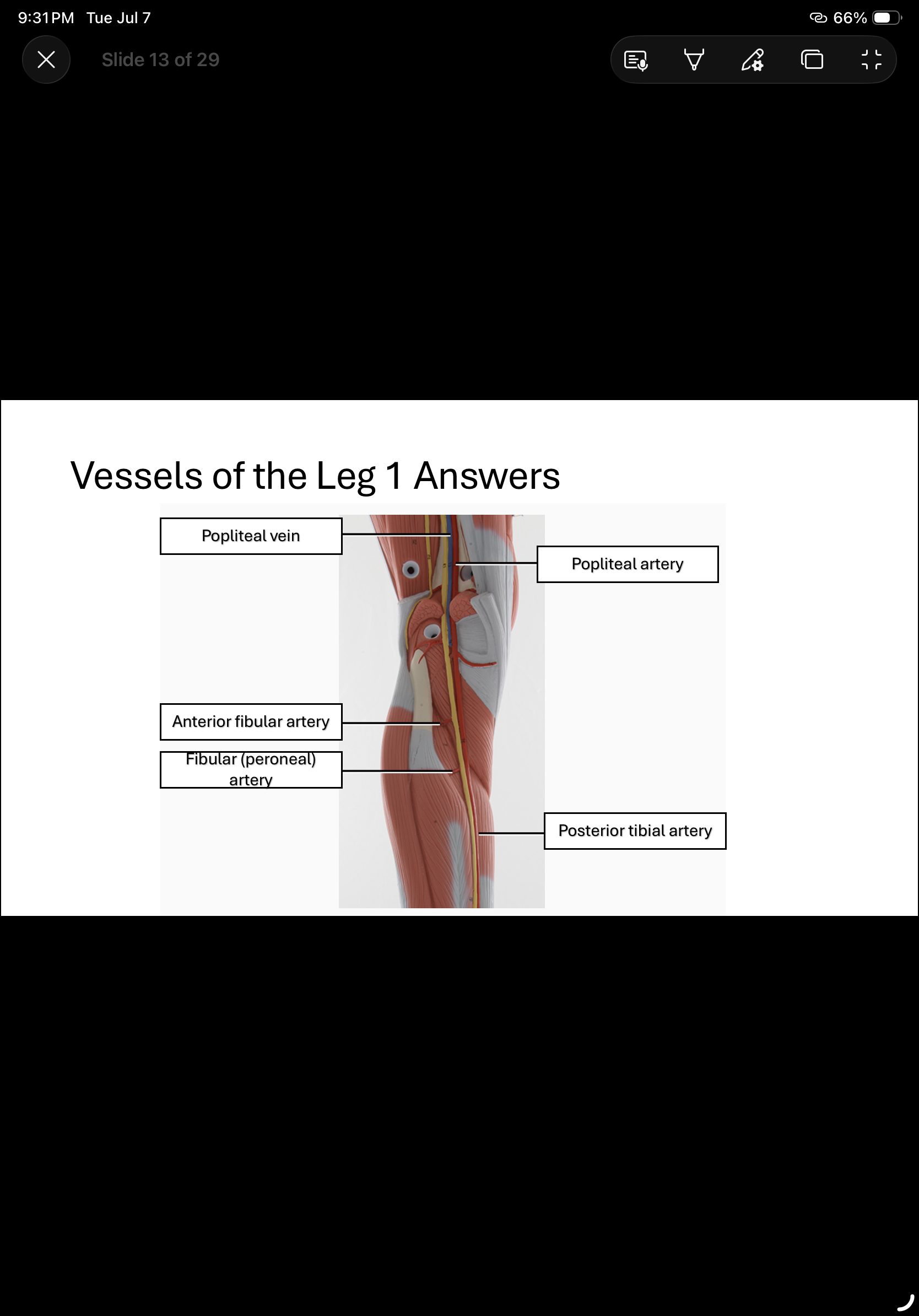
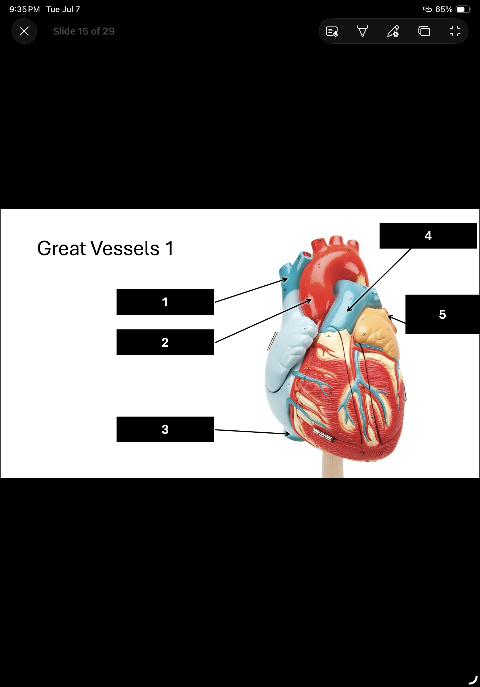
In order.
Superior vena cava
Ascending aorta
Inferior vena cava
Pulmonary trunk
Pulmonary vein
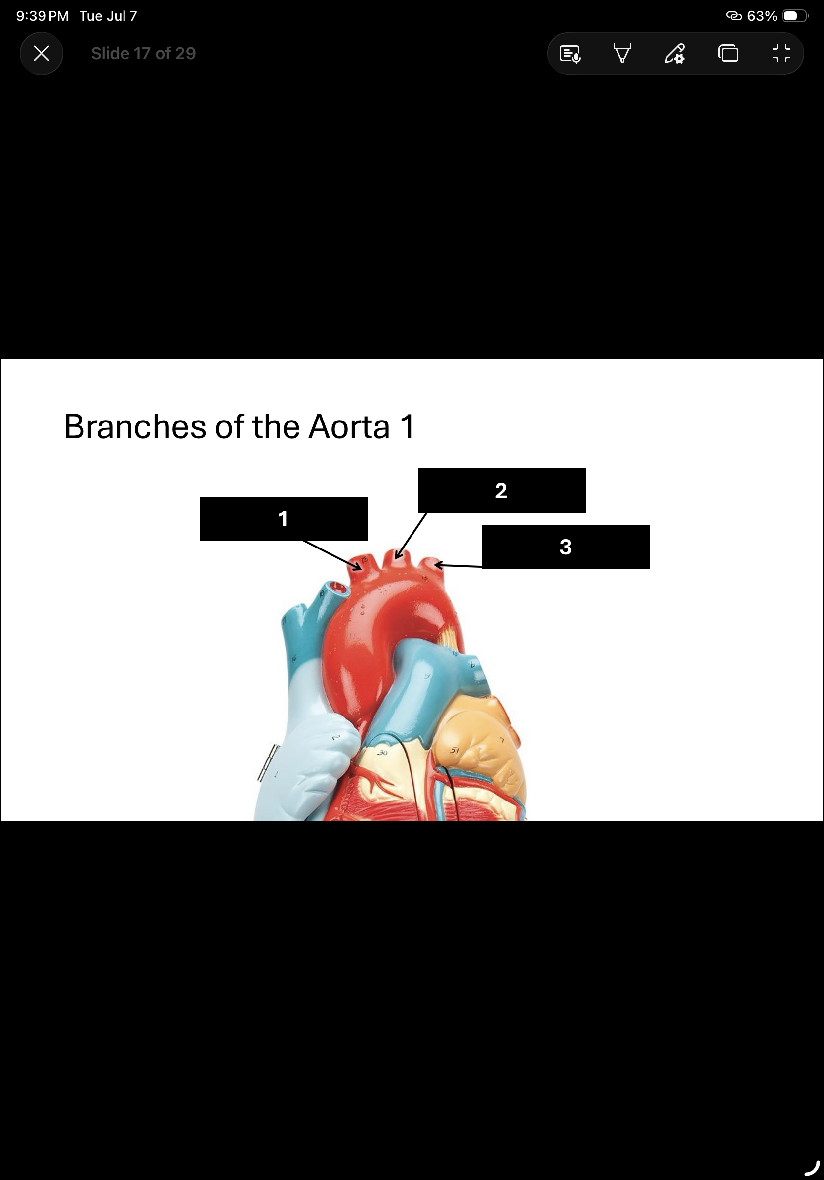
Brachiocephalic trunk
Left common carotid
Left subclavian artery
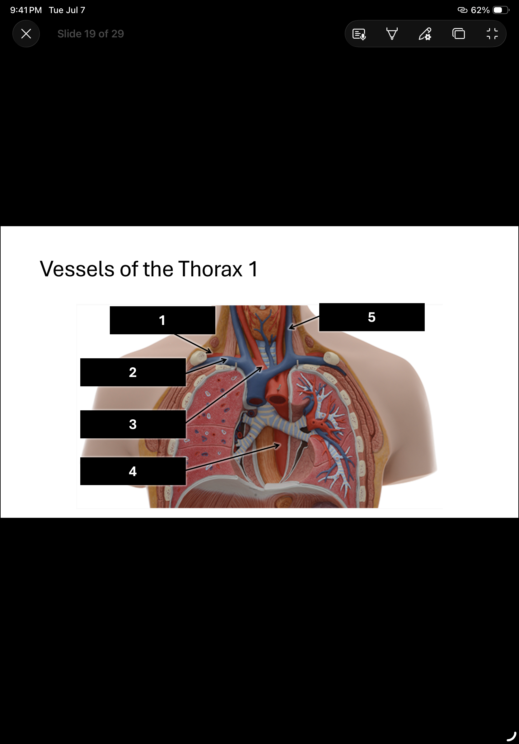
In order.
Right subclavian artery
Right subclavian vein
Brachiocephalic trunk
Thoracic aorta
Internal jugular vein
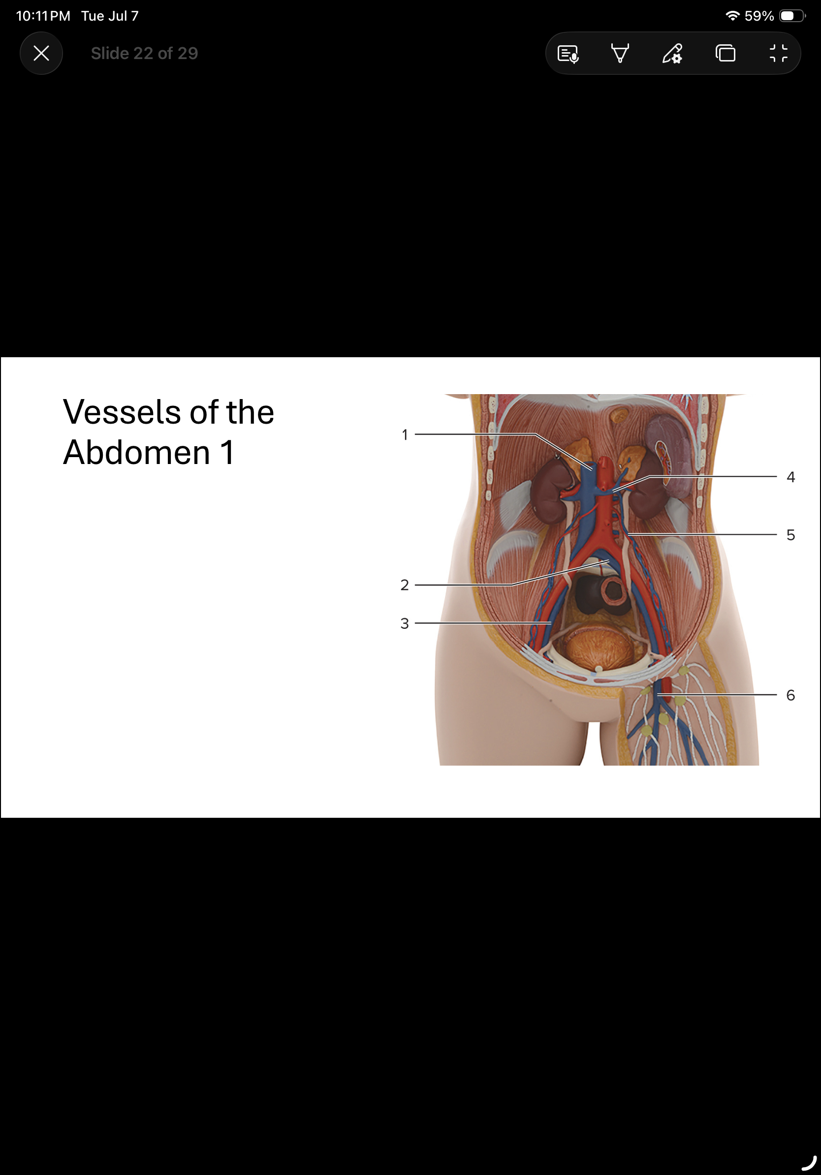
1, 2 & 3.
Ivc
Common iliac vein
External iliac vein
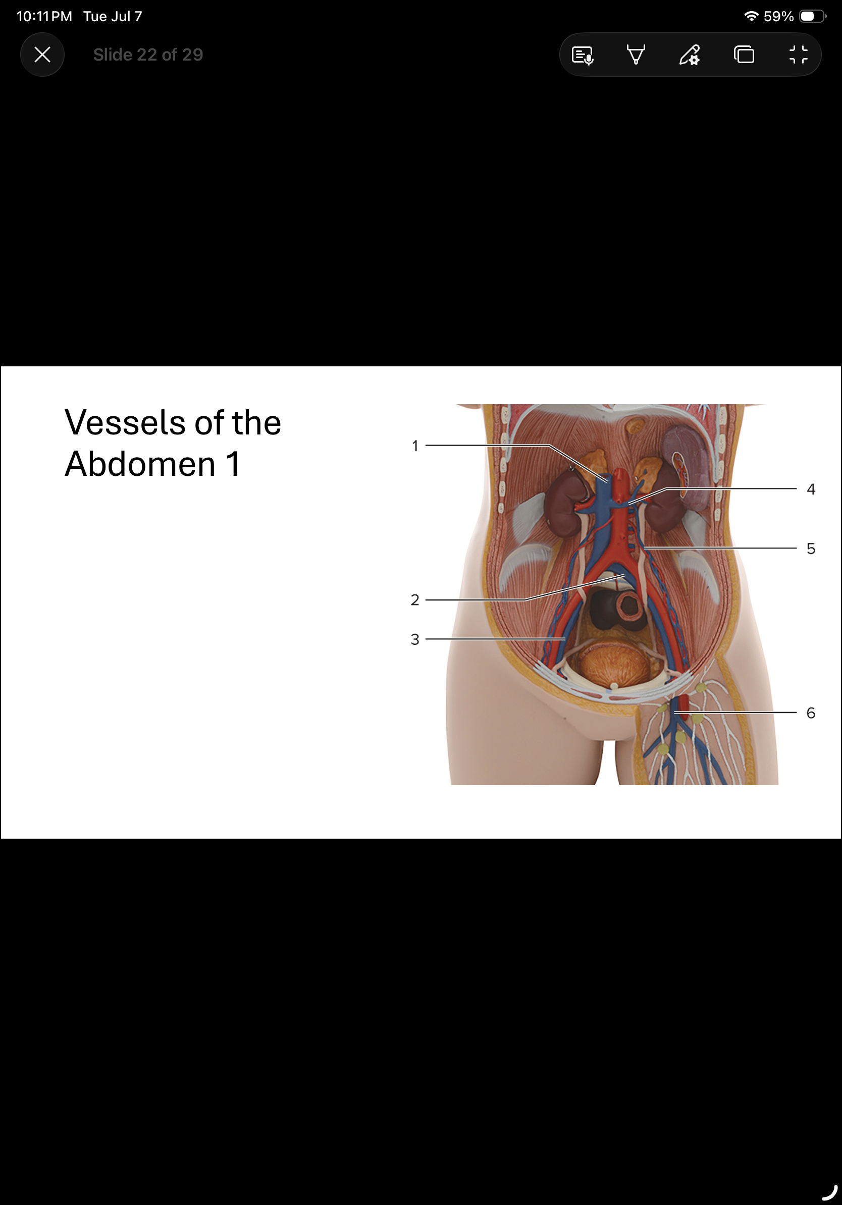
4, 5 & 6.
Renal vein
Gonadal vein
Great saphenous vein

Vertebral arteries

Basillar artery

Vein

Artery
Trace a drop of blood from liver to the spleen.
Liver → Hepatic Veins → Inferior Vena Cava → Right Heart → Lungs → Left Heart → Aorta → Celiac Trunk →Splenic Artery → Spleen.
Trace a drop of blood from the right renal vein to the left renal artery.
Right Renal Vein
Inferior Vena Cava (IVC)
Right side of heart
Lungs (where carbon dioxide is dropped off and oxygen is picked up)
Pulmonary Veins
Left Atrium
Left Ventricle
Aorta ( → Descending Thoracic Aorta → Abdominal Aorta)
Left Renal Artery
Trace a drop of blood from the left medial ankle to the right atrium.
Left Great Saphenous Vein → Left Femoral Vein → Left External Iliac Vein → Left Common Iliac Vein →Inferior Vena Cava (IVC) → Right Atrium
Trace a drop of blood from thoracic cage to rectum.
Intercostal Veins → Azygos Vein → Superior Vena Cava → Heart (Right → Lungs → Left) → Aorta → Inferior Mesenteric Artery → Rectum
How do we find MAP?
MAP = Diastolic pressure +( Pulse Pressure/3 )
If BP is 120/80, what is MAP? Remember find pulse pressure first.
93