Biology OCR A Level Chapter 8 - Transport in Animals
1/69
There's no tags or description
Looks like no tags are added yet.
Name | Mastery | Learn | Test | Matching | Spaced |
|---|
No study sessions yet.
70 Terms
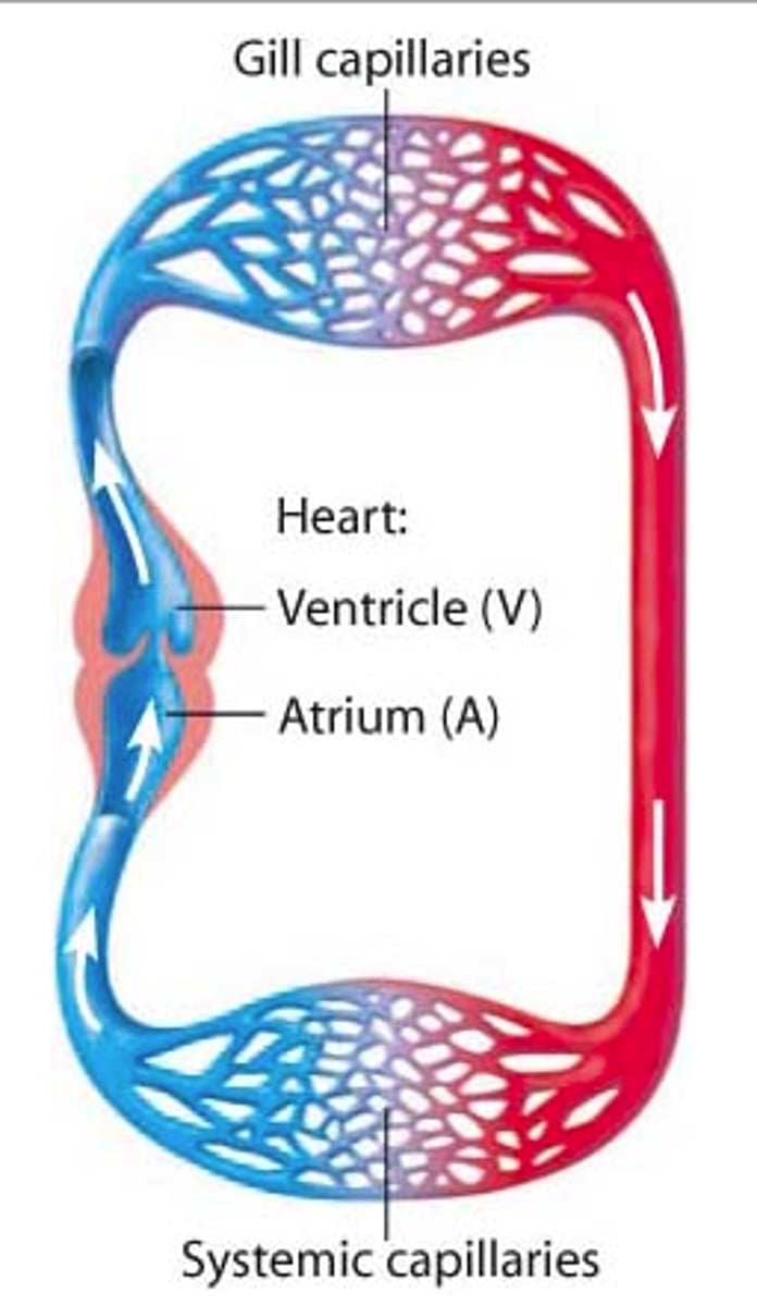
What is a single circulatory system?
-In a single circulatory system, blood only passes through the heart once for each complete circuit of the body.
Reasons for transport systems
- Increasing transport distances > organisms have increased in size thus creating a larger distance for molecules to travel to inner cells and cell would die before it would reach it
- Small SA:V ratio > demand for oxygen cannot be met
- Metabolic rate too high
Give an example of a single circulatory system.
-Fish have a single circulatory system.
- Deoxygenated blood is pumped by the heart to thengills where CO2 is excreted + O2 is absorbed
- Oxygenated blood travels through body to other parts then back to the heart to the gills (only 1 atrium + ventricle)
What are the disadvantages of a single circulatory system?
-low blood pressure ——> after blood is oxygenated in the gills it flows to the rest of the body at low pressure —-> as it loses pressure as it passes through gills
-This limits the efficiency of the exchange processes.
How can fish be so active with a single circulatory system?
-Countercurrent system allows them to take a lot of oxygen from the water.
-Body weight is supported by the water.
-They do not maintain their own body temperature.
-This greatly reduces the metabolic demand on their bodies.
What is a double circulatory system?
-In a double circulatory system, the blood passes through the heart twice for each complete circuit of the body.
Give an example of a double circulatory system.
-Mammals have a double circulatory system.
-The heart is divided down the middle.
-The right side of the heart pumps blood to the lungs to pick up oxygen in the pulmonary system.
-From the lungs it travels to the left side of the heart where is pumped to the rest of the body in the systemic system.
What is the advantage of the mammalian double circulatory system?
-The heart can give the blood an extra pressure thus higher speed of flowbetween the lungs and the rest of the body.
-This makes blood travel faster, so oxygen is delivered to the tissues more quickly + maintains steeper concentration gradient
What is a closed circulatory system?
-All vertebrates have a closed circulatory system.
-In a closed system, the blood is enclosed inside blood vessels.
Give an example of a closed circulatory system.
-In fish, the heart pumps blood into arteries.
-Arteries branch out into millions of capillaries.
-Substances like oxygen and glucose diffuse from the blood in the capillaries into the body cells, but the blood stays inside the blood vessel as it circulates.
What is an open circulatory system?
Where blood isn't enclosed in blood vessels all the time. Instead, it flows freely through the body cavity.
Give an example of an open circulatory system.
- Insects have open circulatory system > blood dnters body cavity
- Insects have one main blood vessel (dorsal vessel) which delivers haemolymph into body cavity pumped by heart
- Hameolymph flows around organs + re enters heart through ostia valvles
- Only supples nutrients, O2 + CO2 exchanged through trachel system
What are the disadvantages of an open circulatory system?
-Steep diffusion gradients cannot be maintained for efficient diffusion.
-The amount of haemolymph flowing to a particular tissue cannot be varied to meet demands.
Why are specialised transport systems needed in multicellular organisms?
-The metabolic demands of most multicellular animals are high so diffusion over the long distances is not enough to supply the quantities needed.
-SA:V ratio gets smaller as organisms get bigger.
-Molecules such as hormones or enzymes may be made in one place but needed in another.
-Food will be digested in one organ system, but needs to be transported to every cell for use in respiration and other aspects of cell metabolism.
-Waste products of metabolism need to be removed from the cells an transported to excretory organs.
What are the arteries?
-Arteries are vessels that carry blood from the heart to the rest of the body.
What is the structure of the arteries?
Tunica intima = Innermost layer of blood vessels + lines the lumen:
- Consists of endothelium > one cell thick w/ smooth surface to reduce friction w/ blood
- Made up of connective tissue + elastic fibres
Tunica media = Thick middle layer in blood vessels
- Consists of smooth muscle + thick layer of elastic tissue
- Elastic layer allows vessels to stretch and recoil to manage pressure fluctations
- Smooth muscle strengthens arerty + prioritises blood flow by contracting + dilating
- Tunica adventitia
Thick layer of collagen
What are the arterioles?
-Arteries branch into arterioles, which are much smaller than arteries.
- Concerned w/ regulating blood flow > mainly made up of smooth muscle
-Smooth muscle constricts: vasoconstriction.
-Smooth muscle relaxes: vasodilation.
What are the structure of arterioles?
-Layer of smooth muscle, but they have less elastic tissue than arteries as they have little pulse surge, but can constrict or dilate to control the flow of blood to individual organs.
-The smooth muscle allows them to expand and contract, thus controlling the amount of blood flowing to tissues.
What are the capillaries?
-Arterioles branch into capillaries, which are the smallest of blood vessels.
-Substances like glucose and oxygen are exchanged between cells and capillaries, so they're adapted for efficient diffusion, e.g. their walls are only one cell thick + are very thin to fit between cells to deliver blood to everybcell
How are the capillaries adapted for efficient gas exchange?
-Large surface area for diffusion.
-The total cross-sectional area of the capillaries is always greater than the arteriole supplying them so the rate of blood flow falls. This relatively slow movement allows more time for the exchange of materials.
-The walls are a single endothelial wall thick, giving a very short distance for diffusion.
What are the venules?
-Capillaries connect to venules, which have very thin walls that contain some muscle cells.
-Venules join together to form veins.
What are the veins?
-Veins take blood back to the heart under low pressure.
- Same structure as but tunica media is thinner > less need for elastic tissue and muscle tissue due to low pressure + larger lumen to inc volume of blood tramsproted to compensate formblood flow loss
How is blood flow through the veins helped?
-Valves in the veins prevent back-flow.
-When muscles of the body contract they squeeze the veins, forcing the blood towards the heart.
-The breathing movements of the chest act as a pump. The pressure changes and the squeezing actions move blood in the veins of the chest and abdomen towards the heart.
What does blood consist of?
-Plasma, which carries a variety of other components including dissolved glucose, hormones and large plasma proteins.
-Red blood cells.
-White blood cells.
-Platelets.
What are the functions of the blood?
-Oxygen to, and carbon dioxide from, the respiring cells.
-Digested food from the
-Hormones.
-Platelets to damaged areas.
-Cells and antibodies involved in the immune response.
What is tissue fluid?
-Tissue fluid is the fluid that surrounds the cells in tissues.
-Cells take in oxygen and nutrients from the tissue fluid, and release metabolic waste into it.
What is tissue fluid made from?
-Substances that leave the blood plasma like oxygen, water and nutrients.
-Does not contain red blood cells or big proteins, because they're too large to be pushed out through the capillary walls.
What causes oncotic pressure?
-Plasma proteins in the blood give fluid low water potential
-As a result, water has a tendency to move into the blood in the capillaries from the surrounding fluid by osmosis.
-The tendency of water to move into the blood by osmosis is called the oncotic pressure.
What is the process of pressure filtration?
-At the start of the capillary bed, nearest the arteries, the hydrostatic pressure inside the capillaries is greater than the hydrostatic pressure in the tissue fluid and the oncotic pressure.
-This forces fluid out of the capillaries and into the spaces around the cells, forming tissue fluid.
-As the fluid leaves, the hydrostatic pressure reduces in the capillaries due to fluid leaving and diffusing- so the hydrostatic pressure is much lower at the end of the capillary bed that's nearest to the venules.
-The hydrostatic pressure now falls below the oncotic pressure.
-Because the water potential in the capillaries is lower than the water potential in the tissue fluid and the oncotic pressure, some water re-enters the capillaries from the tissue fluid at the venule end by osmosis.
What is the lymphatic system?
- Network of vessels + organs that connect to the circulatory system
- Collect in large volume at lymph nodes to fight infections
- oteins found in lymph than tissue fluid due to daded antibodies
-Not all of the tissue fluid re-enters the capillaries at the vein end of the capillary bed - some excess tissue fluid is left over.
-The extra fluid eventually gets returned to the blood through the lymphatic system via subclavian vein - a kind of drainage system, made up of lymph vessels.
-Excess tissue fluid passes into lymph vessels. Once inside, it's called lymph.
What are the smallest lymph vessels called?
-Lymph capillaries.
Where is the lymph returned to the blood?
-Lymph gradually moves towards the main lymph vessels in the thorax.
-Here, it's returned to the blood, near the heart.
Where are red blood cells present?
-Red blood cells are only in the blood as they are too big to get through the capillary walls.
Where are white blood cells present?
-Blood, lymph and few in the tissue fluid.
-Most white blood cells are in the lymph system. They only enter tissue fluid when there's an infection.
Where are platelets present?
-Only in the blood.
-Tissue fluid if the capillaries are damaged.
Where are proteins present?
-In the blood.
-Very few in the tissue fluid.
-Only antibodies in the lymphatic system.
-Most plasma proteins are too big to get through capillary walls.
Where does the right side of the heart pump blood?
-Deoxygenated blood to the lungs.
Where does the left side of the heart pump blood?
-Oxygenated blood to the rest of the body.
Diagram of the heart.
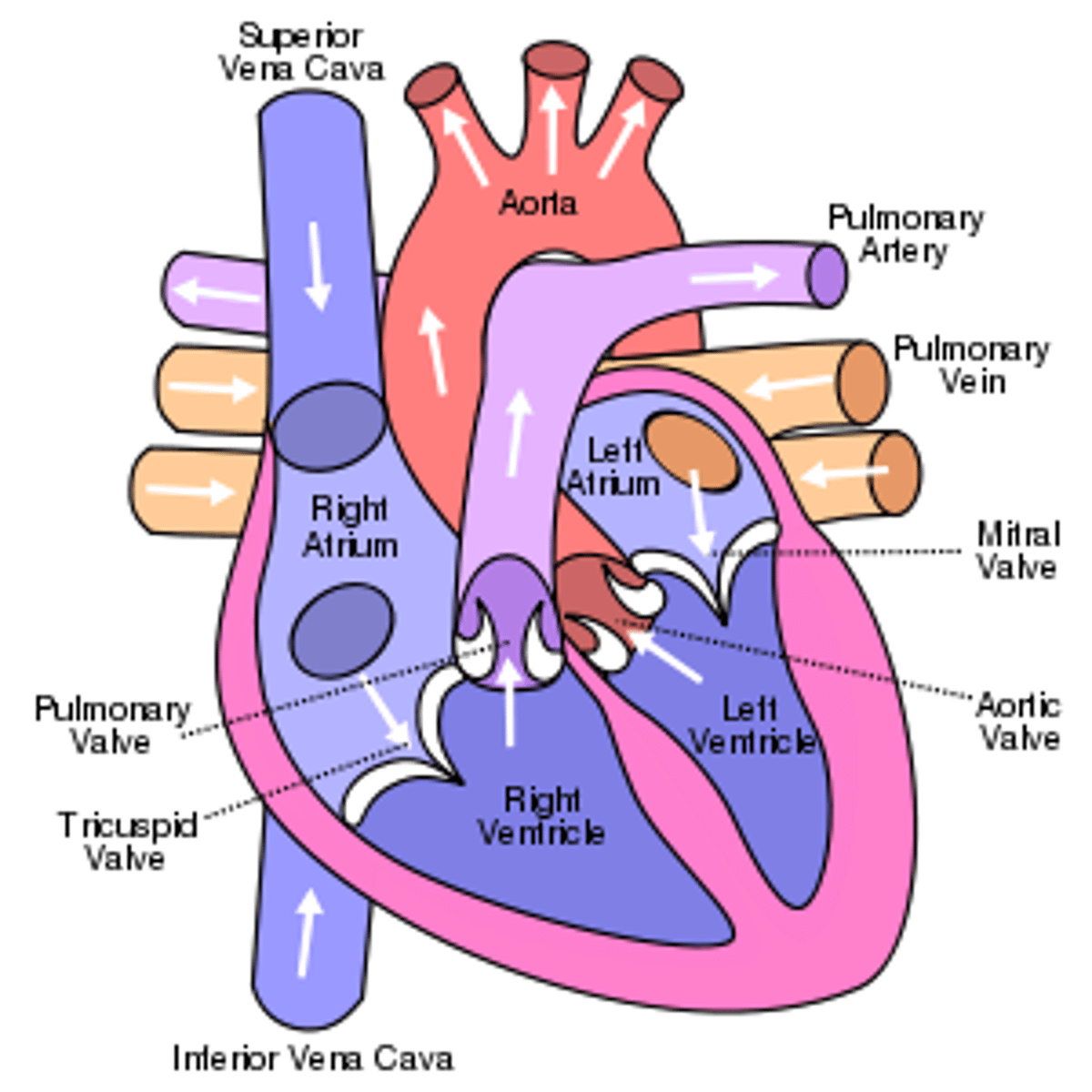
Why are the muscular walls of the heart much thicker on the left side?
-The lungs are relatively close to the heart, and the lungs are also smaller than the rest of the body so the right side of the heart has to pump blood a relatively short distance and only has to overcome the resistance of the pulmonary circulation.
-The left side has to produce sufficient force to overcome the resistance of the aorta and arterial systems of the whole body and move the blood under pressure to all the extremities of the body.
What are the heart valves?
-The atrioventricular valves link the atria to the ventricles.
-The semi-lunar valves link the ventricles to the pulmonary artery and aorta.
What do the heart valves do?
- Prevent backflow
-They only open one way - whether they're open or closed depends upon the relative pressure of the heart chambers.
-If there's higher pressure behind a valve, it's forced open, but if there'e higher pressure in front of the valve it's forced shut.
-Flow of blood is unidirectional.
What is the cardiac cycle?
-The cardiac cycle is an ongoing sequence of contraction and relaxation of the atria and ventricles that keeps blood continually circulating round the body.
-The volumes of the atria and ventricles change as they contract and relax, altering the pressure in each chamber.
-This causes valves to open and close, which directs the blood flow through the heart.
What is diastole?
Relaxation phase
What is systole?
Contraction phase
What is the 'lub-dub' sound?
-The first 'lub' sound is caused by the atrioventricular valves closing.
-The second 'dub' sound is caused by the semi-lunar valves closing.
Cardiac cycle
1. Atrium contracts increasing pressure + decreasing volume as it causes AV valve to open as pressure in atrium > ventricle, blood to flow into ventricles
2. Atrium relaxes causing AV valve to close
3. Ventricle contracts causing semi-lunar valve tomopen + blood flows into aorta
4. Ventricle relaxes, semi lunar valves close due to pressure decrease
5. Aorta walls recoil + pushes blood out
6. Atria fill again
What does myogenic mean?
-Cardiac muscle is myogenic - this means that it can contract and relax without receiving signals from nerves.
-This pattern of contractions controls the heartbeat.
How is the heart beat controlled?
1. Sino-atrial node stimulates electrical impulse to atrial walls + causes them to contract at the same time
- Non conducting collagen tissue stops electrical activity from reaching ventricles
2. Electrical impulse is transferred from SAN to atriroventricular node
3. AVN passes electrical activity to bundle of his > small delay to make sure ventricles have emptied
4. Bundle of his transfaers electrical activity to purkinye tissue which contracts both ventricles from bottom up
What is an electrocardiograph?
-A machine that records the electrical activity of the heart.
-The heart muscle depolarises (loses electrical charge) when it contracts, and repolarises (regains charge) when it relaxes.
-An electrocardiograph records these changes in electrical charge using electrodes placed on the chest.
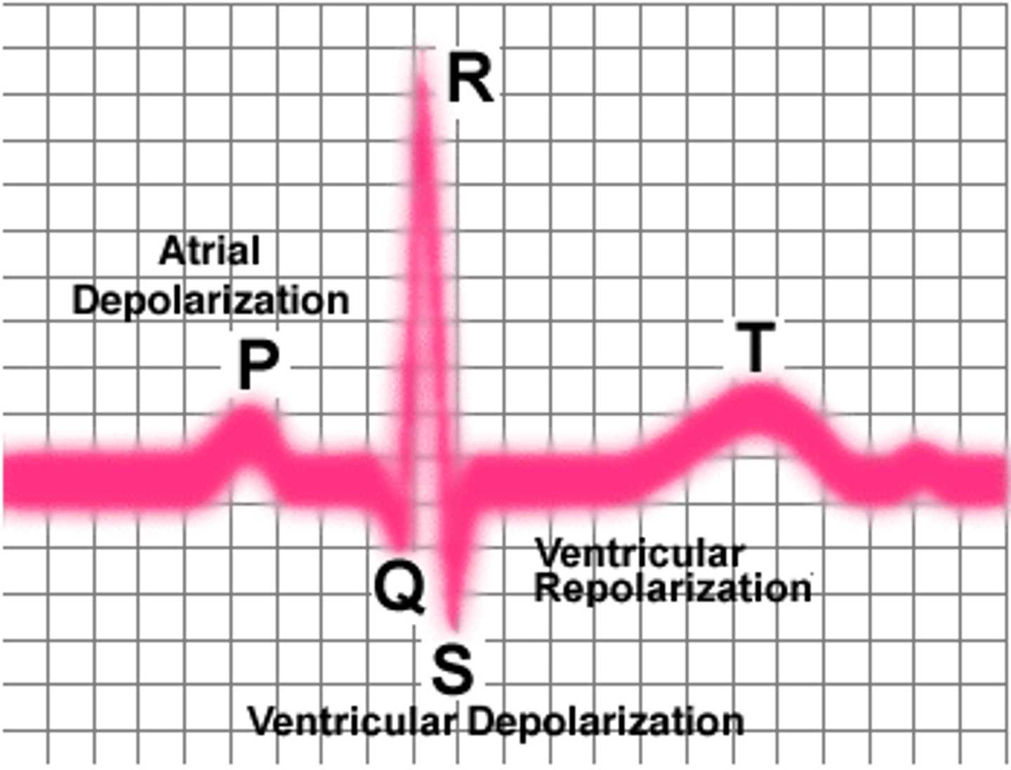
What is an electrocardiogram? (ECG)
-The trace produced by an electrocardiograph is called an electrocardiogram, or ECG.
-The P wave is caused by contraction (depolarisation) of the atria.
-The main peak of the heartbeat, together with the dips either side, is called the QRS complex - it's caused by contraction (depolarisation) of the ventricles.
-The T wave is due to relaxation (repolarisation) of the ventricles.
-The height of the wave indicates how much electrical charge is passing through the heart - a bigger wave means more electrical charge, so (for the P and R waves) a bigger wave means a stronger contraction.
How do you calculate heart rate?
-Your heart rate is the number of beats per unit time - usually beats per minute.
-Heart rate (bpm) = 60 / time taken for one heart beat (s)
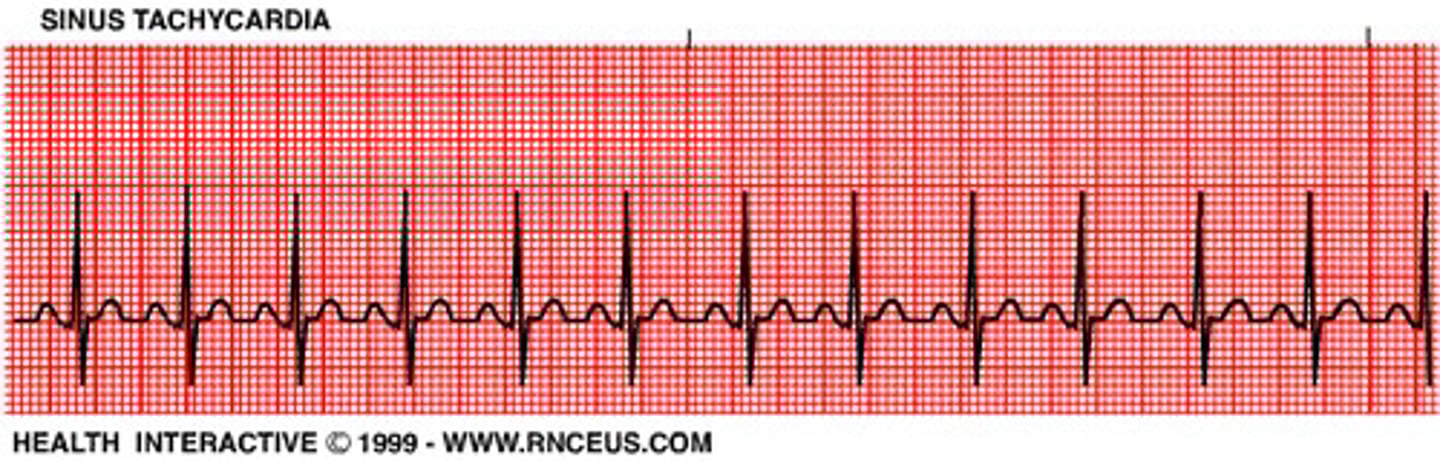
What is tachycardia?
-The heartbeat is too fast - around >100 beats per minute. At rest it shows that the heart isn't pumping blood efficiently.
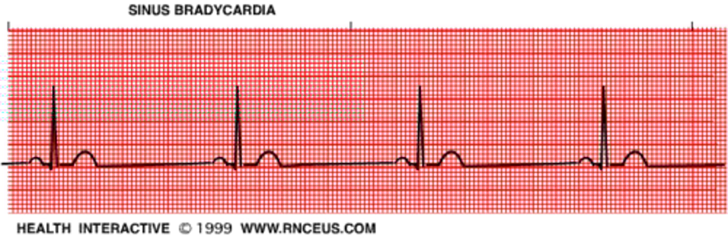
What is bradycardia?
-The heartbeat is too slow < 60 beats per minute.
-A heartbeat this slow is normal in some people (e.g. trained athletes) but in others it can indicate a problem with the electrical activity of the heart, e.g there may be something preventing impulses from the SAN being passed on properly.
What is ectopic heartbeat?
-An 'extra' heartbeat that interrupts regular rhythm.
-E.g. the atria or ventricles contract too early.
-Occasional ectopic heartbeats in a healthy person don't cause a problem.
What is fibrillation?
-Fibrillation is a really irregular heartbeat.
-The atria or ventricles completely lose their rhythm and stop contracting properly.
-It can result in anything from chest pain and fainting to lack of pulse and death.
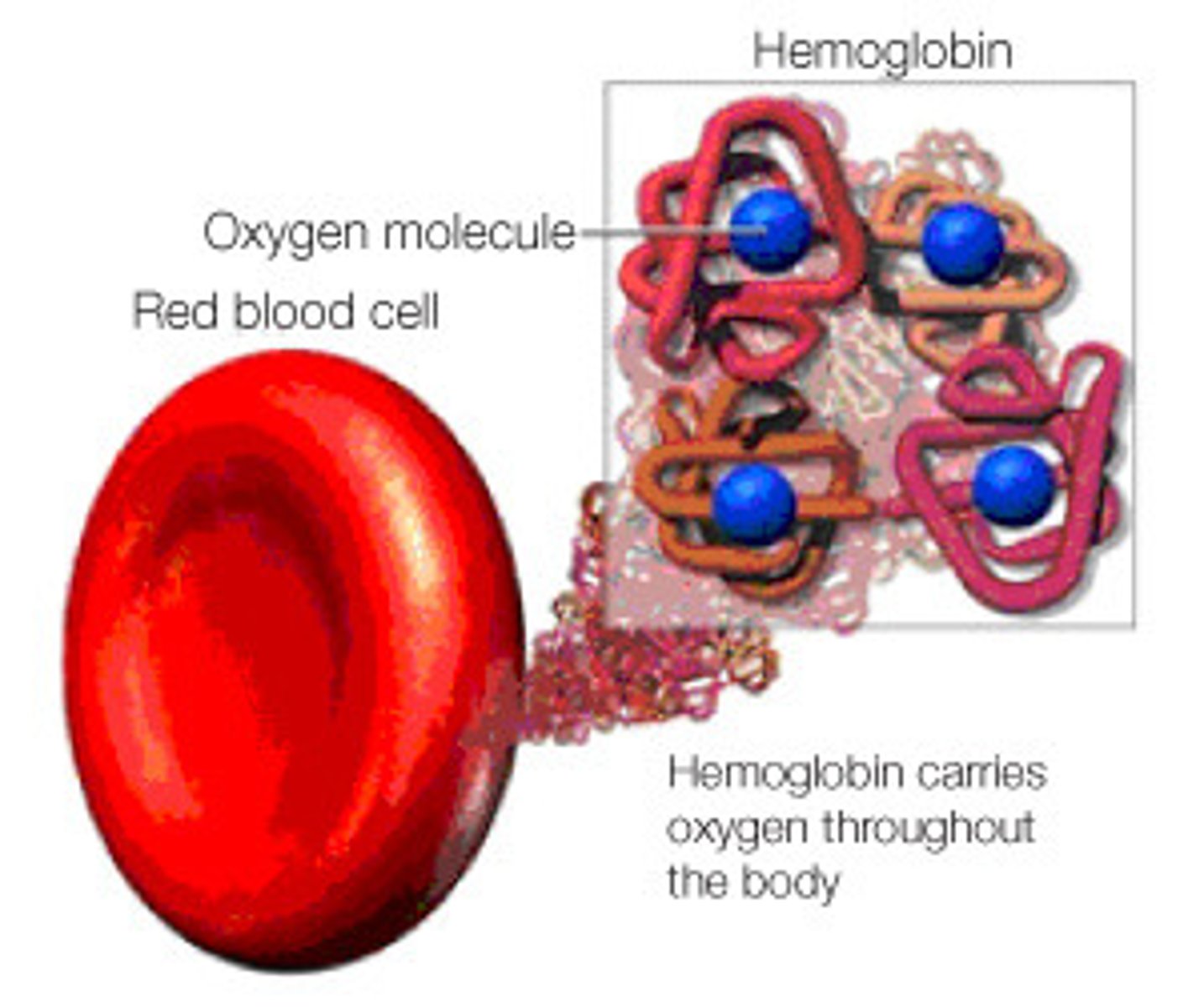
What is haemoglobin?
-Found in red blood cells.
-It's role is to carry oxygen around the body.
-Large protein with a quaternary structure - it's made up of four polypeptide chains.
-Each chain has a haem group which contains iron and gives haemoglobin its red colour.
-Each molecule of human haemoglobin can carry four oxygen molecules.
-In the lungs, oxygen joins to the iron in the haemoglobin to form oxyhaemoglobin. This is a reversible reaction.
-Near the body cells, oxygen leaves oxyhaemoglobin and it turns back to haemoglobin.
-When an oxygen molecule joins to haemoglobin it's referred to as association or loading, and when oxygen leaves oxyhaemoglobin it's referred to as dissociation or unloading.
What does affinity for oxygen mean?
-The tendency a molecule has to bind with oxygen.
-Haemoglobin's affinity for oxygen varies depending on the condition's it's in - one of the conditions that affects it is the partial pressure of oxygen (pO₂).
Haemoglobin oxygen chemical reaction
Hb + 4O2 > HbO8
How does haemoglobin increase its affinity for oxygen
- Everytime a haem group absorbs an O2 atom, it alters the structure of its molecule to make it easier to absorb a other molecule
What is partial pressure of oxygen?
-pO₂ is a measure of oxygen concentration.
-The greater the concentration of dissolved oxygen in cells, the higher the partial pressure.
How does pO2 affect hb affinity and unloading/loading?
-As pO₂ increases, haemoglobin's affinity for oxygen also increases:
-Oxygen loads onto haemoglobin where there's a high pO₂
-Oxyhaemoglobin unloads its oxygen where there's a lower pO₂.
What is a dissociation curve?
-An oxygen dissociation curve shows how saturated the haemoglobin is with oxygen at any given partial pressure.
Why is the graph 's-shaped'?
-The saturation of haemoglobin can have an affect on the affinity.
-When haemoglobin combines with the first O₂ molecule, its shape alters in a way that makes it easier for other molecules to join too.
-But as the haemoglobin starts to become saturated, it gets harder for more oxygen molecules to join.
-As a result, the curve has a steep bit in the middle where it's really easy for oxygen molecules to join, and shallow bits at each end where it's harder.
-Where the curve is steep, a small change in pO₂ causes a big change in the amount of oxygen carried by the haemoglobin.
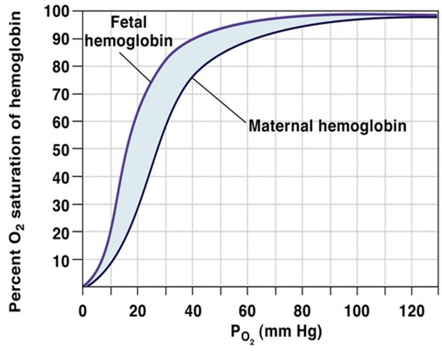
What different about fetal hb to usual hb?
- Fetal hb has a higher affinity to oxygen than usual hb .
- placenta has lower pO2 so its adapted
CO2 methods of transport
- Dissolved CO2 in plasma
-HCO3 (main)
- Bound to carboaminohaemoglobin
What is the partial pressure of carbon dioxide?
-A measure of the concentration of CO₂ in a cell.
-pCO₂ also affects oxygen unloading.
How does pCO2 affect oxygen unloading/loading?
-Haemoglobin gives up its oxygen more readily at a higher pCO₂.
-When cells respire they produce CO₂, which raises the pCO₂.
-This increases the rate of oxygen unloading thus lowers affinity of hb to oxygen
What is the Bohr effect?
- Increasing CO2 concentration lowers the affinity for haemoglobin to oxygen (due to formation of haemoglobinic acid, lower pH inc unloading)
What is the chemical process of the Bohr effect?
- CO2 diffuses into red blood cell
- CO2 + H2O is converted into carbonic acid catalysed by carbonic anhydrase to form H+ + HCO3- ions
- HCO3- ions diffuse out of the cell down a conc gradient + combine w/ Na+ to form NaHCO3
- To maintain electrochemical neutrality, Cl- ions diffuse in to replace HCO3- ions > chloride shift
- H+ ions combine with Hb to form haemoglobinic acid which causes:
1. Increase O2 release due to increase of pH (bohr effect)
2. Acts as a buffer