Lameness (TV4101, Lecture 15-19 Notes)
 Call Kai
 Learn
Learn Practice Test
 Spaced Repetition
 Match
Match Flashcards
 Knowt Play
1/43

There's no tags or description
Looks like no tags are added yet.
Name | Mastery | Learn | Test | Matching | Spaced | Call with Kai | Chat |
|---|
No analytics yet
Send a link to your students to track their progress
44 Terms
Lecture 15 - Lameness examination and diseases of the interdigital skin
Describe the weight-bearing surface of the claw

Discuss claw horn growth
- Claw horn grows at a rate of 5 mm (0.5 cm) per month
- Wall 75mm long = 15 months for horn produced at the coronary band to come into wear
- Sole 10-15 mm thick = 8-12 weeks to grow out
List the 8 steps of a lameness examination
1. Which leg is the cow lame in? Observe the cow walking = head up (forelimb) or head down (hindlimb)
2. Is the leg swollen and painful above the claw? yes (infection) or no (examine claws)
3. Examine the external surface of both claws and compare these with the other leg - are there any vertical or horizontal cracks present?
4. Restrain the cow, lift the leg and thoroughly clean external surface - check the interdigital space for swelling, foreign body, pain, smell, discharge or damaged skin (if no pare weight bearing surface of the claw)
5. Use hoof testers over the weight-bearing surface to search for any sensitive areas - bruising only (rest) or abscess/under-run sole (trim, pare or grind to remove all loose horn, drain pus, etc.)
6. Carefully pare or sand away superficial outer surface of sole - check for abnormal discolouration of the sole horn
7. Carefully check the white line - is it disintegrated and filled with dirt, gravel or stones?
8. No abnormality detected in either claw - examine upper leg
Discuss interdigital necrobacillosis (footrot)
- Fusobacterium necrophorum penetration of preexisting soft-tissue lesions (i.e. secondary invader)
- Acute, severe lameness with swelling of interdigital space
- Antibiotic treatment (may be associated with septic arthritis) with procaine penicillin (72hr WHP) or ceftiofur (nil WHP) for 3 days

Discuss interdigital fibroma (interdigital hyperplasia)
- Fleshy growth (excess epidermal and hypodermal tissue) 'hanging down' from skin between the claws of the foot
- May be associated with intermittent lameness (possibly inherited)
- Intravenous regional anaesthesia or interdigital nerve block for growth excision

Discuss digital dermatitis
- Infectious, superficial inflammation of the epidermis
- Common on plantar aspect of the hind feet near the skin-horn junction bordering the interdigital cleft
- Associated with prolonged exposure to slurry (i.e. dairy facilities or feedlots), multifactorial disease
- Present as red, moist, exudative, circumscribed ulcerative dermatitis (strawberry footrot) or proliferative, wart-like lesions (hairy-foot wart)
- Treat with topical oxytetracycline or formalin/copper sulphate footbaths with environmental hygiene

Compare and contrast intravenous regional anaesthesia and interdigital nerve block

Lecture 16 - Claw lesions 1
Discuss thin sole
- Due to excessive wear (common in pasture-fed dairy cattle)
- Associated with long walking distances, time spent on concrete, moist conditions or nutritional/health concerns
- Present with shuffling gait and sole can be depressed with thumb
- Address with rest

Discuss bruised sole
- Associated with flat/thin soles or rough surfaces
- Hind claws are more commonly affected in older cows and front claws are most commonly affected in heifers
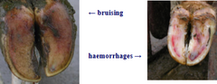
- Haemorrhage visible through the surface of the sole, may cause sole under-running
- Address with rest (i.e. consider block application)

Discuss sole abcess
- Due to penetrating wound or underrun sole
- Present with severe lameness (affected cows first slide affected foot along the surface, before fully bearing weight)
- Assess with hoof testers and foot pairing
- Establish drainage (i.e. follow tract), remove all under-run horn, reduce weight bearing (e.g. apply block to unaffected claw or pair strategically), avoid pain/bleeding and administer antibiotics with soft tissue swelling

Discuss white line disease/abscess
- Associated with long walking distances, time spent on poor surfaces, twisting/turning in yards, wet conditions, claw deformities or laminitis
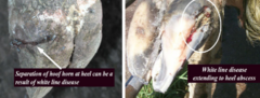
- Haemorrhage, fissure and abscess formation along the white line
- Pare out the cracks between the wall and sole, apply foot block to sound claw (antibiotics generally not required)
Lecture 17 - Claw lesions 2
Discuss sole ulcer
- Due to excess pressure compressing the horn immediately distal to the flexor tuberosity of the distal phalanx
- Granulating lesion 1 cm in diameter, concealed below layer of horn that may be discolored
- Associated with heavy cows, prolonged time on concrete/confined conditions and claw overgrowth
- Present with severe lameness with reduced milk yield, poor body condition and delayed oestrus
- Render sole concave, slope/thin horn around any protruding corium and remove excess granulation tissue

Discuss axial wall crack
- Lameness presents with impaction (common after heavy rainfall) and the two edges of the crack move, and pull/push the sensitive lamellae during walking
- Remove debris from crack and carefully pare horn edges adjacent, removing under-run horn and excess granulation tissue (antibiotic treatment with corium or coronary band infection)

Discuss vertical fissure of the claw wall
- Associated with dry conditions, coronary band damage or nutritional deficiencies
- Loss of continuity of horn tubules (split) in dorsal claw wall, extending distally from the coronary band for a variable distance
- If causing lameness trim out to a U-shaped groove that won't accumulate any grit/dirt

Discuss horizontal fissure (hardship grooves) of the claw wall
- Due to interruption of claw horn production (e.g. illness, laminitis, toxins or transport)
- Pare, grind or sand detached claw wall away (may require heavy sedation)

Discuss digit amputation
Amputation through the distal 1/3 of P1;
1. Incise with scalpel the interdigital space and extend 3 cm proximally on the cranial side 2.5 cm on the plantar side
2. Insert embryotomy wire and cut at an oblique angle through P1
3. Dress stump with topical antibiotics and bandage, change dressing after 3-4 days and leave for 8-10 days

Discuss laminitis
- Due to a disturbance in the micro-circulation of the corium (e.g. degeneration or inflammation)
- Acute laminitis, accidental 'carbohydrate overload' with rapid onset of pain on all feet, hyperaemia at the coronary band and increased pulsation of digital arteries
- Chronic laminitis, development of claw deformity with elongated/concave dorsal border, and a flattened/broadened sole
- Subclinical laminitis, poor horn quality due to impaired keratogenesis and weaking of suspensory apparatus of distal phalanx (i.e. multifactorial disease, including subacute rumen acidosis), predisposing to other claw deformities

Lecture 18 - Upper limb lameness
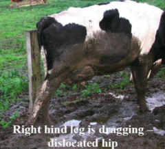
Discuss hip dislocation
- Dairy cows during parturition and early lactation (i.e. calving paralysis or milk fever)
- Dislocates craniodorsally or caudoventrally into obturator foramen
- Causes relative shortening of the upper limb with the hock more proximal
- Deep palpation when limb is rotated and alternatively flexed and extended with abnormal movement of head of femur can be felt and 'loud' creaking noises heard
- Treat with traction under heavy sedation in lateral recumbency (reluxation = poor prognosis)
i.e. fix cow's body to a solid object, using a heavy rope situated through inguinal area, apply block and tackle (or wire strainers) to affected limb and secure this to another traction point, apply traction while at same time rotating the femur by pushing down on stifle and lifting the hock
Discuss sacro-iliac luxation/subluxation
- Partial or complete separation of the fibrocartilaginous joint surfaces
- Due to dystocia or milk fever
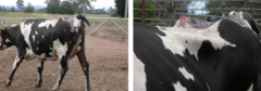
- Present with slight ataxia and weakness in the hind quarters, with slight knuckling of the fetlock or back (loin area) appears to be dropped and both tubercoxae are very pronounced
- Encourage rest and NSAIDs with salvage slaughter
Discuss pelvic fractures
- 'Knocked' or 'dropped' hips
- Lameness is usually minor
- Requires no treatment (unless ilium protruding through skin) with good prognosis
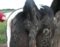
Discuss upward fixation of the patella
- Patella becomes temporarily (or permanently) fixed on medial aspect of upper part of the femoral trochlea
- Present with a jerky walk and prolonged hind-limb extension
- Treatment requires medial patella desmotomy in the standing sedated animal, using local anaesthesia

Discuss femoro-tibial/stifle instability
- Due to trauma in old animals
- Present with severe lameness due to rupture of cruciate ligaments, injuries of one or both menisci or rupture of collateral ligaments
- May feel excess joint fluid in stifle joint

Discuss leg fractures
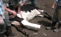
- Fractures below the hock respond well to casting for 5-6 weeks to stabilise joints above and below, incorporate fetotomy wire in the cast to facilitate its removal
- Upper limb fractures difficult to address
Discuss septic arthritis
- Due to direct penetration of a joint by a foreign body, extension of cellulitis from a decubital lesion, spread of pathogens from a localised focus adjacent to a joint or haematogenous spread (e.g. joint-ill)
- Bacteria produces excess fluid, inducing pain with cartilage/capsule destruction and fibrin production (i.e. perform joint tap)
- Sudden onset of lameness with marked muscle atrophy of affected limb (may involve deep flexor tendon rupture and joint ankylosis) and swollen, hot, painful claw
- Early treatment of calves (adults often unrewarding) with long antibiotic course (e.g. procaine penicillin, ampicillin, amoxicillin, ceftiofur or oxytetracycline), NSAIDs and joint lavage
- Amputate digit
Discuss spastic syndrome/periodic spasticity
- Intermittent spasmodic contraction of hindlimb muscles
- Uncommon in bulls and adult cows (hereditary component)
- Presents with 'saw horse' stance and hindlimbs extended caudally

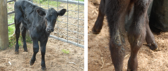
Discuss spastic paresis
- Progressive condition affecting one or both hindlimbs in growing cattle (3-6 months old)
- Excessive tone and spastic contracture of the muscles with gross over-extension (straightening) of the hock
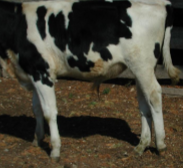
Discuss peripheral nerve injuries
- Common in cattle
- Limb dysfunction (not lameness) with weakness, paralysis, poor muscle tone, and decreased/absent reflexes causing muscle atrophy
- Associated with calving, metabolic disease or trauma
Discuss brachial plexus injury/paralysis
- Radial, ulnar and musculocutaneous nerves
- Calves, due to excessive traction on calf in anterior presentation
- Older cattle, damage due to compression of plexus (haematoma, abscess) between scapula and ribs
- Paralysis of all muscles of the forelimb with complete inability to support weight, and scapula, humerus and distal limb appear to 'hang' from the thoracic girdle

Discuss radial paralysis
- Associated with recumbency in milk fever, cast on hard surfaces or blunt trauma to the shoulder area
- Damage at high level, shoulder and elbow will drop and animal is unable to extend its limb (i.e. drag on ground)
- Damage distal to mid-shaft of humerus, only extensors of the digits will be affected animal is able to support/bear weight on affected leg

Discuss sciatic nerve paralysis
- Due to severe intra-pelvic pressure associated with dystocia (both hindlimbs may be affected) or when injections are administered into the gluteal muscles of thinly muscled, often young animals, or into the muscles of the caudal femur area
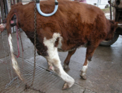
Discuss sciatic nerve paralysis grading
Grade 1 - loss of motor function (proprioception) but full sensation at the pastern, few day recovery.
Grade 2 - loss of function of peroneal branch and loss of sensation to anterior pastern, 7-10 day recovery.
Grade 3 - loss of sensation to tibial nerve as well, no superficial sensation below fetlock, 2-4 week recovery.
Grade 4 - loss of deep pain reflexes, no sensation at pastern, at least 4 week recovery.
Discuss femoral paralysis
- Due to pressure or nerve overstretching (i.e. attempt to rise with recumbency)
- Loss of function to extensors of stifle with inability to support weight (i.e. dog-sitting) and limb collapses when weight-bearing
- Absent patellar reflex

Discuss femoral paralysis grading
Grade 1 - legs go out behind when trying to stand, can stand and walk normally when lifted, good prognosis.
Grade 2 - absent patella reflex, legs behind when trying to stand, walk a few paces before falling after lifting, good prognosis.
Grade 3 - no patellar reflex, unable to support weight when lifted, poor prognosis.
Grade 4 - cow sits in frog leg position and is unable to correct it, euthanasia.
Discuss obturator paralysis
- Due to dystocia (especially hip-lock presentation)
- Paralysis of adducting muscles of the hindlimb with normal sensation of lower limb
- Unilateral or bilateral paralysis (poor prognosis)

Discuss peroneal paralysis
- Causes knuckling of fetlock and extension of hock, with prolong recumbency on hard surfaces
- Associated with milk fever
- Rapid recovery if able to stand

Discuss tibial paralysis
- Paralysis of extensors of the hock and flexors of the digits
- Stifle dropped, hock over-flexed, fetlock partly flexed (knuckled forward) but weight bearing surface of claws remains firmly in contact with the ground
- Skin sensation to plantar surface of limb below the hock may be decreased or lost
- Partial or complete rupture of gastrocnemius muscle may follow tibial paralysis
Lecture 19 - Lameness risk factors and control
List risk factors for lameness
- Cow comfort (e.g. maintenance of farm tracks, patience of farm employees, concrete, holding yard, milking shed or housing)
- Nutrition (e.g. carbohydrate/energy, fibre or protein)
- Cow factors (e.g. age, lactation stage, genetics/conformation or claw maintenance)
- Environment
Discuss cow behaviour when walking
- Organised movement with dominant cow leading group
- Heads down for careful/planned foot placement
- Hind feet follow front feet
- Increased pressure causes unplanned foot placement (i.e. increased lameness risk)
Describe the ideal track
- Straight, < 1km long and 5-6 metres wide (even with no congestion points)
- Crowned surface, ideally 3-5% slope
- Non-abrasive, 'foot friendly' top surface
- Drained towards outside of the fence with no shaded/slow-drying areas
- Widens out before it enters the holding yard with concrete skirt (transition area) where cows turn in
- Holding yard large enough for herd (i.e. ≥1.3 m^2/cow for Jerseys and ≥1.8 m^2/cow for Holsteins) and leading straight into dairy with non-skid concrete
- Pipework, rails and gates safe to prevent injury