1
1/31
There's no tags or description
Looks like no tags are added yet.
Name | Mastery | Learn | Test | Matching | Spaced |
|---|
No study sessions yet.
32 Terms
which is the pump?
heart
what are you hearing with the ‘lub’ and ‘dub’?
-closures of heart valves
-both together is one heart beat
difference between lub and dub (normal)
L: lower tone/freq, longer
definition:
biphasic
-having two phases
(in cardio) lub, dub
blood flow basics:
which atrium receives de-oxygenated blood?
R
blood flow basics:
which atrium receives oxygenated blood?
L
blood flow basics:
which chamber does the aorta come off from?
LV
which chamber does the pulmonary a come off from?
RV
blood flow basics:
where does the pulmonary a go to ?
-lungs
blood flow basics:
how does the blood circulate?
-based on a high/low P circuit
high P: body (larger part)
low P: lungs
what is the main artery leaving the L side circulation?
-aorta
What is the main artery leaving the right side circulation?
Pulmonary artery
how many views of the heart do you take? why?
2
lateral and DV/VD (dorsoventral/ventrodorsal)
-to view size and any other abnormalities
which is the auricular surface?
generally L thorax view
-side where you see auricles
which is the atrial surface?
generally R thorax view
-can see both atria
why can’t you use L/R when talking about the heart?
-because during development, the heart twists
basics:
how to read xrays (black vs white)
– White will generally be bone and stuff
– Black will be (empty), gas
Note: in thorax a lot of black is good

turquoise oval
aorta
(auricular surface)

light oval
pulmonary trunk
(auricular surface)

bright blue oval
pulmonary a
(auricular surface)
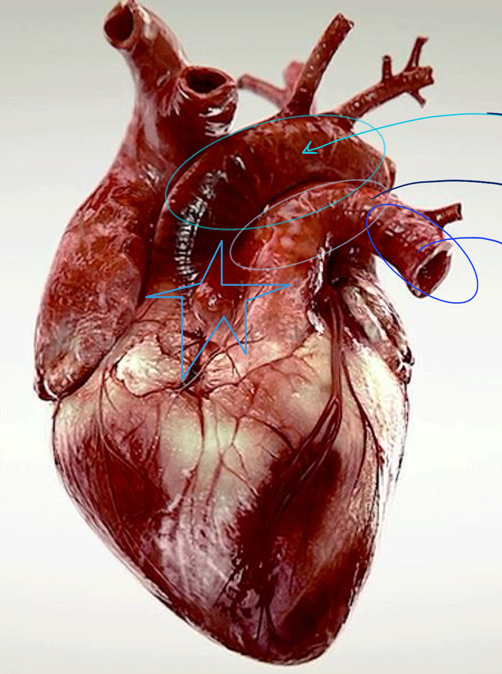
star (general note)
-pulmonary a and aorta generally come from A
differences between conducting, distributing, and arterioles (aa)?
c: elastic to support large amt of blood coming from aorta
d: muscular to contract/expand depending on blood flow needs to certain parts of the body
a: v fine and attach to capillaries
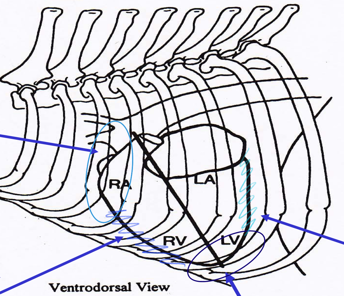
bright blue oval
base

navy oval
apex

bright blue edge
cranial border

light blue edge
caudal border

bright rectangle (L corner)
-trachea (can tell because outlined by cartilage)

turqouise oval (top L)
base

light oval (R bottom corner)
apex
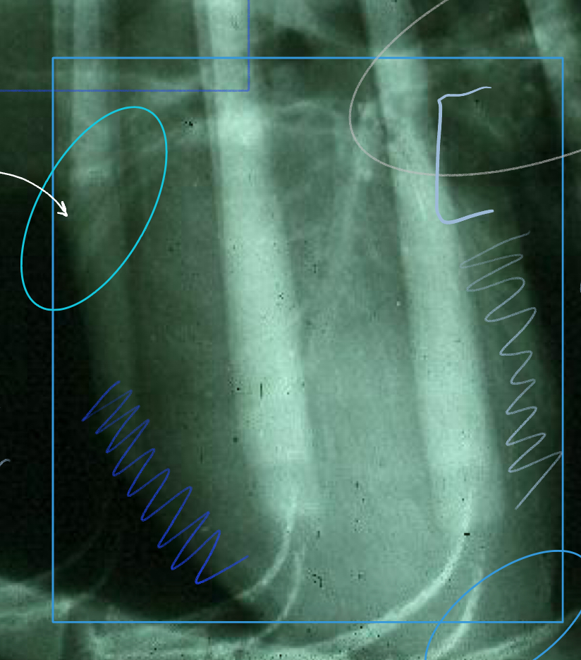
large rectangle
heart
-can’t tell it’s LHS or RHS just based on lateral view)

Right blue edge
Cranial border

Gray edge
caudal border