7- Sensory Transduction + Reflexes
1/35
There's no tags or description
Looks like no tags are added yet.
Name | Mastery | Learn | Test | Matching | Spaced | Call with Kai |
|---|
No analytics yet
Send a link to your students to track their progress
36 Terms
definition of transduction
sensory systems are made up of receptors that convert various forms of energy into electrical signals
4 forms of transduced energy
mechanical: touch, pressure, joint
chemical: smell + taste
electromagnetic: light on retina
thermal
what are proprioceptors
provide info about the positions of different body parts, needed to coordinate movement
monitor stretch in locomotory organs
3 types of proprioceptors
muscle spindles
Golgi tendon organs
joint kinesthetic receptors
what are muscle spindles
measures changing length of a muscle
imbedded in perimysium between muscle fascicles
what are Golgi tendon organs
monitors tension within tendons
located near muscle-tendon junction
what are joint kinesthetic receptors
sensory nerve endings within joint capsules
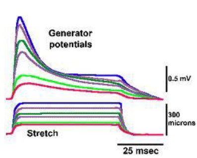
what’s a generator (receptor) potential
depolarization after reception of signals by sensory neurons
describe the mechanism of a generator potential
sensory signal received → Na+ channels open
T/F: generator potentials are graded
true, because increasing a stimulus produces an increase in generator potential amplitude
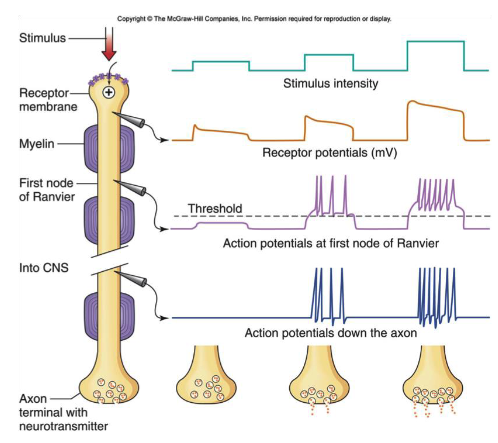
what’s a coding (spiking-generating) region
region adjacent to receptor region, where voltage-gated channels are located + action potentials are generated if receptor potential exceeds threshold
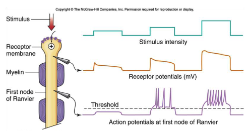
what is frequency modulated coding
when increased sensory stimulus intensity → increased frequency action potentials
what is adaptation
prolonged stimulus → generator potential gradually decreases → action potential frequency decreases (aka accommodation)
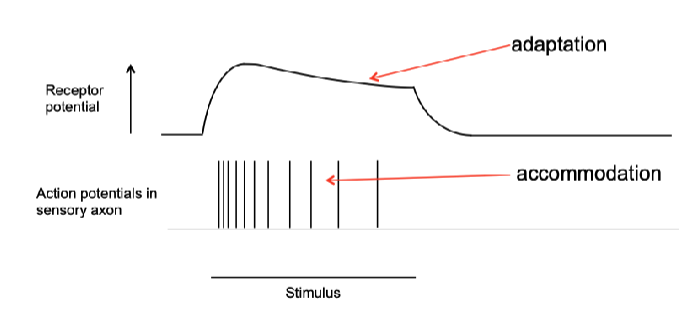
T/F: accommodation can still occur even if generator potential remains the same
true
describe sensory input at the spinal cord level
neurons within the cord are arranged into neuronal pools w/ varying numbers of cells + each pool receives input from primary afferent neurons
a neuron in a pool can receive excitatory, inhibitory stimulation or both
what is facilitation
a spinal cord neuron can become transiently more excitable to incoming stimulation after previous stimulation that did not go to threshold → increases probability of postsynaptic firing if another excitatory stimulus comes
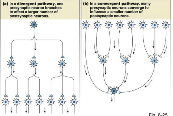
2 types of neuronal pools
divergent
convergent
divergent vs. convergent neuronal pools
divergent: single presynaptic neuron branches to affect multiple postsynaptic neuron
convergent: many presynaptic neurons converge to a smaller # of post synaptic neurons
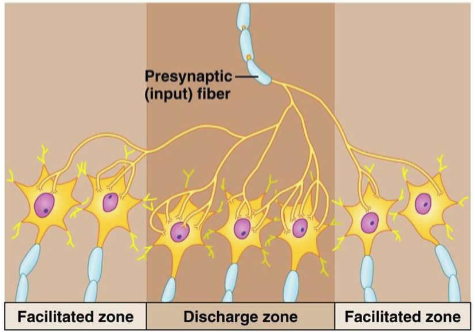
2 types of zones within a neuronal pool
discharge zone: center neurons that’re more likely to reach threshold b/c they have multiple synaptic connections w/ the presynaptic neuron
facilitated zone: peripheral neurons that may be depolarized but not to threshold, more excitable to future stimuli
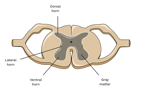
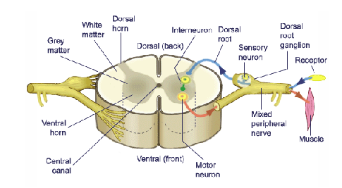
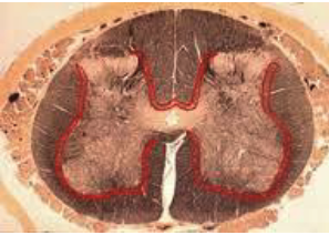
what are the 2 layers of matter in the spinal cord
white matter- outer
grey matter- inner
what’s in white matter
myelinated neurons carrying signals up + down the cord
what’s in grey matter
mostly non-myelinated neurons called interneurons
grey matter has dorsal + ventral horns
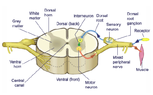
each spinal nerve communicates w/ the spinal cord via
2 pathways:
dorsal root
ventral root
describe how the mechanism of flexion (withdrawal) reflex starts
pain stimulus carried to spinal cord via 1o afferent (sensory) neuron → afferent neuron enters cord via dorsal root

what happens once the afferent neuron enters the cord
afferent synapses w/ interneuron → interneuron synapses w/ α-motor neuron (efferent) neuron in ventral horn

where does the efferent neuron go after it synapses w/ interneuron
efferent leaves cord via ventral root + terminates in effector organ → muscle contracts to withdraw from pain stimulus

describe the location of the 1o afferent (sensory) neuron
soma located in dorsal root ganglion (an enlarged region in the dorsal root containing the somas of many afferent neurons)
axon terminates in dorsal horn of grey matter
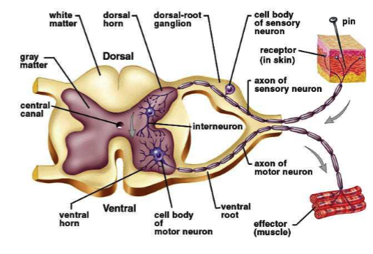
the arrangement of afferent, efferent + interneuron in the flexion (withdrawal) reflex is called
reflex arc
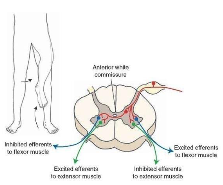
what’s reciprocal inhibition (innervation)
within peripheral muscle pairs (ex: biceps brachii + triceps):
the agonist is stimulated while the antagonist is simultaneously inhibited by excitatory + inhibitory interneuron activated by the same afferent neuron
interneurons create
neural circuits, enabling communication between sensory + motor neurons + w/ the CNS
are interneurons excitatory or inhibitory
both
3 types of interneurons
ipsilateral: connect afferents to efferents on the same side of cord
contralateral: connect afferents to efferents on the opposite side of cord
intersegmental: ascending/descending, carries afferent signals to efferents located 1 or more spinal segments above/below its origin
2 types of intersegmental interneurons
short: typically travel entirely within gray matter of cord
long: originate in gray matter but may leave gray matter + travel to distant cord segments within the fasciculus proprius, a region of white matter adjacent to the gray (red in figure)
when long intersegmental interneurons are in the fasciculus proprius they’re called
propriospinal neurons, which are myelinated + re-enter gray matter when they reach their destination
T/F: same afferent signal elicits responses on both sides of the body
true
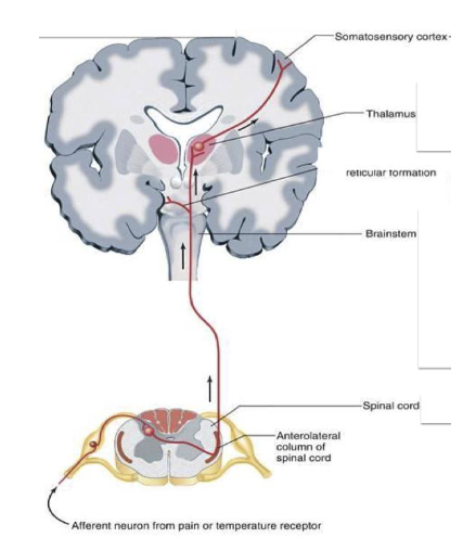
what are second order (secondary) afferent fibers
they carry sensory info from primary afferent in cord to cognitive centers in brain
are myelinated, have rapid conduction velocities
run in “tracts” in white matter of the cord