respiratory and cardiovascular
 Call Kai
 Learn
Learn Practice Test
 Spaced Repetition
 Match
Match Flashcards
 Knowt Play
1/217

There's no tags or description
Looks like no tags are added yet.
Name | Mastery | Learn | Test | Matching | Spaced | Call with Kai | Chat |
|---|
No analytics yet
Send a link to your students to track their progress
218 Terms
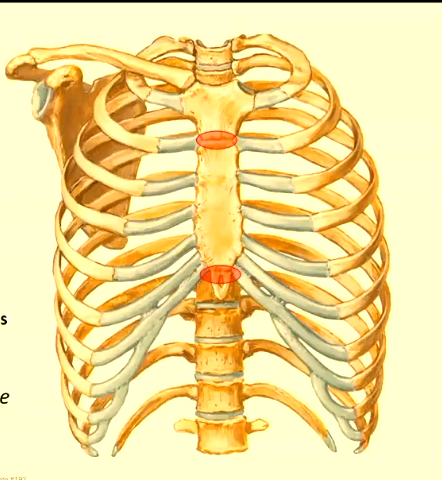
sternum
three bones, from top to bottom:
manubrium (jugular notch above this)
body
xiphoid process (xiphisternum)

ribs
12 pairs
true ribes (1-7)
flase ribs (8-10)
floating ribs (11-12)
all attach to vertebrae at the back (all thoracic)
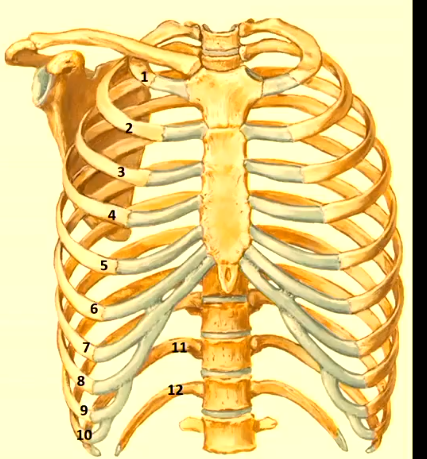
true rib
direct attachment to sternum via on costal cartilage
false ribs
indirect attachment via cartilage above (join up to the cartilage of other ribs)
floating ribs
no anterior attachment
costal margin
formed by costal cartilages of ribs 7-10
upside don v shape
diaphragm attaches here
boundary beteen thoracic cage and abdomen
made of hyaline cartilage
thoracic vertebrae
12, joined together by intervertebral discs
secondary fibrocartilaginous joints: hyaline on either side of the vertebral bodies and a disc in the middle
thoracic vertebrae articulate with ribs to form thoracic cage stability
apertures in the sternum
superior thoracic aperture (trachea and oesophagus pass through, arteries pass through)
inferior thoracic aperture (sealed by diaphragm, but diaphragm has holes)

intercostal spaces
gaps beteen the ribs
costal cartilage
hyaline cartilage that joins ribs to sternum
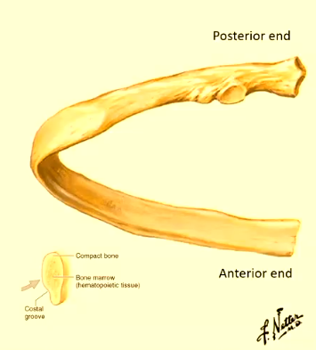
typical ribs
3-9
head
neck
tubercle
shaft (body)
atypical ribs
1, 2, 10-12
modified shape or articulation based on function
head of a rib
articulates ith the thoracic vertebra
tubercle
bump close to the thoracic vertebrae
articular part: facet for transverse process of corresponding vertebra
non-articular part: attachment for costotransverse ligament
joint important for breathing
costal groove
groove in the inferior side of the shaft
vein, artery and nerve run through here on the inner surface of the bone

clinical applications of the costal groove
fluid sometimes pools in the lung, have to be careful here the aspirator needle goes to avoid the vein, artery and nerve here, clinicians ill alays put the needle at the top of the rib
head
edge shaped
to articular facets:
superior facet —> vertebra above
inferior facet —> corresponding vertebra
middle of the head joins to the disc
neck
narro region beteen head and tubercle
shaft
long, curved main portion of rib
costal angle
point of greatest curvature of the shaft
thoracic vertebrae
vertebral body
heart shaped
to costal demifacets
transverse processes
contain a transverse costal facet
articulate ith tubercle of corresponding rib
spinous process points don
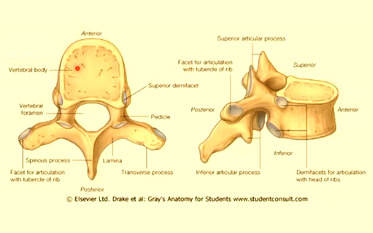
costal demifacets
articulate ith head of a typical rib
inferior and superior, join to to different ribs
hy does the transverse process of the thoracic vertebrae point don?
rotation alloed
bending backards is restricted to avoid compression of lungs and heart
joints of the thoracic cage
sternal joints
sternocostal joints
costochondral joints
interchondral joints
costovertebral & costotransverse joints
sternal joints
manubriosternal joint
xiphisternal joint
manubriosternal joint
secondary cartilaginous (symphysis): hyaline on either side of the joint and a disc in the middle
slightly mobile
forms the sternal angle (transverse thoracic plane/angle of Louis) at T4-5 intervertebral level
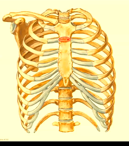
importance of the transverse thoracic plane
arch of aorta, trachea bifurcates into the bronchi
xiphisternal joint
primary cartilaginous (synchondrosis)
slightly mobile in youth
ossifies in adulthood —> becomes a synostosis (usually after 4th decade)
becomes one bone
sternocostal joints
rib 1
ribs 2-7
rib 1 sternocostal joints
costal cartilage —> manubrium
primary cartilaginous joint (synchondrosis)
almost immobile, protect important vessels and trachea, etc

ribs 2-7 sternocostal joints
synovial plane joints
costal cartilages —> sternum
permit small gliding movements during respiration
supported by anterior and posterior sternochondral ligaments
costochondral joints
junction beteen rib and its costal cartialge
primary cartilaginous (synchondrosis)
immobile

interchondral joints
here false ribs join true ribs

interchondral joints of costal cartilages 6-9
synovial plane joints
beteen costal cartilages of ribs 6-7, 7-8, 8-9
allo small gliding movements
interchondral joints of costal cartilages 9-10
fibrous joint
more rigid connection
minimal to no movement
costovertebral joints
red crosses: here rib 5 joints vertebral body 4, vertebral body 5 and intervertebral disc
synovial plane joints
head of rib articulates ith thoracic vertebrae
allo small gliding movements for espiration
ribs 1, 10, 11, 12 —> single vertebral articulation

costotransverse joins
synovial plane joints
tubercle of rib ←→ transverse process of corresponding vertebra
present in ribs 1-10 (absent 11-12)
allow small gliding movements
bottom red circle (the top red circle is the costovertebral joint)

thoracic cage movement
different shape sof costotransverse joints allo rib movments that expand the thoracic cage in different direcitons during inspiration
movement in ribs 2-7
shape of costotransverse joint is rounded
pump handle movement
elevation of ribs and sternum
increases antero-posterior (AP) diameter
sternum moves upard and forard

movement in ribs 8-10
shape of costotransverse joint is flat
bucket handle movement
elevation of lateral rib shafts
increases transverse (lateral) diameter
ribs move outard and upard

muscles of the thoracic cage
exctinsic muscles
intrinsic muscles (intercostal)
diaphragm
extrinsic muscles
ith attachments to ribs
come from somewhere that is not the thoracic cage
recruited during exercise

pectoralis minor
from scapula to ribs
pectoralis major
to the sternum
sternomastoid
to the manubrium
examples of extrinsic muscles
abdominal muscles
pectoralis minor and major
sternomastoid
intercostal muscles
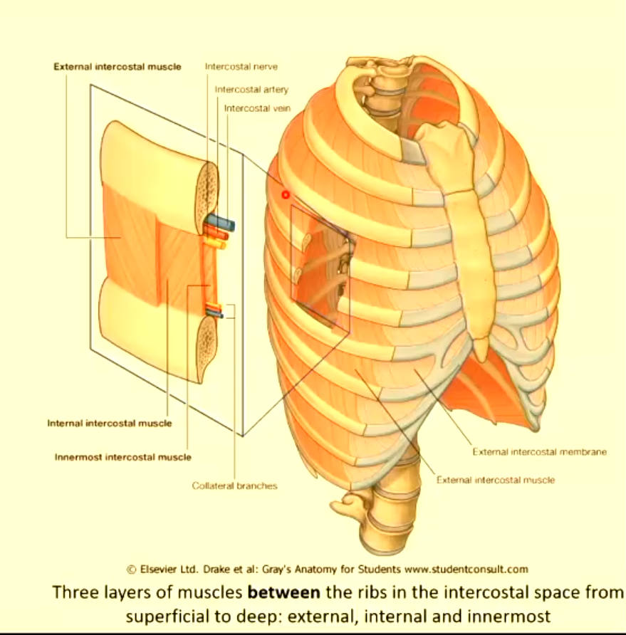
external, internal, innermost

external intercostal
donard and forard direction
external intercostal membrane anteriorly, external is see-through (aponeurosis) anteriorly
elevate ribs = muscles of inspiration
internal intercostal
perpendicular to external intercostal
internal intercostal membranes (aponeurosis) posteriorly
depress ribs = muscles of expiration
innermost intercostal
same direction as internal intercostal
not a continuous sheet of muscle
here are the vein artery and nerve in comparison to the intercostal muscles?
in beteen the internal and innermost intercostal
diaphragm
double dome-shaped muscle forming the floor of the thoracic cage and separation the thorax from the abdomen
right dome higher due to underlying liver
attaches to xyphoid process anteriorly (T9) and L1-3 posteriorly (therefore the back is a bit lower don)
muscle fibres radiate from the peripheral margins of the thoracic cage and converge into a central tendon

central tendon
heart sits on central tendon
rigid structure that can’t contract, avoids movement of the shape of the heart
muscles surrounding this tendon contract
attachments of the diaphragm
sternal (anterior)
costal (lateral)
lumbar (posterior)
sternal attachment of the diaphragm
xiphoid process
costal attachment of the diaphragm
internal surfaces of ribs 7-12 and their costal cartialges (costal margin for ribs 7-10)
lumbar attachment of the diaphragm
lumbar vertebrae via left and right crura and arcuate ligaments

contraction of the diaphragm
diaphragm goes flat during inspiration (concentric contraction)
natural recoil for breathing out
mechanism of inspiration
create a low pressure environment in lungs so that air rushes in
do this by increasing volume by stretching thoracic cage
pump handle increases front-to-back diameter
bucket handle increases side-to-side diameter
diaphragm contracts to increase top-to-bottom diameter
forced breathing
inspiration: sternocleidomastoid, scalenes, pectoralis minor
expiration: abdominal muscles, internal intercostals
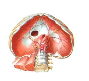
three major diaphragmatic openings
caval opening (blue)
oesophageal hiatus (middle)
aortic hiatus (red)

caval opening
inferior vena cava
through the central tendon
T8
oesophageal hiatus
oesophagus goes through the right cruus (in the muscle, functional sphincter)
therefore, hen the muscle contracts, the oesophagus is closed to prevent food regurgitating
T10

aortic hiatus
abdominal aorta
T12
not a true hole, passes next to the vertebrae and the diaphragm pins it to the lumbar vertebrae

intercostal neurovascular bundle
located in costal groove beteen internal and innermost intercostal muscles
intecostal vein (superior)
intercostal artery (middle)
intercostal nerve (inferior)
supplies thoraacic all (muscle, skin and pleura)
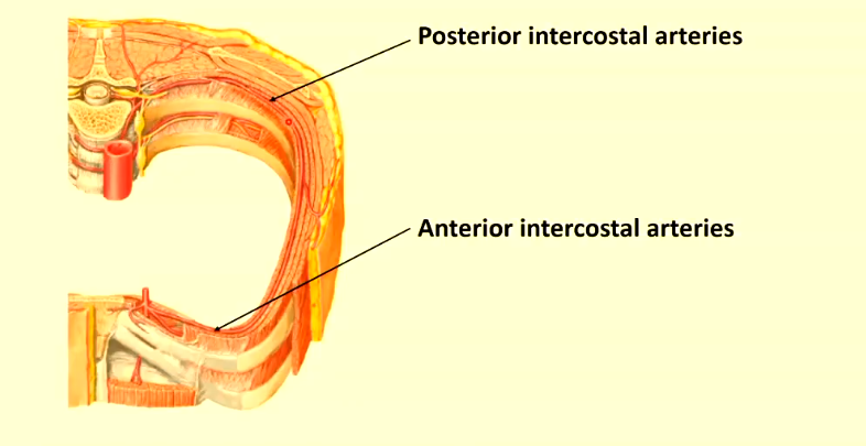
arteries of the thoracic cage
artery gives off posterior intercostal arteries
anastomoses ith anterior intercostal arteries, hich are given off by internal thoracic artery
veins of the thoracic cage
anterior intercostal veins
drain into the internal thoracic vein, then brachiocephalic vein, then SVC
posterior intercostal veins
drain into the azygos system (vein next to aorta)
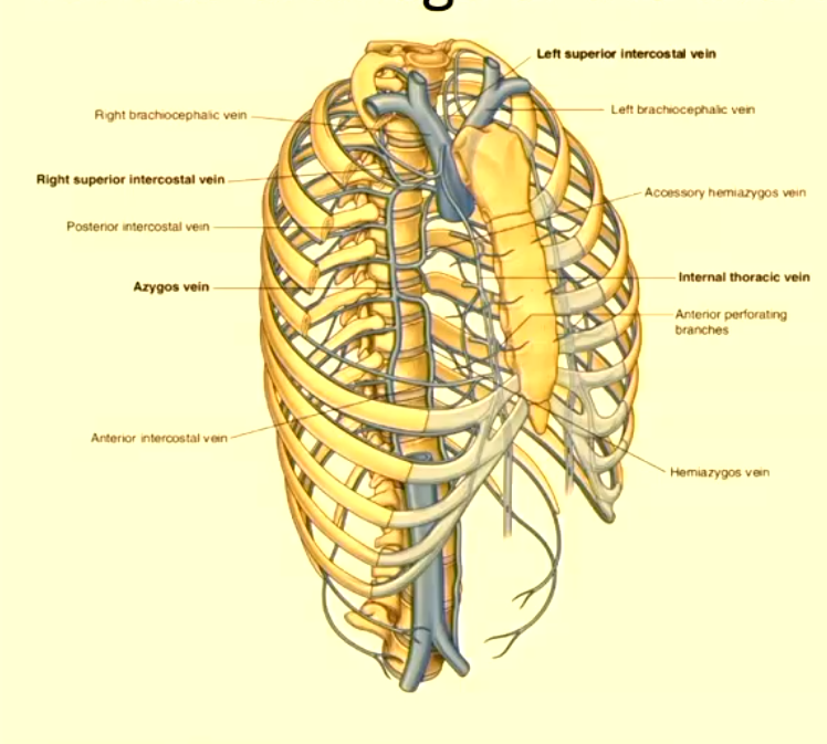
azygos venous system
variable
azygos vein
hemiazygos
accessory hemiazygos
drains posterior thoracic all and abdomen

azygos vein
right
T12→T5
drains into SVC, hich drains into right atrium
receives right hand side of posterior intercostal veins

hemiazygos
left loer
T12→T9
crosses to azygos at T9
drains loer posterior intercostal veins on the left
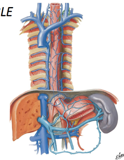
accessory hemiazygos
left upper
T5→T8
crosses to azygos at T8
drains top fe posterior intercostal veins on the left

innervation of the thoracic cage
intercostal nerves
most superior structure of the intercostal bundle is vein, then arteries, then nerves
no posterior/anterior distinction

intercostal nerves
arise from ventral rami T1-T11
T12 = subcostal nerve
functions of intercostal nerves
motor → intercostal muscles
sensory → skin of thoracic all + parietal pleura
contribution to peripheral diaphragm sensation
innervation of the diaphragm
motor: phrenic nerve (C3-5 ventral rami), pierces the diaphragm and pierces it from underneath
sensory: phrenic nerve (central diaphragm - central tendon) and intercostal nerves (peripheral diaphragm - muscular part)
heart pain can be referred to ja and neck area as C3-5 innervation

compartments of the thoracic cavity
pleural cavities (right and left)
mediastinum (centrally)

mediastinum
the heart lies ithin the fibrous pericardium in the middle mediastinum (space in the midde)

subdivisions of the mediastinum
superior + inferior → (anterior, middle, posterior)
heart occupies the largest part (middle mediastinum)
anterior mediastinum is the gap in the front, posterior mediastinum contains the oesophagus, etc

middle mediastinum
pericardiac sac & heart lies obliquely, 1/3 to the right of the midsternal line and 2/3 to the left
2nd rib to 5th intercostal space (apex of heart)
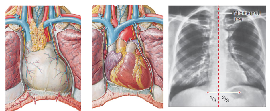
pericardium
layer 1: fibrous pericardium
layer 2: serous pericardium
fibrous pericardium
tough external layer, continuous with diaphragm, anchors and protects the heart
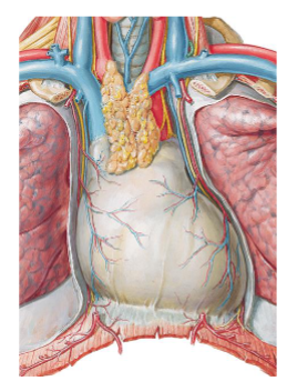
serous pericardium
thin double-layered membrane (parietal and visceral layers), lines fibrous pericardium and covers the heart, secretes serous fluid into pericardial cavity to reduce friction during cardiac movement
parietal (right image): somatic innervation, continuous over the heart and the vesels of the heart, stuck to fibrous layer
visceral: autonomic innervation (left image lining heart), continuous ith parietal layer at the great vessels
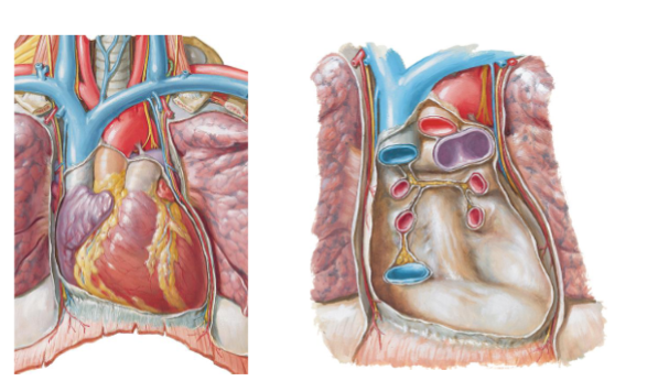
pericardial cavity
the space beteen parietal and visceral layers, contains serous fluid, enables heart to beat in a frictionless environment
in image can see ho parietal and visceral layer are continuous

close up of pericardium
serous fluid secreted into the pericardial cavity
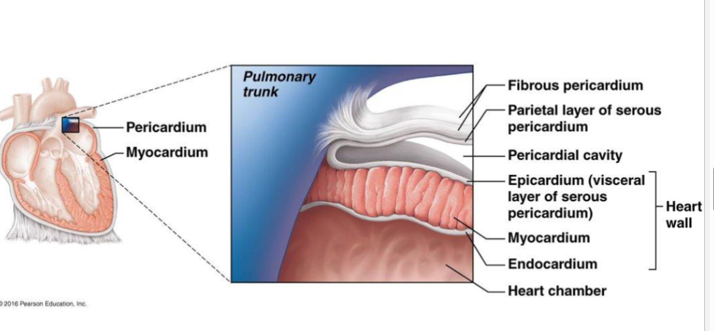
development of the heart and pericardium
heart starts as a tube and then bends
red: right chambers, blue: left chambers
front of the heart is predominantly right chamebrs

shape of the heart
apex should line up ith mid clavicle on the left

external features of the heart
atrioventricular groove
interventricular grooves

atrioventricular groove
coronary sulcus
separates atria from ventricles
interventricular grooves
separate right and left ventricles
anterior and posterior
arteries and veins run through them
right atrium
SVC - blood from upper body
IVC - loer limb, pelvis, gut
pectinate muscles: run parallel
crista terminalis:
fossa ovalis
atrioventricular orifice
right auricle
coronary sinus
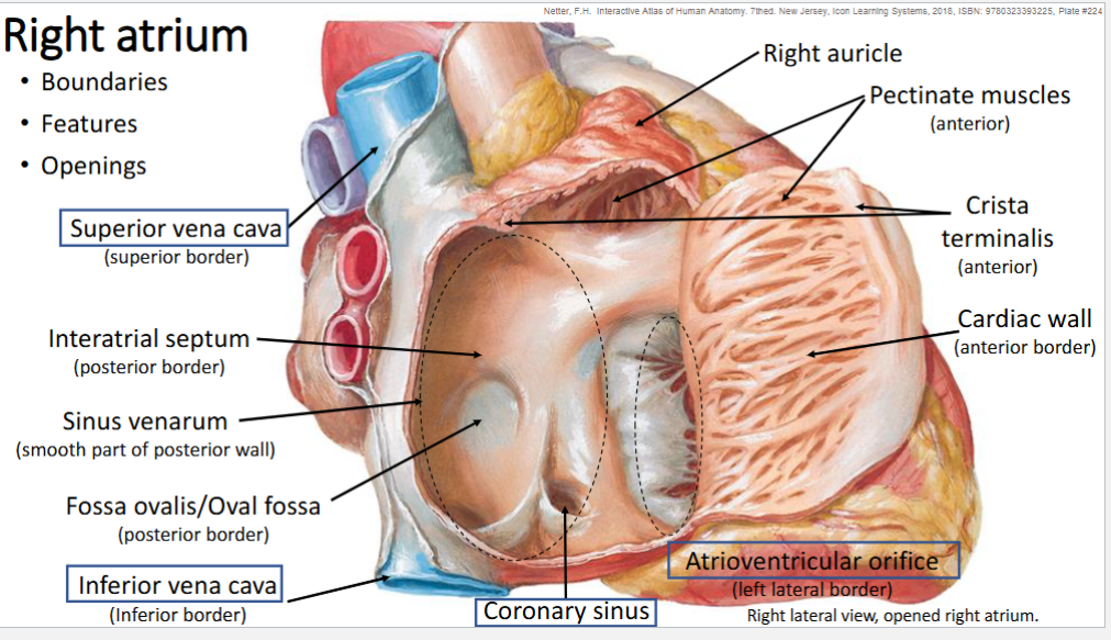
fossa ovalis
open in a foetus (foramen ovalis), direct connection between atria, as foetus’s don’t need to send their blood to pulmonary circulation as it’s already oxygenated
crista terminalis
bump corresponding to demarcator beteen rough pectinate muscles and smooth muscles
right auricle
gives more room for the chamber to expand hen blood enters
extension of the chamber
coronary sinus
receives deoxygenated blood from the heart itself
drained from cardiac veins
right ventricle
atrioventricular valve
trabeculae carnae
pulmonary valve

right atrioventricular valve
cusps (3)
chordae tendinae
papillary muscle (3)
called the tricuspid valve

trabeculae carnae
irregular muscle hich the papillary muscles are connected to
pulmonary valve
semilunar valve
three cusps
no chordae tendinae or papillary muscles

right atrioventricular valve
cusps (2)
chordae tendinae
papillary muscle (2)
called the bicuspid valve