Anatomy Lab Exam #3
1/47
There's no tags or description
Looks like no tags are added yet.
Name | Mastery | Learn | Test | Matching | Spaced | Call with Kai |
|---|
No analytics yet
Send a link to your students to track their progress
48 Terms

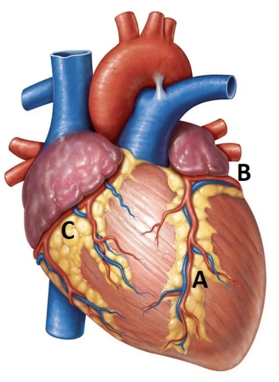
“A” represents the ___ of the heart.
Apex

“C” represents the ___ of the heart.
Aorta

“A” is most likely labeling the ____.
Right ventricle

“B” is most likely labeling the ___.
Left ventricle

“C” is most likely labeling the ___.
Right atrium

“D” is most likely labeling the ____.
left atrium

What are the blind pouches that extend the heart chambers labeled C & D?
Auricles
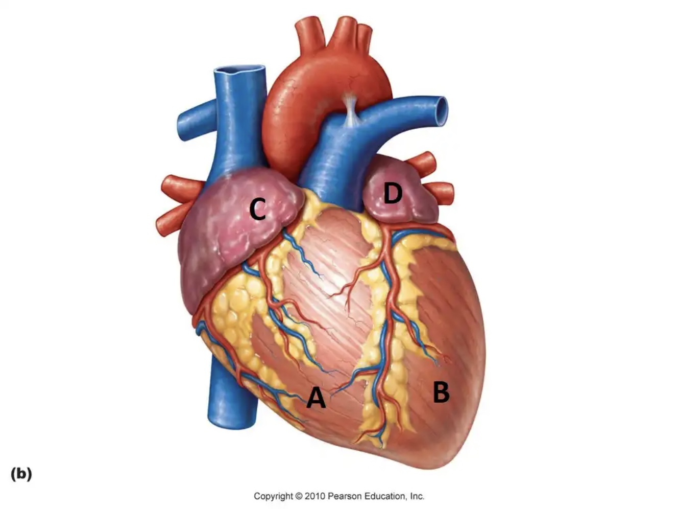
What is the primary purpose of the structures that are lying on the heart chambers labeled C & D?
Hold blood
Which structure surrounds the heart and provides protection, while reducing friction?
Pericardium

Which structure is located at “C?”
Interventricular septum
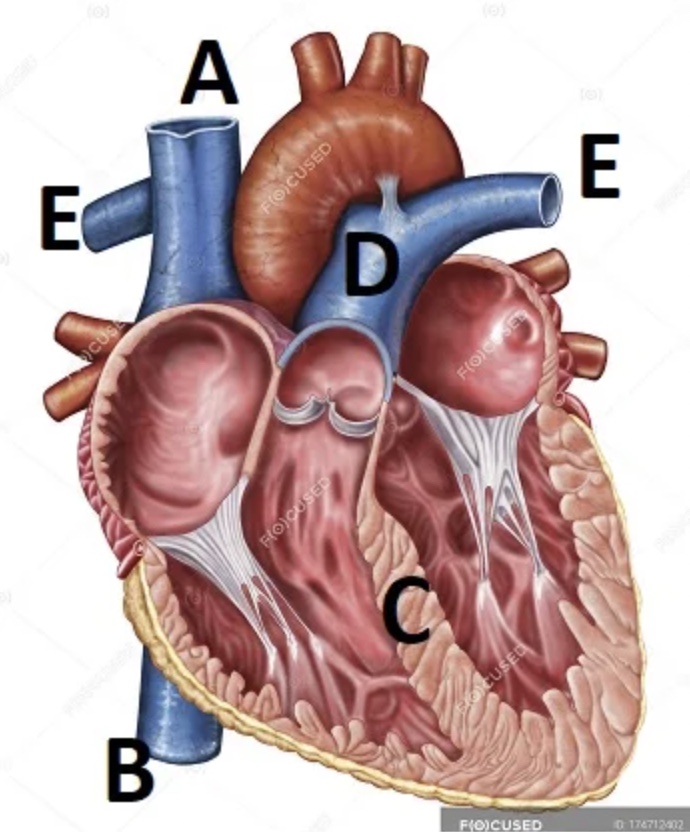
Which structure is located at “D,” which splits off into the E’s: ____?
Pulmonary trunk; pulmonary arteries
Which valve is located at A?
Right atrioventricular valve

Which valve is located at B?
Left atrioventricular valve

Which valve is located at C?
Pulmonary valve

Which valve is located where D meets the heart?
Aortic valve
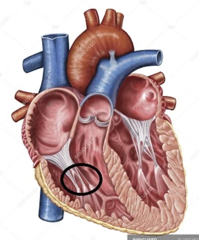
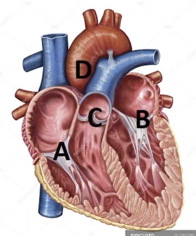
The thin structures inside the heart, circled here, are called ____, and are held in place by ____ muscles.
Which vessels are circled?
Pulmonary veins

What is located at “A?”
Subsinuosal interventricular sulcus

What is located at “B?”
Circumflex artery
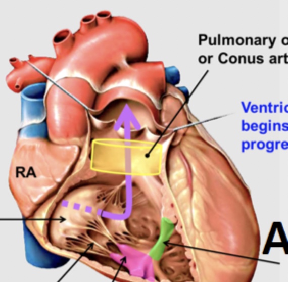
What is located at “A,” and is shown in green?
Trabeculae septomarginalis
What tissue separates the left and right atria?
Interatrial septum
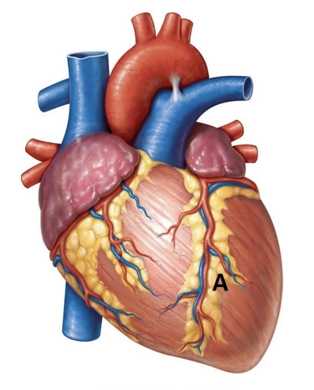
Which artery is located at A?
Paraconal interventricular artery
Which arterial branch is located at A?
Subsinuosal interventricular artery
Which artery is located at “C?”
Right coronary
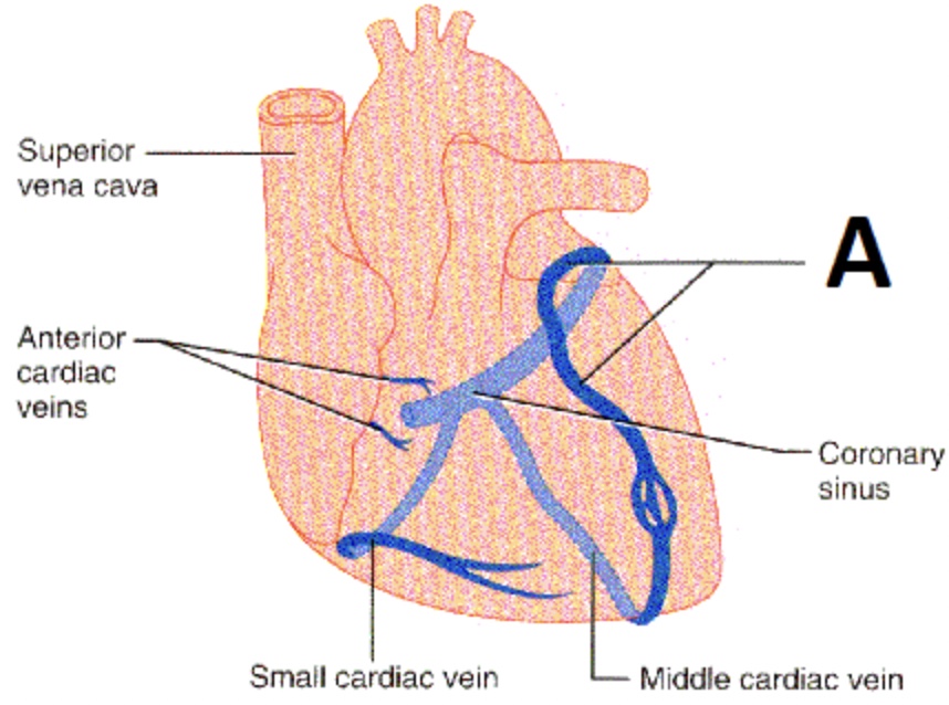
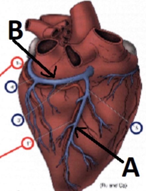
Which blood vessel is located at A?
Great cardiac vein
Which species possesses an os cordis, and what type of tissue is it?
Bovine; bone
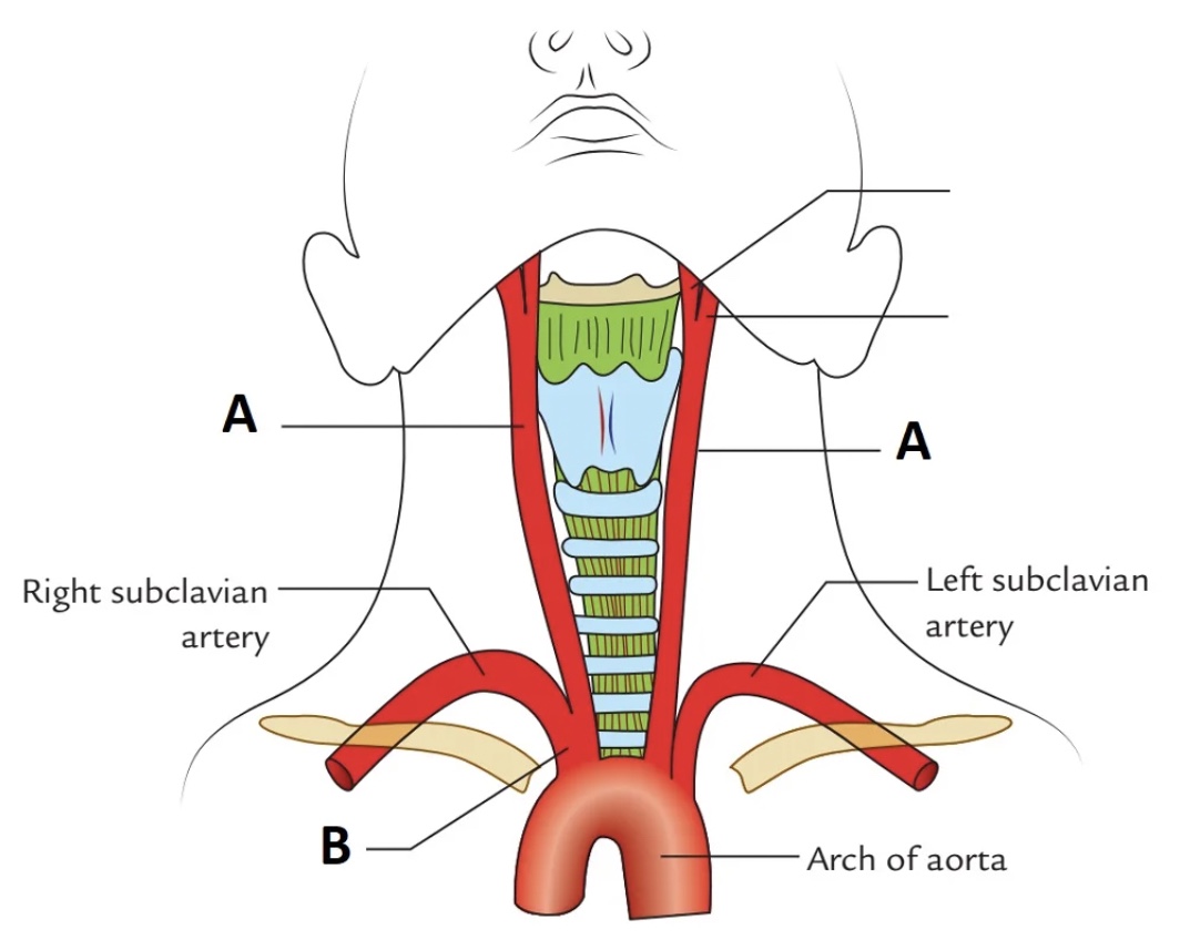
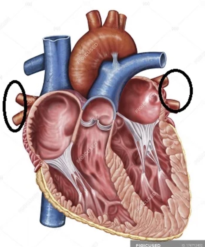
Which arteries are located at the “A’s?”
Common carotid
Where is the foramen ovale located in a fetus?
Passageway between right and left atria
Post-birth, the foramen ovale scars shut. What is this new depression called?
Fossa ovalis
PDA ocurs when the ductus arteriosus (a ____) fails to fully form the ligamentum arteriosus after birth.
Passageway between the pulmonary artery and aorta
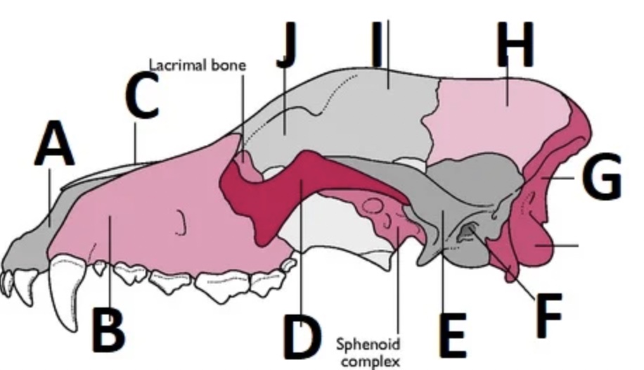
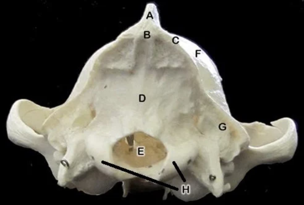
Which bone is labeled “A?”
Incisive

Which bone is labeled “H?”
Parietal

Which bone is labeled “E?”
Temporal

Which bone is labeled “G?”
Occipital

Which bone is labeled “B?”
Maxillary

Which bone is labeled “C?”
Nasal

Which bone is labeled “I?”
Frontal

J is pointing to the ____ (the space between bone I and bone D).
Orbit

Which bone is labeled “D?”
Zygomatic arch

Which opening is labeled “F?”
External acoustic meatus

The “bony bulbs” next to structure F are called ____.
Tympanic bullae
Structure A is known as the ____.
External sagittal crest
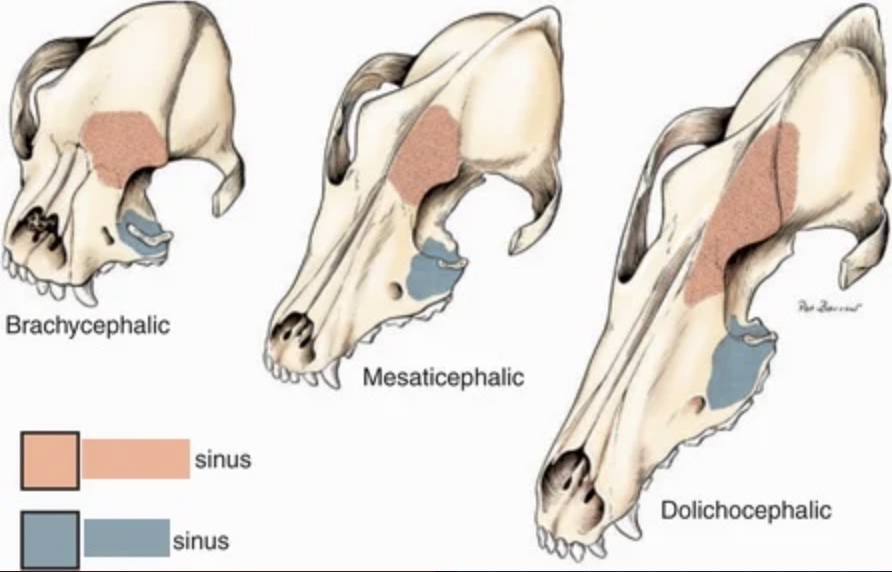
The peach color represents the ____ sinus, while the teal color represents the ____ sinus.
Frontal; maxillary

The feature at B is called the ____ in this particular species.
Facial crest
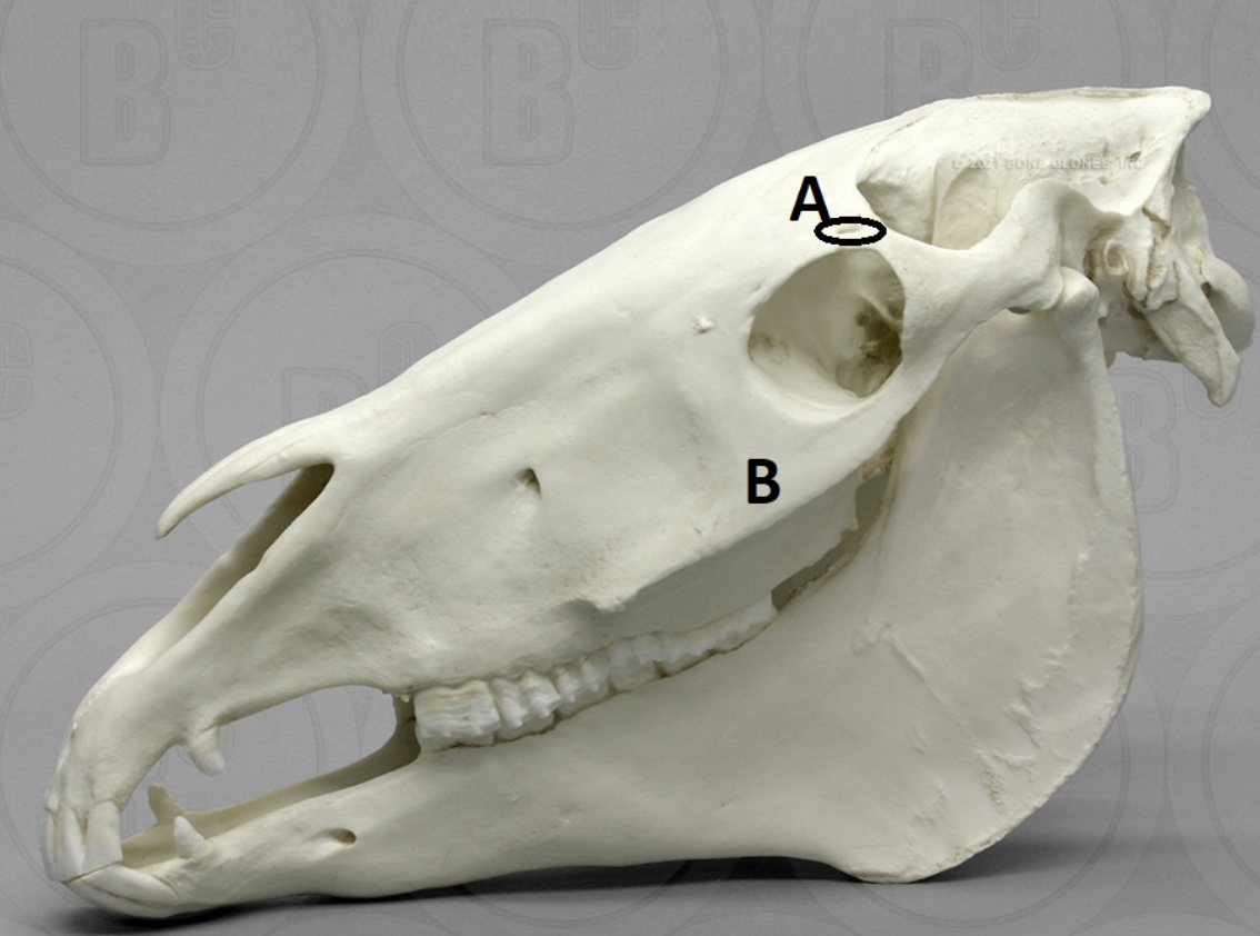
The opening at A is called the ____.
Supraorbital foramen

List the muscles shown below in order (A-E).
A- Masseter
B- Temporalis
C- Buccinator
D- Levator Nasolabialis
E- Digastricus
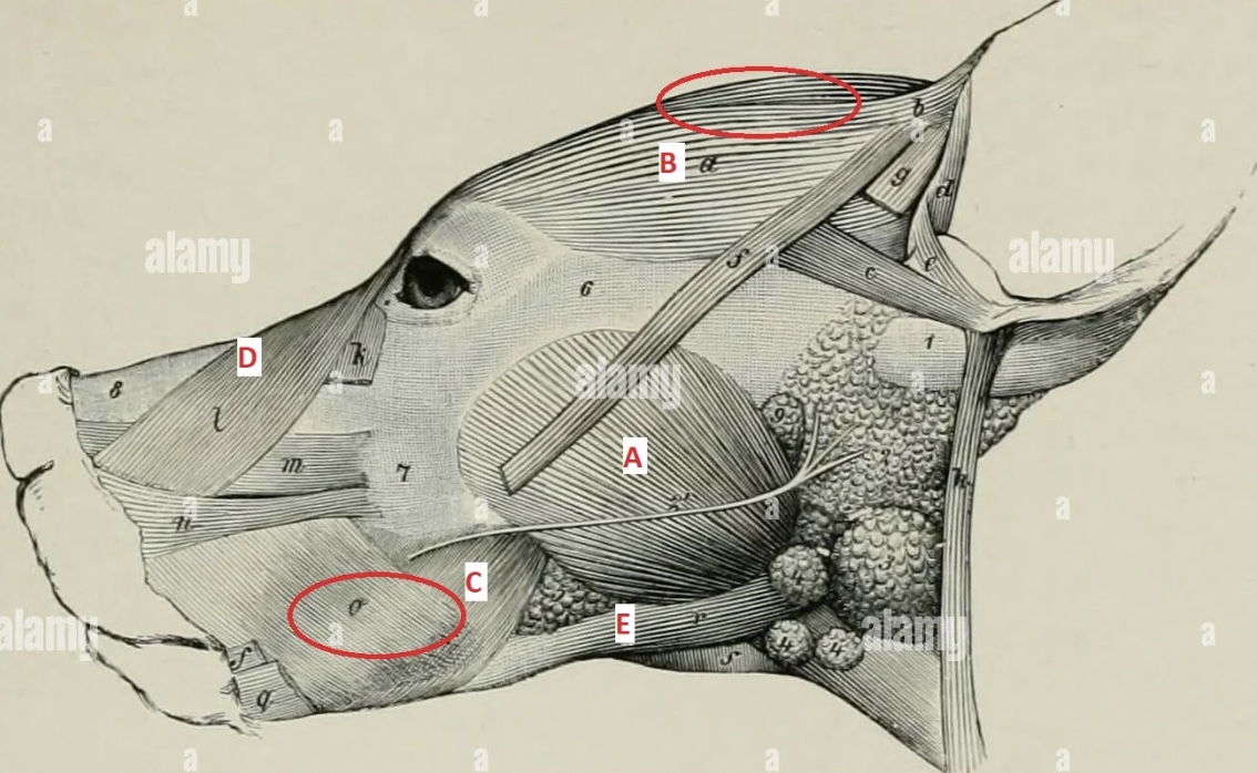
What is muscle D responsible for?
Snarling
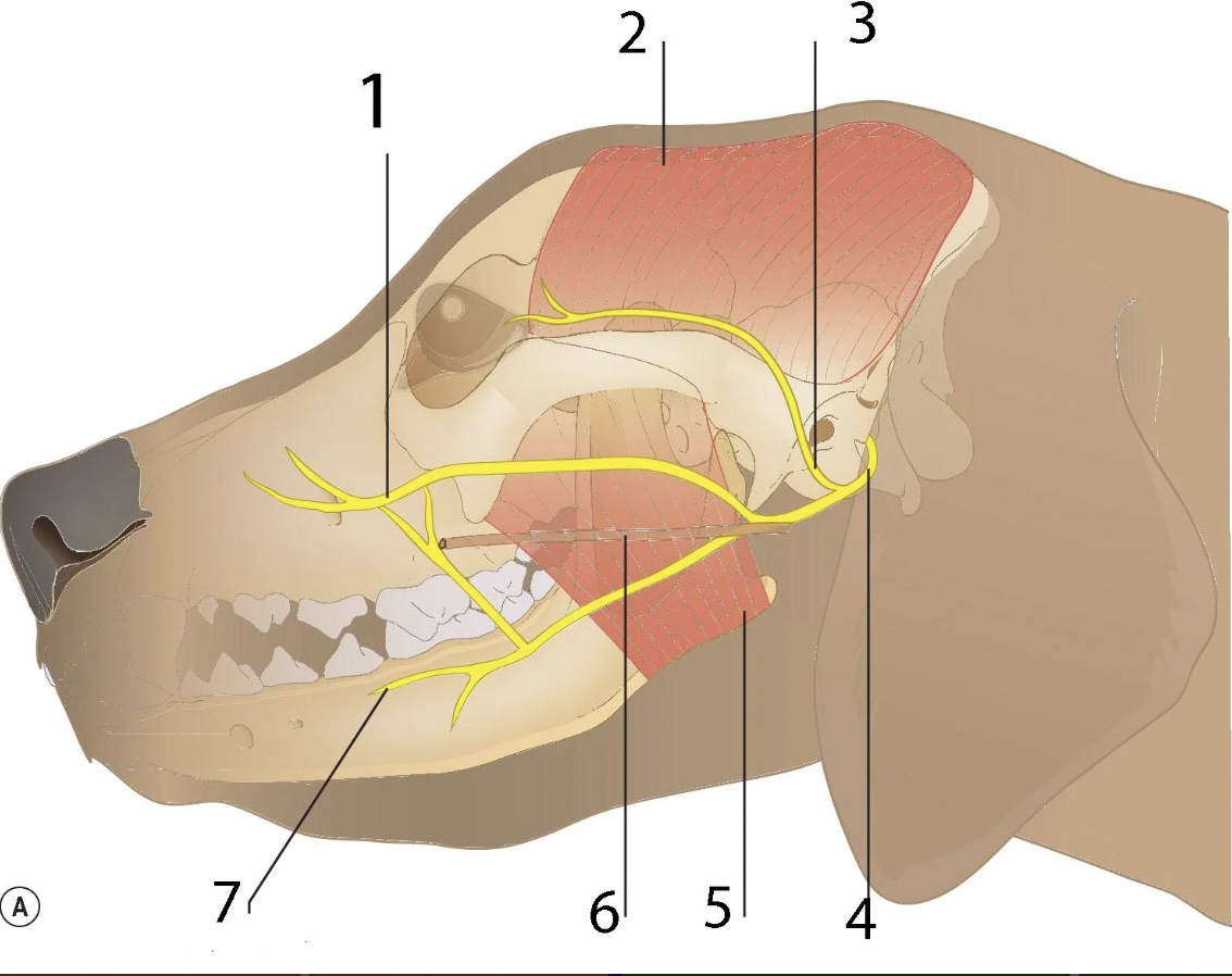
What structures are located at 1, 6, and 7 (in order)?
Dorsal buccal branch; parotid salivary duct; ventral buccal branch