lab practical 1 a+p 1 - tissue types to study
1/24
There's no tags or description
Looks like no tags are added yet.
Name | Mastery | Learn | Test | Matching | Spaced | Call with Kai | Chat |
|---|
No analytics yet
Send a link to your students to track their progress
25 Terms
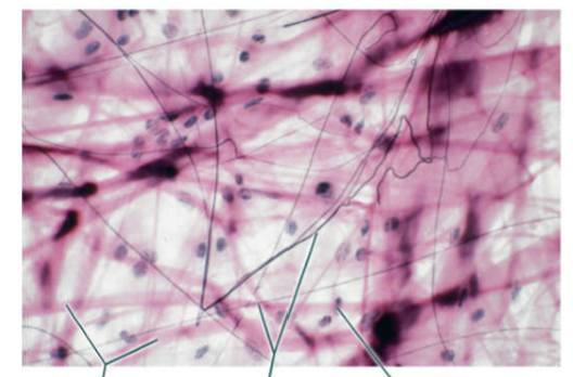
Loose Areolar Connective Tissue
- collagen fibers, elastic fibers, fibroblast nuclei
collagen fibers = pink strands
elastic fibers = black strands
fibroblast nucleus = black dots
looks like the tissue samples I used last summer my internship, and my internship was loose + all over areolar the place **
(connective tissue)
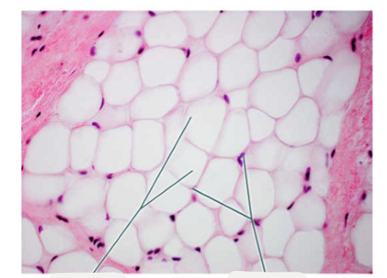
Adipose Tissue
adipocytes (fat cells) = white cells
nuclei = dark dots
more white than pink thyroid iodine cuboidal cells
(connective tissue)

Dense Irregular Connective Tissue
- collagen fibers, fibroblast nuclei
collagen fibers = pink swirls
fibroblast nuclei = purple dots
dense irregular = all swirly bc that’s how my insides feel bc mine is irregular
(connective tissue) (thick skin)
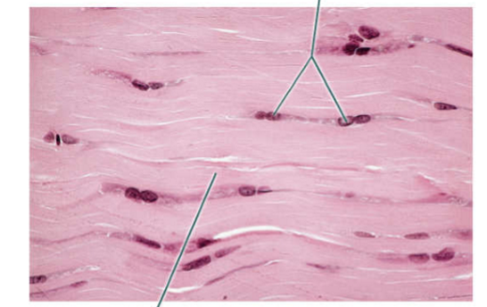
Dense Regular Connective Tissue
- collagen fibers, fibroblast nuclei
collagen fibers = pink waves
fibroblast nuclei = dark pink big dots
dense regular looks normal **
(connective tissue) (tendon)
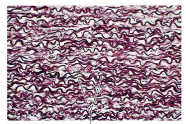
Dense Elastic Connective Tissue
- elastic fibers
elastic fibers = dark curls/waves/worms
you need dense elastics to take care of such curly hair **
(connective tissue) (aorta)
Reticular Connective Tissue
- medulla with reticular tissue, cortex
- (capsule, lymphoid follicles, trabecula)
medulla with reticular tissue = whole lower big chunk
cortex = top part of tissue (includes capsule, lymphoid follicles, + trabecula)
reticular is ridiculously complicated **
(connective tissue) (lymph node)
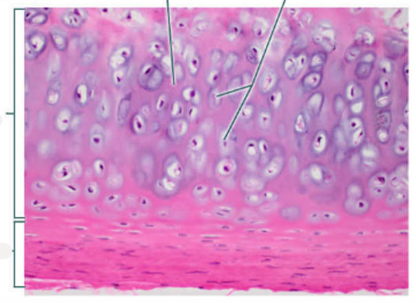
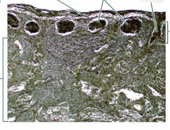
Hyaline Cartilage
- matrix, chondrocytes (cartilage cells)
- (hyaline cartilage, perichondrium, matrix, chondrocytes in lacunae)
matrix = pink background
chondrocytes = purple cells with nuclei
looks like ocean floor if you were high (HYaline)**
(connective tissue) (trachea)
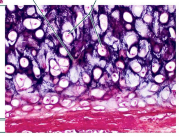
Elastic Cartilage
- matrix, chondrocytes
- (elastic cartilage, perichondrium, matrix, chondrocytes in lacunae)
matrix = dark purple background
chondrocytes = white/pink blobs with nuclei
stretches the mind even further when hy than hyaline —> ocean floor if it were even more crazy when hy**
(connective tissue) (epiglottis)
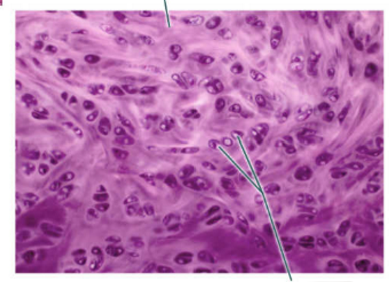
Fibrocartilage
- matrix, chondrocytes (in lacunae)
matrix = light purple/white spider webs **
chondrocytes = dark purple circles with light purple nuclei
fibrocartilage = discs = plates with spider webs on them = spider webs **
(connective tissue) (pubic symphysis)
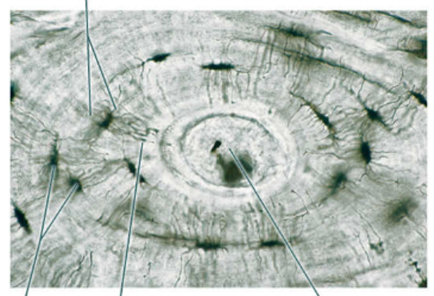
Bone
- lacunae, lamella, central canal, canaliculi
from center —> outside: central canal —> lamella —> canaliculi —> lacunae
central canal = middle middle
lamella = wirey into middle
canaliculi = wirey into sideways
lacunae = black dots on outside
CC L C L
can’t can’t lie, can lie **
(connective tissue) (compact bone)
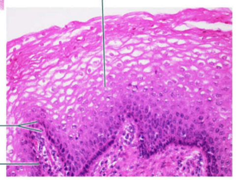
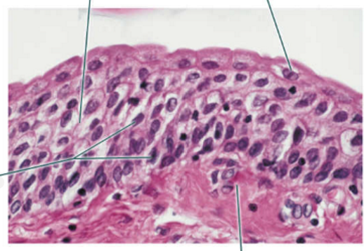
Non-keratinized Stratified Squamous Epithelium
- epithelium, basement membrane, nuclei
basement membrane = darker purple, bottom, zig zag
nuclei = dark purple spiked dots, layer inbetween basement membrane + epithelium
epithelium pink upper park
looks like an asian fruit net wrap** non-keratinized because you haven’t eaten the fruit yet **
(epithelial tissue) (vagina)
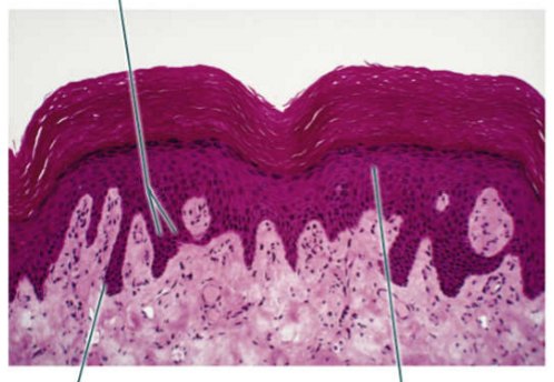
Keratinized stratified squamous epithelium
- epithelium, nuclei, basement membrane
basement membrane = light pink seeded area on bottom
epithelium = dark pink seeded area in middle
nuclei = seeds in dark pink epithelium
looks like a dragonfruit! dragonfruit = vitamins = better nutrition = more keratin*
(epithelial tissue)
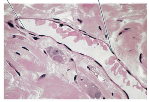
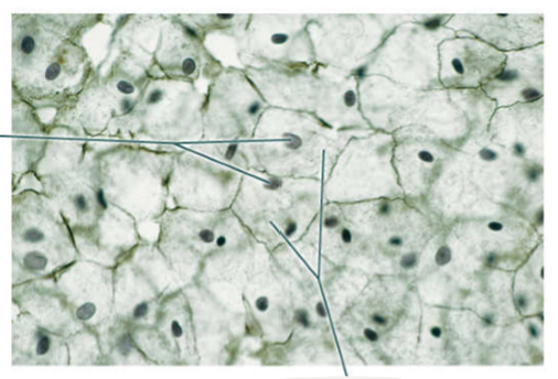
Simple Squamous Epithelium
- epithelium, nucleus
epithelium = thin layer right on edge of finger, light pink
nuclei = black dots (*cancer) along thin epithelial layer
looks like a witch finger = squamous sounds like those skin cancer cells —> warts —> witch finger **
(epithelial tissue) (venule)
Simple Squamous Epithelium
- simple squamous cells, nuclei
simple squamous cells = jagged edges like I drew them = squamous squirmy edges *
nuclei = black dots
looks like leaf layers/foil **
jagged edges like I drew them = squamous squirmy edges **
(epithelial tissue) (mesothelium)
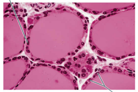
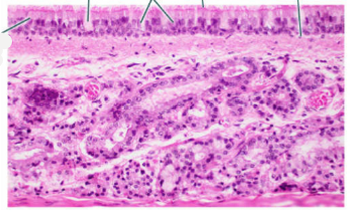
Simple Cuboidal Epithelium
- epithelium, nuclei
simple cuboidal epithelium = pink layer around thyroid iodine reservoirs
nuclei = dark pink dots in epithelium
looks like transitioning fish pebbles but pink —> fish —> cuboidal + Ryan —> endo class —> thyroid
(epithelial tissue) (thyroid gland!!!!!!**) (don’t mix up with adipose tissue)
Simple Cuboidal Epithelium
- epithelium, basement membrane, nuclei
basement membrane = dark purple baseline
epithelium = white space in little circle cells
nuclei = black dots of eyeballs
looks like a bunch of little fish eggs or eyeballs ** my eyes so cube/tbi apparatus is a cube = fish eggs**
(epithelial tissue) (kidney)
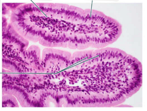
Simple Columnar Epithelium
- epithelium, basement membrane, nuclei
basement membrane = white little underpart inside the middle that the columns start from
epithelium cells = tall light purple outward strands
nuclei = dark purple dots
looks like a kiwi!!!** - kiwis irritate the column of my throat with allergic itchiness **
(epithelial tissue) (small intestine, duodenum)
Simple Columnar Epithelium
- basement membrane, epithelium, nuclei, goblet cells, microvilli
basement membrane = separating layer between kitchen-sink brownie underneath + tall columns above
epithelium = tall cells
nuclei = dark purple/pink circles
goblet cell = random white pocket circles = synthesize and secrete gel-forming mucins to protect from pathogens/irritants
microvilli = outside layer
kiwi one on its side*
(epithelial tissue) (small intestine)
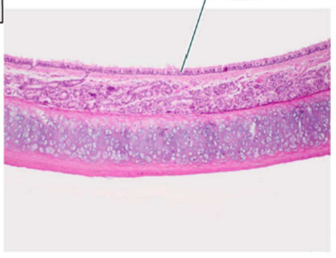
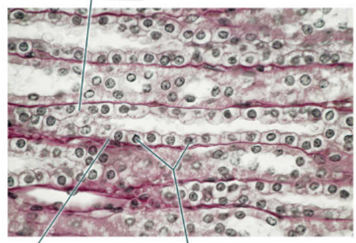
Pseudostratified Columnar Epithelium
- epithelium
epithelium = outmost layer of snake
looks like a movie monster snake passing by ** —> but it’s actually a cell so it’s a pseudosnake, not real
(epithelial tissue) (trachea) (far away picture)
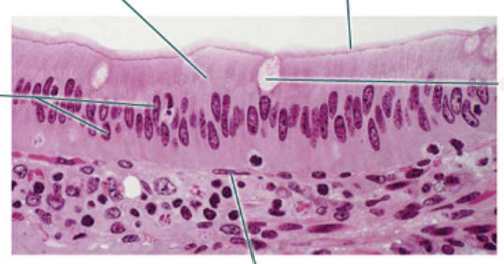
Pseudostratified Columnar Epithelium
- basement membrane, epithelium, nuclei, goblet cells, cilia
basement membrane = bottom of top little inch of tissue
epithelium = columns (look like pomelo strands **)
nuclei = dark purple dots
goblet cells = white circles randomly placed
cilia = outside layer
looks like an ant hill’s insides underneath ** —> ants make columns in the ground ** —> but this isn’t ants so it’s pseudostrantified
(epithelial tissue) (trachea) (closer picture)
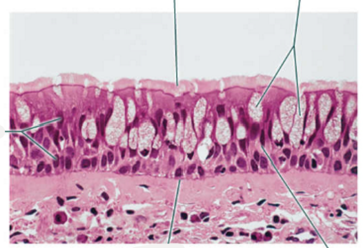
Pseudostratified Columnar Epithelium
- basement membrane, epithelium, nuclei, goblet cells, cilia
basement membrane = bottom of top little inch of tissue
epithelium = columns (look like pomelo strands **)**
nuclei = dark purple dots
goblet cells = white circles randomly placed, shiny
cilia = outside layer, pink little hairy guys
(epithelial tissue) (trachea) (even closer picture)
Transitional epithelium
- basement membrane, epithelium, nuclei, umbrella cells
basement membrane = pink meaty tissue at bottom
epithelium = actual cell shapes
nuclei = dark purple dots in cells
umbrella cells = white cells at edges
(epithelial tissue) (urinary bladder)
perichondrium
a dense, fibrous membrane that covers the external surface of most cartilage in the body
goblet cells
in columnar tissue
specialized, cup-shaped epithelial cells found in the respiratory, gastrointestinal, and reproductive tracts
primary function is to synthesize and secrete gel-forming mucins, which combine with water to form a protective mucus layer that lubricates tissues and traps pathogens or irritants.
lubricates tissues and traps pathogens or irritants
umbrella cells
in transitional tissue
highly specialized, terminally differentiated cells that form the outermost layer of the urinary tract's lining
get your umbrella out to protect from all your pee**