Cellular Adaptations
1/73
There's no tags or description
Looks like no tags are added yet.
Name | Mastery | Learn | Test | Matching | Spaced |
|---|
No study sessions yet.
74 Terms
what is pathology?
The study of understanding the cause of disease and the changes in cells, tissues, and organs that are associated with disease
what is etiology?
Underlying causes and modifying factors that are responsible for initiation and progression of disease
what is pathogenesis?
Mechanisms of development and progression of disease, which account for the cellular and molecular changes that give rise to the specific abnormalities that characterize any particular disease
what is homeostasis?
• Steady state of normal cells
• Equilibrium between the cells and their environment
• Cells actively interact with their environment, constantly adjusting their structure and function to accommodate changing demands and extracellular stresses
what happens when homeostasis is disturbed?
If disturbed, the cell can be predisposed for onset of pathology
what are adaptations to environmental stress?
reversible changes in the number, size, phenotype, and metabolic activity cellular functions in response to change in their environment
Cell injury within limits is reversible. If the stressor is severe, persistent, or rapid in onset it
will result in …?
irreversible injury and death of the affected cell
what are causes of cell injury?
• Hypoxia and ischemia
• Toxins
• Infectious agents
• Immunologic reactions
• Genetic abnormalities
• Nutritional imbalances
• Physical agents
• Aging
what is reversible cell injury?
The deranged function and morphology of the injured cells can return to normal if the
damaging stimulus is remove
what are the 2 main morphologic correlates of reversible cell injury?
cellular swelling
fatty change
what is cellular swelling?
Injury associated with increased permeability of the plasma membrane
Cells and organelles take in water due to failure of the energy dependent ion pumps
Pallor due to compression of capillaries
Increased organ weight
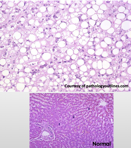
what is fatty change?
Appearance of triglyceride containing lipid vacuoles in the cytoplasm
Nucleus is displaced and the cell swells
Seen frequently in liver, heart, and kidney
Ex: secondary to alcoholism, diabetes mellitus, malnutrition, obesity, and poisoning
Imbalance among uptake, utilization, and secretion of fat
what are the 2 pathways of irreversible injury?
apoptosis
necrosis
what is apoptosis?
programmed cell death (How we eliminate potentially harmful cells or cells that have outlived their usefulness (physiologic apoptosis)
No inflammatory cell response
Eliminates cells damaged beyond repair (apoptosis in pathologic conditions)
Ex: exposed to radiation, cytotoxic drugs, certain infections or viruses
what is necrosis?
accidental cell death (Cell death in which cellular membranes fall apart, and cellular enzymes leak out and ultimately digest the cell)
inflammatory cell response
Due to severe disturbances like loss of oxygen or nutrients
Rapid and uncontrollable
how does apoptosis work?
Fragments of the apoptotic cells break off (“apoptotic bodies”)
Apoptotic bodies are consumed by phagocytes
plasma membrane remains intact = liitle leakage of cellular contents so no inflammatory process
what is physiologic apoptosis?
During normal development, some cells die and are replaced by new ones
In these situations, cell death is always via apoptosis
Unwanted cells are eliminated without eliciting potentially harmful inflammation
In the immune system, apoptosis eliminates excess leukocytes and lymphocytes left at the end of an immune response
what is pathologic apoptosis?
Cells damaged beyond repair
Ex: DNA damage after radiation or cytotoxic drugs
Misfolded proteins
Certain infectious agents like viruses elicit apoptosis
what are morphologic characteristics of apoptosis?
Cells shrink
Chromatin condensation and aggregation
Fragmentation of DNA
Cells form cytoplasmic buds and fragment into apoptotic bodies
Composed of membrane bound pieces of cytosol and organelles
Rapid extrusion of fragments and phagocytosis
No inflammatory response
necrosis involves nuclear changes resulting from…?
breakdown of DNA and chromatin
Pyknosis- shrinkage of nucleus into a basophilic (darker) mass due to DNA condensing
Karyorrhexis- fragmentation of nucleus into multiple small pieces
Karyolysis- fading of nucleus, less and less basophilic as DNA is digested
what are some cytoplasmic changes that occur during necrosis?
Increased pink cytoplasm – eosin dye binding to denatured proteins
Glassy, homogenous appearance
Vacuolated or “moth eaten” once the organelles have been digested by enzymes
normal
early necrosis
necrosis (irreversible)
what are types of necrosis?
• Coagulative
• Liquefactive
• Gangrenous
• Caseous
• Fat
• Fibrinoid
what is the most common type of necrosis?
COAGULATIVE NECROSIS
what is COAGULATIVE NECROSIS?
Sudden loss of blood supply to an organ (ischemia)
Does not occur in the brain
Denaturation of proteins and enzymes resulting in blockage of proteolysis of
dead cellsUnderlying tissue architecture is preserved for a few days after death of the cells in the tissue
what is liquefactive necrosis?
Seen in focal bacterial and occasional fungal infections
Microbes stimulate rapid accumulation of inflammatory cells
Enzymes of the leukocytes digest (“liquefy”) the tissue
Hypoxic death of cells in the CNS
If initiated by acute inflammation like in bacterial infection the liquid will be pus
liquefactive necrosis
what is GANGRENOUS NECROSIS?
A clinical descriptor, not a distinct pattern of necrosis
Usually refers to a limb that has undergone coagulative necrosis involving multiple
tissue layers
what is “dry” gangrene?
Coagulative necrosis without liquefaction
what is “wet” gangrene?
Bacterial infection superimposed
Results in liquefactive necrosis
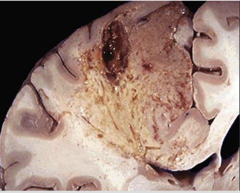
CASEOUS NECROSIS is most often seen in?
tuberculosis
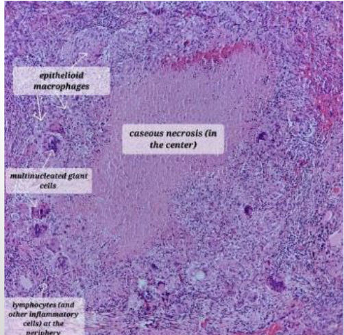
CASEOUS NECROSIS
what is the histopathology of CASEOUS NECROSIS?
architecture is not preserved
Fragmented or lysed cells
Amorphous pink and granular
Often surrounded by a collection of macrophages and other inflammatory cells (granuloma)
describe the histopathology of fat necrosis
Shadowy outlines of necrotic fat cells
Basophilic calcium deposits
Inflammatory reaction
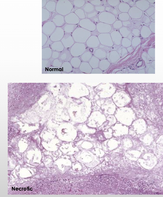
FAT NECROSIS
what is traumatic FAT NECROSIS?
severe injury to areas with high fat content
• Breast- post-cancer procedures and radiation
• Thigh and buttock- following medical and cosmetic procedures
what is enzymatic FAT NECROSIS?
complication of acute pancreatitis
• Pancreatic enzymes leak out of pancreas and destroy fat cells in peritoneum
• Triglyceride esters within the fat are split releasing fatty acids
• Fatty acids combine with calcium to produce chalky white areas (fat saponification)
what is FIBRINOID NECROSIS?
Can only be seen by microscopic examination
• Often associated with complexes of antigens and antibodies deposited in the walls of blood vessels
• Deposited immune complexes and plasma proteins that leak into the wall of the damaged vessels produce a bright pink amorphous appearance on H&E staining that pathologists call fibrinoid (fibrin-like
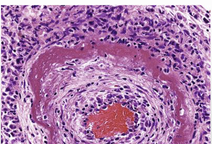
FIBRINOID NECROSIS
Adaptations are reversible changes in cells in response to changes in the environment such as
• Number
• Size
• Phenotype
• Metabolic activity or
• Functions of cells
what are physiologic adaptions?
responses to normal stimulation by hormones or endogenous chemical mediators or to demands of mechanical stress
what are pathologic adaptations?
responses to stress.
Cells modulate their structure and function to escape injury, but at the expense of normal
function
what is hypertrophy?
Increase in the size of cells resulting in increase in size of the organ
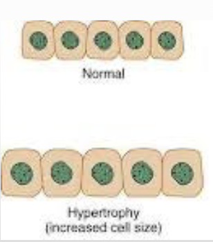
when does hypertrophy occur?
when there is a limited capacity to divide
hypertrophy is caused by?
increased functional demand or by growth factor or hormonal stimulation (physiologic and pathologic causes)
what are some physiologic causes of hypertrophy?
smooth muscle of uterus during pregnancy
skeletal muscles during weightlifting
what are some pathologic causes of hypertrophy?
cardiac enlargement due to hypertension or aortic valve disease
what is hyperplasia?
Increase in the number of cells in an organ that stems from increased proliferation (Can be physiologic or pathologic)
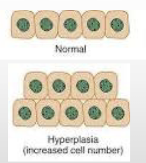
hyperplasia takes place if…?
if the tissue contains cell populations capable of replication (May occur concurrently with hypertrophy and often in response to the same stimuli)
what is an example of physiologic hyperplasia?
Hormonal hyperplasia- glandular epithelium in breast tissue at puberty and during pregnancy
Compensatory hyperplasia- residual tissue grows after removal or loss of part of an organ as in liver resections
what is an example of pathologic hyperplasia?
Excessive hormonal or growth factor stimulation
what characteristic distinguishes pathologic hyperplasia from cancer?
responsiveness to normal regulatory contro (hyperplastic process remains controlled; if the signals that initiate it abate, the hyperplasia disappears)
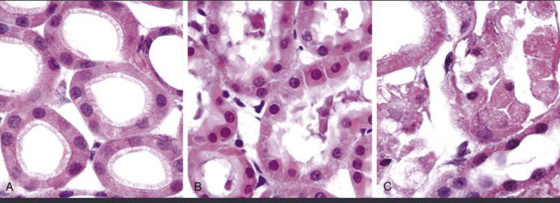
what is atrophy?
The shrinkage in the size of cells by the loss of cell substance
If significant number of cells are involved, the entire tissue or organ is reduced in size
Cells may have diminished function, but are not dead
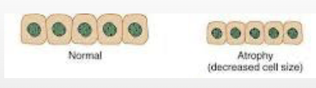
what are some causes of atrophy?
• Decreased workload
• Loss of innervation (pathologic)
• Diminished blood supply
• Inadequate nutrition
• Loss of endocrine stimulation
• Aging
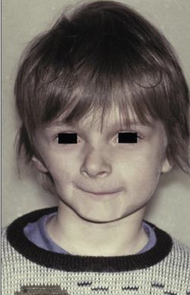
Progressive Hemifacial Atrophy
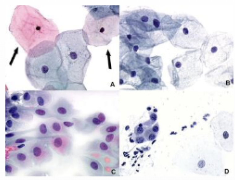
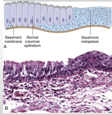
what is metaplasia?
Change in which one adult cell type is replaced by another adult cell type
A cell type sensitive to a particular stress is replaced by another cell type better able to withstand the adverse environment
Example- respiratory epithelium in habitual cigarette smokers
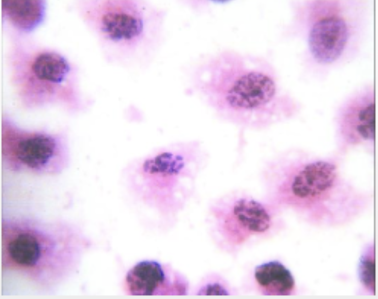
what are intracellular accumulations?
cells may accumulate abnormal amounts of various substances (harmless or harmful)
in cytoplasm, within organelles, or in nucleus
may be synthesized by affected cells, or it may be produced elsewhere
what are some examples of intracellular accumulations?
• Fatty change (steatosis)
• Cholesterol and cholesteryl esters
• Proteins
• Glycogen
• Pigments
what are the main pathways of abnormal intracellular accumulation?
• Inadequate removal or degradation of the substance
• Excessive production of an endogenous substance
• Deposition of abnormal exogenous material
fatty change can be due to
alcohol abuse
obesity
(can affect many organs, but commonly the liver)
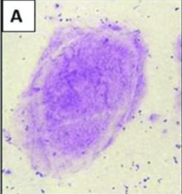
example of protein intracellular accumulation
amyloidosis: Proteins are misfolded and deposited into the tissue. This can rarely be seen in the oral cavity
what are some pigments that can be intracellular accumulations?
• Carbon- most common, pollution
• Melanin
• Hemosiderin- local or systemic excess of iron
• Drug or drug metabolites
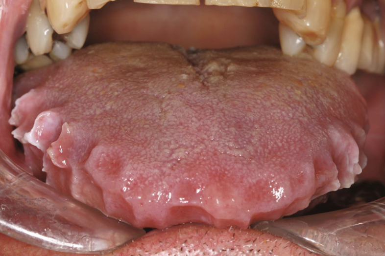
oral amyloidosis
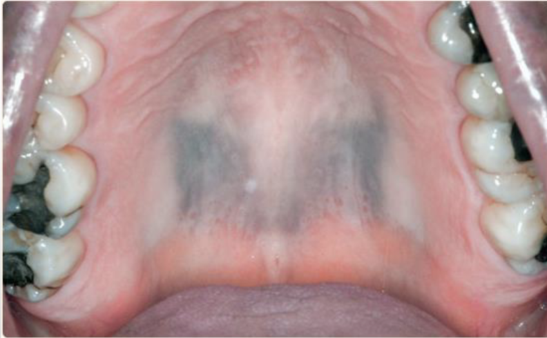
drug-related discoloartion (pigment)
what is pathologic calcification?
Abnormal deposition of calcium salts, together with smaller amounts of iron, magnesium, and other minerals
pathologic calcification can occur as: (2 types)
Dystrophic calcification- calcium metabolism is normal, but it deposits in injured or dead tissue
Metastatic calcification- associated with hypercalcemia and can occur in normal tissues
dystrophic calcification can cause
organ damage
For example: calcification in aging or damaged heart valves resulting in severely compromised valve motion
dystrophic calcification is common in areas of
aseous necrosis in tuberculosis
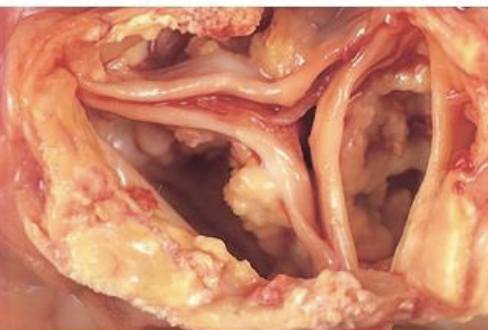
calcific stenosis of aortic valve
metastatic calcifcation is associated with
hypercalcemia
what are the main causes of hypercalcemia?
Increased secretion of parathyroid hormone
Destruction of bone
Vitamin D-related disorders (sarcoidosis)
Renal failure