Canine Orthopedic Lameness Overview
 Call Kai
 Learn
Learn Practice Test
 Spaced Repetition
 Match
Match Flashcards
 Knowt Play
1/12

Earn XP
Description and Tags
Vocabulary flashcards covering the clinical signs, radiographic diagnosis, and treatment of common canine orthopedic conditions including hip dysplasia, panosteitis, HOD, OCD, and Legge-Perthes disease.
Name | Mastery | Learn | Test | Matching | Spaced | Call with Kai | Chat |
|---|
No analytics yet
Send a link to your students to track their progress
13 Terms
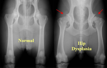
Hip dysplasia
A condition characterized by a "bunny hopping" gait
difficulty rising
a positive Ortolani sign indicating hip laxity.
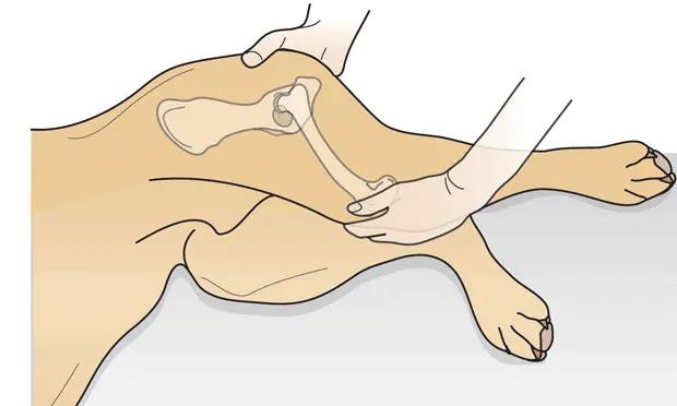
Ortolani sign
A clinical physical exam finding that indicates hip laxity, commonly associated with the diagnosis of hip dysplasia.
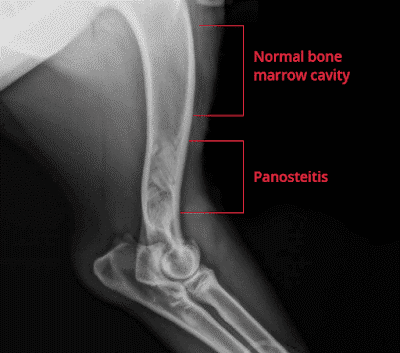
Panosteitis
An orthopedic disease in young, medium to giant breed dogs characterized by
acute lameness
fever
long bone pain
increased medullary opacity on radiographs.
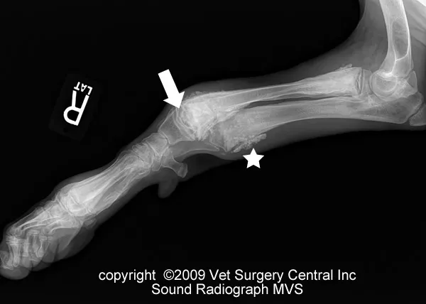
Hypertrophic osteodystrophy (HOD)
A condition in young large breeds causing
swollen and warm distal limbs
metaphyseal pain
a characteristic "double physeal line" on radiographs.
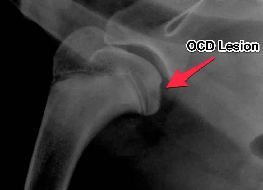
Osteochondrosis dissecans (OCD)
A joint disorder involving joint effusion and decreased range of motion, radiographically appearing as flattening of subchondral bone or a "joint mouse."
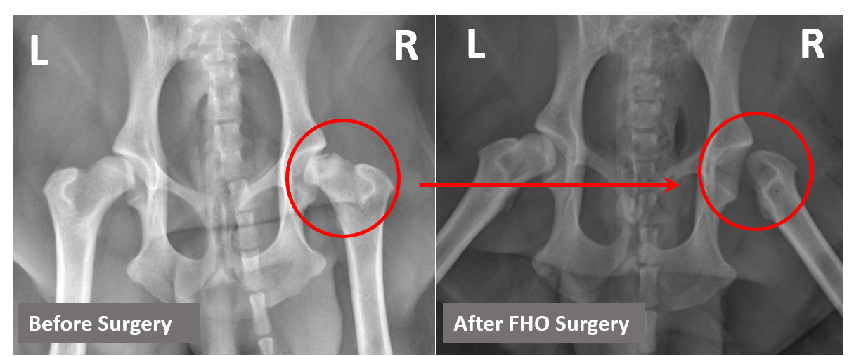
Legge-Perthes disease
Avascular necrosis of the femoral head affecting 3- to 12-month-old small or toy breeds
resulting in pelvic limb lameness.
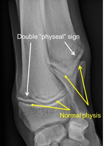
Double physeal line
A specific radiographic sign found in cases of Hypertrophic osteodystrophy (HOD).
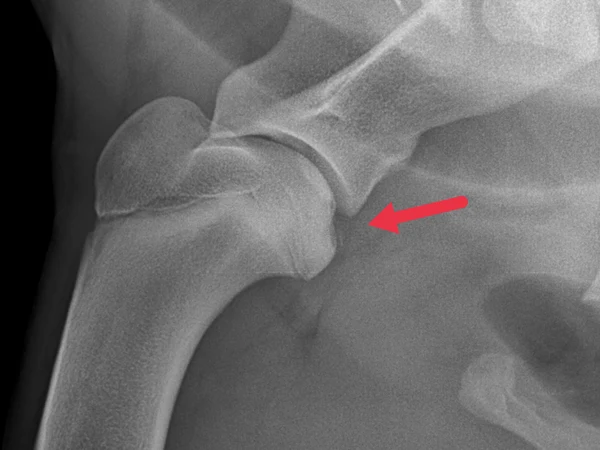
Joint mouse
A radiographic finding associated with OCD representing a piece of bone or cartilage that has become detached within the joint.
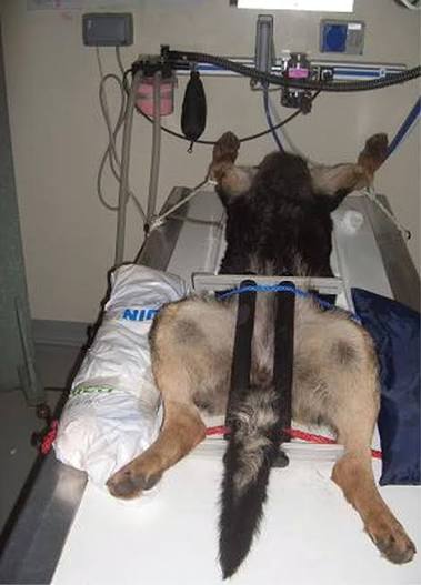
PennHIP
A diagnostic method for hip dysplasia that is considered more accurate for diagnosis at an earlier age than the OFA method.
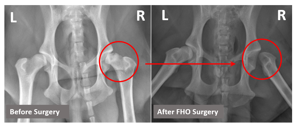
Legge-Perthes Radiographic Dx
Radiographic findings include loss of bone opacity at the femoral epiphysis and a moth-eaten appearance of the femoral neck and head.
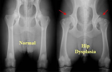
Hip Dysplasia Radiographic Dx
Characteristic findings include a shallow acetabulum, flattening of the femoral head, and a thickened femoral neck.
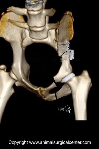
Triple pelvic osteotomy
A surgical treatment option specifically listed for the management of hip dysplasia.
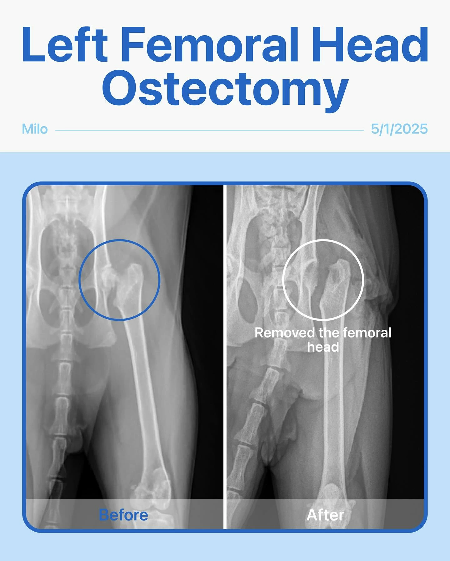
Femoral head ostectomy
A surgical procedure used to treat both Hip dysplasia and Legge-Perthes disease.