Anatomical Directions and Terminology
 Call Kai
 Learn
Learn Practice Test
 Spaced Repetition
 Match
Match Flashcards
 Knowt Play
1/43

There's no tags or description
Looks like no tags are added yet.
Name | Mastery | Learn | Test | Matching | Spaced | Call with Kai | Chat |
|---|
No analytics yet
Send a link to your students to track their progress
44 Terms
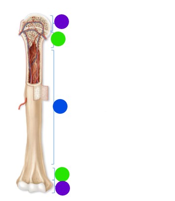
What is purple pointing at
Epiphysis (proximal and distal)
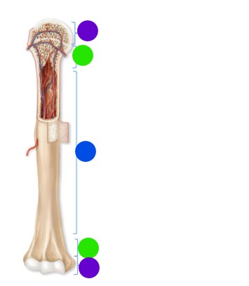
What is green pointing at
Metaphysis
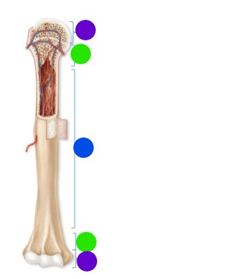
What is blue pointing at
diaphysis
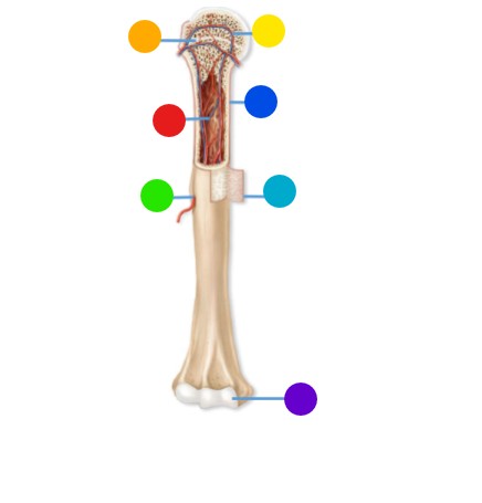
What is orange pointing at
epiphyseal line
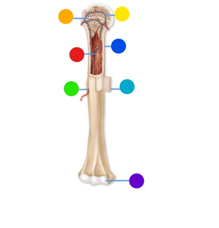
What is yellow pointing at
trabecular bone
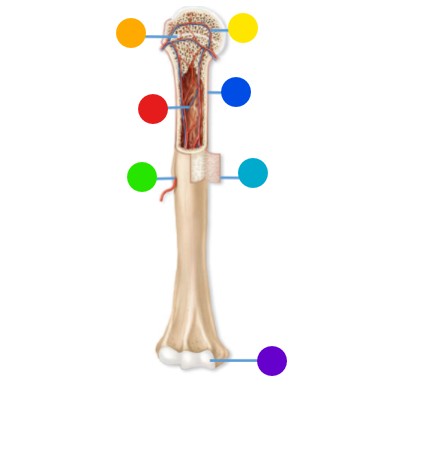
What is red pointing at
Medullary cavity
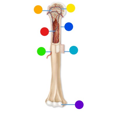
What is green pointing at
nutrient foramen
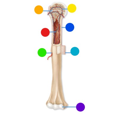
What is blue pointing at
compact bone
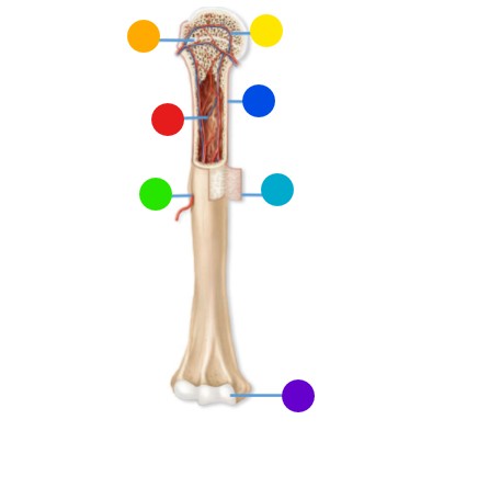
What is aqua pointing at
Periosteum
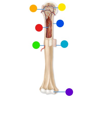
What is purple pointing at
Articular cartilage
What is the structural unit of a compact bone
osteon
Describe an osteon
structural unit of compact bone
Cylindrical structures arranges parallel to the long axis of the bone
Each contains a haversian canal that contains blood vessels and nerves
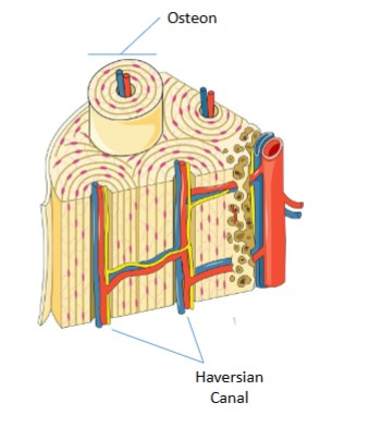
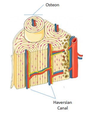
Label the two arrows
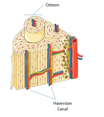
How many layers are in periosteum
2
Outer fibrous layer
Inner cellular layer
describe the outer fibrous layer of periosteum
Fibroblasts and dense connective tissue
Describe the cellular layer of periosteum
contains progenitor cells that can become osteoblasts
Endosteum
similar in structure to periosteum but found on the INNER surface of bones
Structural unit of a spongy bone
trabecular
describe a trabecular
thin bony plates
Made of lamella containing osteocytes in lacunae
receives nutrients via diffusion from blood vessels in bone marrow
Describe short bones and give examples
cube shaped; carpals and tarsals
describe sesamoid bones and give an example
bones found within tendons
Which type of bone is more metabolically active
trabecular (spongy) → more surface area
Primary osteoporosis
age relates bone loss
type 1: post-menopausal
type 2: elderly
Secondary osteoporosis
loss due to secondary causes (lifestyle, medication, disease)
What are the three main types of joints
Fibrous
Synovial
Cartilaginous
Describe fibrous joints
joints connected by ligaments
what type of joint is connected by ligaments
fibrous joints
What are the types of fibrous joints
Sutures, syndesmosis, Gomphosis
Define suture joints
Fibrous
Connects flat bones of skull
Define syndesmosis joints
fibrous
connects shafts of adjacent long bones
Radius and ulna
Tibia and fibula
Define gomphosis joints
fibrous
Holds teeth in their sockets
Define cartilaginous joints
joints connected by cartilage
Define a symphysis joint
cartilaginous joint
comprised of fibrous cartilage
Pubic symphysis
Intervertebral discs of spine
Define synchondrosis joints
cartilaginous joint
comprised of hyaline cartilage
Epiphyseal growth plates
What are the features of a synovial joint
Joint capsule
Fibrous capsule surrounding fluid filled cavity
Synovial membrane
Secretes a lubricative fluid into cavity
Joint cavity
Filled with synovial fluid
Articular cartilage
Hyaline cartilage covering the ends of long bones
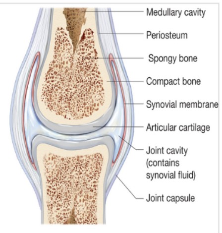
Diarthrosis
joint with high mobility (synovial)
Amphiarthrosis
joint with slight mobility (synchondrosis, symphysis)
Synarthrosis
joints with very low to no mobility (suture, gomphosis)
Agonist (muscles)
prime mover of a joint or structure
Synergist
muscles that work together (with agonist) to produce movement
Fixator
muscles that hold the joint steady so that agonists and synergists can produce movement
Antagonist
performs the OPPOSITE action as the agonist to control the speed of a movement
Composition of fascia (cells, fibers, fluids)
Cells
Mostly fibroblasts
Secrete fibers and ground substance
Fibers
Collagen
Gives structural integrity and make up largest proportion of tissue
Elastin
gives stretch and recoil
Reticular
Gives support to cells
Ground substance
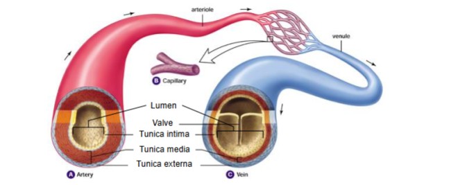
Arteries vs veins
Arteries
Narrower lumen
No valves
Tunica media thickest
More elastic
Veins
Larger lumen
Valves
Tunica externa thickest
less elastic