Vet systems unit 2
 Call Kai
 Learn
Learn Practice Test
 Spaced Repetition
 Match
Match Flashcards
 Knowt Play
1/130

There's no tags or description
Looks like no tags are added yet.
Name | Mastery | Learn | Test | Matching | Spaced | Call with Kai | Chat |
|---|
No analytics yet
Send a link to your students to track their progress
131 Terms
Slide deck 1 ( CARDIAC AND CIRCULATORY MICROANATOMY LC 19)
…
What are the 3 layers of the heart wall?
OUTER
Epicardium (Outer layer):
Mesothelium & connective tissue & fat
Contains coronary vessels and nerves
Continuous with visceral pericardium
Myocardium (Middle Layer):
• Thickest layer = cardiac muscle cells
• Branching fibers, central nuclei, intercalated discs
• Responsible for contractile force
Endocardium (inner layer):
• Endothelium & connective tissue
• Smooth surface for blood flow
• Subendocardial region: conduction (Purkinje Fibers)
INNER
What is the primary function of the endocardium? (Outer Layer)
The endocardium provides a non- thrombogenic, non-turbulent surface for blood flow
What are the layers/components of the endocardium?
→ Endothelium + subendothelial CT + subendocardial layer
What type of epithelium makes up the endocardium?
Simple squamous epithelium (endothelium)
What can be found in the subendocardial layer of the endocardium?
→ Purkinje fibers, which conduct cardiac impulses
What is the myocardium? What is it composed of?
The myocardium is the thickest layer of the heart
Composed of cardiac muscle cells
Branching fibers with central nuclei
Connected by intercalated disk
Rich capillary network
What is the primary function of the myocardium? (Outer Layer)
Generates contractile force to pump blood
What is the epicardium?
→ The outer covering of the heart, consisting of mesothelium, connective tissue, and adipose tissue.
What type of epithelium covers the epicardium?
→ Simple squamous mesothelium
What structures are found within the epicardium?
→ Coronary vessels, nerves, connective tissue, and adipose tissue
continuous with the visceral pericardial sac
What is the basic structure of a heart valve (cusps/leaflets)?
endocardium covering a fibrous connective tissue core
What is the function of heart valves?
Ensure unidirectional blood flow.
What is the difference between AV and semilunar valves?
→ AV valves: atria → ventricles
→ Semilunar valves: ventricles → aorta/pulmonary artery
Which valves have chordae tendineae?
→ AV valves only
Semilunar valves (DO NOT)
→ AV valves only
Semilunar valves (DO NOT)
What do chordae tendineae connect?
→ AV valve leaflets to papillary muscles.
What is the cardiac skeleton?
→ A dense fibrous connective tissue framework forming the structural core of the heart.
What are the major functions of the cardiac skeleton?
Supports the valves and myocardium and provides electrical insulation between atria and ventricles.
How does the cardiac skeleton affect cardiac conduction?
→ It electrically insulates the atria from ventricles so conduction occurs through the AV node.
What is the general difference between arteries and veins?
Arteries: thicker tunica media
for pressure and flow regulation
Veins: thicker tunica adventitia
For support and capacitance
What are the 3 basic layers of a typical blood vessel wall?
Tunica intima
Tunica media
Tunica adventitia
What is the main function of the tunica intima?
Maintains a smooth, non-thrombogenic blood–tissue interface and helps regulate permeability, vascular tone, and clotting.
What makes up the tunica intima?
Endothelium
Simple squamous epithelium
subendothelial connective tissue
Thin connective tissue layer
internal elastic lamina (IEL)
Distinct in arteries (wavy elastic sheet)
(Less prominent or absent in veins)
What is the main function of the tunica media?
Regulates vascular diameter (vasoconstriction & vasodilation through smooth muscle), blood pressure, and blood flow distribution
What is the tunica media primarily composed of?
Concentric layers of smooth muscle cells with variable elastic fibers, reticular fibers, and proteoglycans.
What is the tunica adventitia primarily composed of?
Loose connective tissue
What are the 2 special structures in the tunica adventitia?
Vasa Vasorum
Small blood vessels that supply the outer walls of large arteries and veins.
Nervi varorsum
Autonomic nerves regulating smooth muscle tone
What is the function of the tunica adventitia?
Anchors the vessel to surrounding tissues and provides nutritional and neural support.
What is the primary function of elastic arteries?
Maintain continuous blood flow by storing energy during systole and releasing it during diastole.
Give an example of an elastic artery.
Aorta or pulmonary arteries, major branches (e.g. carotid artery)
What is the major structural feature of elastic arteries?
→ Numerous elastic lamellae in the tunica media.
Tunica intima: Prominent endothelium & internal elastic lamina
Tunica adventitia: Relatively thin with vasa vasorum & nerves
What is the primary function of muscular arteries?
→ Act as resistance vessels, controlling systemic vascular resistance and organ perfusion.
What is the major structural feature of muscular arteries?
(List what the other vessel layers do as well)
→ A thick tunica media composed mainly of smooth muscle.
Tunica intima: endothelium & prominent internal elastic lamina (IEL)
Tunica adventitia: collagen, elastic fibers, vasa vasorum (large arteries
What are arterioles?
→ The smallest arteries leading into capillary beds.
How many layers of smooth muscle are typically found in arterioles? what key function do these layers provide?
(recognize what the other vessel layers do as well)
Tunica Media: 1–3 layers of smooth muscle
Key site of resistance control
Tunica intima: Endothelium with thin subendothelial layer; IEL may be present in larger arterioles
• Tunica adventitia: Minimal, blends with surrounding connective tissue
What is the major function of arterioles?
→ They are the primary resistance vessels, regulating blood pressure and capillary perfusion through vasoconstocution/dialitation
What is the basic structure of a capillary?
→ Endothelium only (simple squamous cells), with possible pericytes (for support repair and regulation)
56. What are the 3 types of capillaries?
Continuous: tight junctions
Fenestrated: pores allow higher exchange
Sinusoidal (discontinuous): wide gaps incomplete basement membrane
What is the function of capillaries?
• Site of gas, nutrient, and waste exchange
• Regulate fluid balance and permeability depending on type
~5–10 μm (just wide enough for red blood cells to pass in single file)
What is the major functions of venules?
Drain blood from capillary beds
Regulate exchange & immune cell trafficking
(postcapillary venules)
Serve as capacitance vessels, holding much of
body’s blood volume
What is a major function of postcapillary venules besides blood drainage? how are they structured?
• Structure: Endothelium + thin connective tissue (little or no smooth muscle)
• Function: Major site of leukocyte migration (diapedesis) and inflammation
Collecting venules & small veins add________ of smooth muscle in the ____________layer
Add 1–2 layers of smooth muscle in
tunica media (discontinuous and very little
relative to lumen size)
What are examples of medium and large veins?
Femoral, jugular, portal, vena cava
What prevents backflow in many veins?
Valves (folds of intima) prevent backflow
What structure in medium and large veins helps return venous blood to the heart?
→ Venous valves and skeletal muscle pumps
Act as capacitance vessels
What is the function of lymphatic vessels?
• Collect interstitial fluid (lymph) & return it to venous circulation
• Transport immune cells (lymphocytes, antigen-presenting cells)
• Maintain tissue fluid balance
How are the lymphatic vessels walls structured?
• Very thin walls
• Endothelium with incomplete basal lamina
• Thin-to-no tunica media
• Tunica adventitia blends with CT
• Valves present for unidirectional flow
slide deck 2 ( Electrocardiophysoilogy)
How do cardiomyocytes conduct cell signaling?
cardiomyocytes rely on direct electrical connection through gap
junctions (intercalated discs) that allow an action potential in one cell to travel to the next cell
Why are intercalated disk (gap juntions) important in cardiomyocyte excitation?
This is important, because it means that the cardiomyocytes will depolarize and contract in unified waves
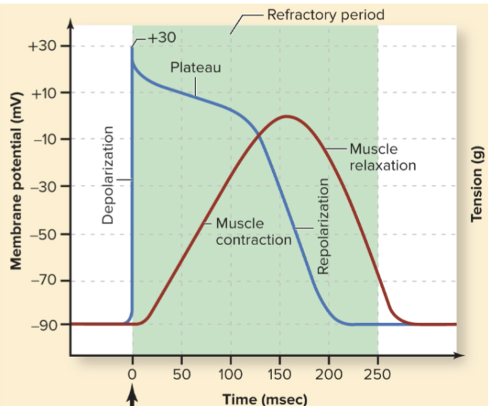
Why is the phase 2 plateau important in cardiac muscle?
It prolongs the action potential and refractory period, allowing the cardiac cell to completely contract before relaxing and helping prevent tetanic contraction.
How does an action potential cause cardiomyocyte contraction?
Depolarization opens L-type Ca²⁺ channels → Ca²⁺ enters the cell → triggers additional Ca²⁺ release from the sarcoplasmic reticulum → Ca²⁺ binds troponin C → allows actin-myosin cross-bridge formation → contraction.
Trace the normal pathway of electrical excitation through the heart.
SA node → atrial myocardium → AV node → bundle of His → bundle branches → Purkinje fibers → ventricular myocardium.
Key thing to memorize:
👉 SA starts → AV delays → His/branches/Purkinje spread → ventricles contract.
What are the intrinsic pacemaker rates of the major cardiac conduction tissues, and why does the SA node normally control heart rate?
SA node: ~100/min
AV node: ~40/min
Purkinje fibers: ~25/min
The SA node dominates because it depolarizes spontaneously at the fastest rate. (Without any nervous system input.
What sends the pacemaker cells towards threshold?
During phase 4, the HCN ("funny") current gradually depolarizes the cell toward threshold. Calcium channels then contribute to the action potential, while K⁺ channels contribute to repolarization.
What determines how quickly the SA node reaches threshold and therefore heart rate?
How quickly the SA node depolarizes depends on how steep the phase 4 current is
Q: How do sympathetic and parasympathetic stimulation affect SA-node activity?
Steeper phase 4 → threshold reached faster → ↑ heart rate
Flatter phase 4 → threshold reached slower → ↓ heart rate
How do sympathetic and parasympathetic stimulation affect SA-node activity?
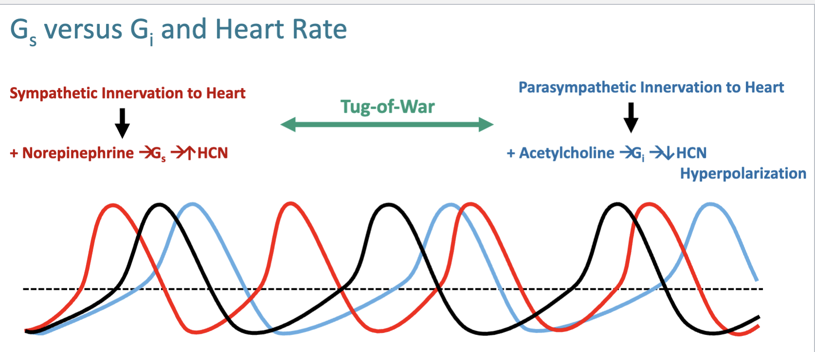
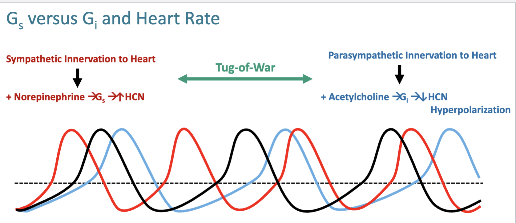
Sympatheticinnervation releases norepinephrine and activates β-
adrenoceptors (Gs/↑cAMP) on the SA node, AV node, and bundle branches to increase heart rate and contractility
(Gs/↑cAMP): bind to HCN and makes the heart beat faster do to increased depolarization
Parasympathetic innervation releases acetylcholine and activates M2 receptors (Gi/↓cAMP) on the SA node, AV node, and bundle branches to decrease heart rate and contractility
Why Does Sympathetic Stimulation Increase Heart Rate?
Norepinephrine → β₁ receptor → Gs → ↑ adenylyl cyclase → ↑ cAMP → ↑ HCN activity → steeper phase 4 → faster threshold → ↑ heart rate.
Why Does Parasympathetic Stimulation Decrease Heart Rate?
Acetylcholine → M₂ receptor → Gi → ↓ cAMP → ↓ HCN activity, plus activation of GIRK K⁺ channels, causing hyperpolarization → slower phase 4 → ↓ heart rate.
How do β-blockers decrease heart rate?
They block β₁-adrenergic receptors, reducing sympathetic signaling → ↓ Gs/cAMP signaling → ↓ HCN activity → less/ slower SA-node depolarization → ↓ heart rate. They are classified as Class 2 antiarrhythmics.
What are the major antiarrhythmic classes and their primary targets?

What is the function of an ECG? (ELECTROCARDIOGRAM)
Allows us to visualize the electrical activity occurring in the heart and aids in diagnosing electrical abnormalities
ECG is measured as ______ vs time
voltage
Voltage is a vector: it has both ________ and ________ information
Voltage is a vector: it has both magnitude and direction information
What is magnitude and direction in an ECG defined as?
Magnitude: Voltage measured is proportional to the mass of tissue
depolarizing
• Direction: Voltage measured is affected by the angle of
measurement
the highest voltage will be recorded when the angle of measurement = the angle of current propagation
How are leads placed on the four limbs?
Lead 1: RA (-), LA (+)
Lead 2: RA (-), LL (+)
Lead 3: LA (-), LL (+)
** White on the right, snow over grass
** Smoke over fire
One lead is designated the reference lead (-) and one lead is designated the recording lead (+) and the potential difference from reference to recording is measured (- to +)

The three ECG leads create __________ triangle
Einthoven’s triangle
If the SA Node is in control of the heart, there should be:
• A _____for every ______ – the ventricles shouldn’t depolarize unless the ______ depolarized first
• A QRS for every ____ – when the atria depolarize the ________ always depolarize
If the SA Node is in control of the heart, there should be:
• A P for every QRS – the ventricles shouldn’t depolarize unless the atria depolarized first
• A QRS for every P – when the atria depolarize the ventricles always depolarize
If there is not a P for every QRS what does this suggest?
If there is not a P for every QRS, this
suggest that the ventricles are depolarizing on their own
Vice president is conducting business
What direction should the angle of the mean electrical axis point?
left ventricle
The left ventricle should be significantly larger than the right
ventricle in the healthy heart, so the angle should point toward the left ventricle
Why should you always confirm with your patients physically and not rely solely on the ECG?
ECG only shows us what is going on electrically in the heart, it
DOES NOT show us whether the heart is actually beating
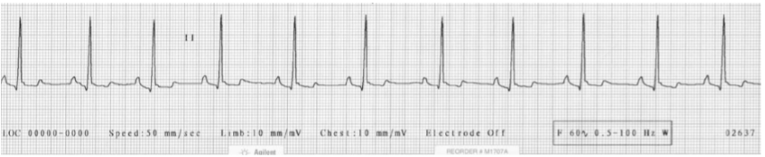
This ECG is an example of a ________________
(give the definition and features)
Respiratory Sinus Arrhythmia
If it is more than a 10% deviation between QRS complexes it is a sinus Arrhythmia
Can be caused by normal respiration – change in
intrathoracic pressure changes vagal tone
• Very common in brachycephalic dogs
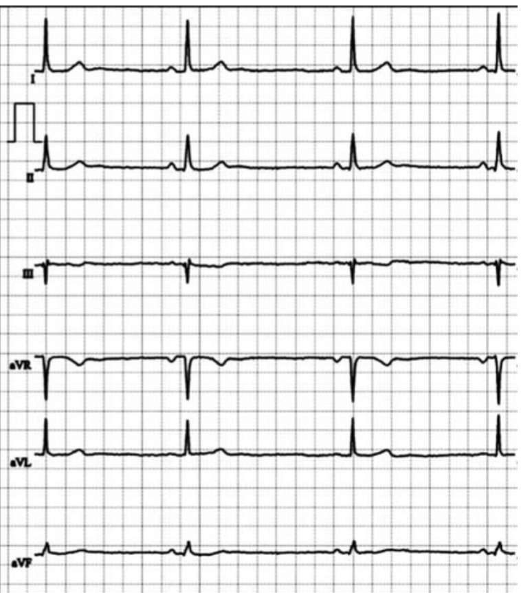
This ECG with prolonged RR intervals in an example of a______________
(give the definition and features)
Sinus Bradycardia
Sinus rhythm with a heart rate below normal species range
ECG features: Regular sinus rhythm, normal P-
QRS-T morphology, but prolonged RR intervals
(slow rate)
What causes sinus bradycardia and what is the treatment?
Causes:
• Physiologic: High vagal tone (athletic dogs, sleep)
• Pathologic: Hypothermia, hypothyroidism, hyperkalemia
• Drug-induced: β-blockers, Ca²⁺ channel blockers, digoxin, opioids
Treatment:
Often none if asymptomatic; treat underlying cause or atropine (speed up heart) if unstable
This ECG is an example of ________________
(give the definition and features)
Sick Sinus Syndrome
• Definition: Dysfunction of the SA node causing intermittent failure of impulse initiation or conduction.
• ECG features: Highly irregular sinus rhythm; pauses/sinus arrest; may alternate with atrial tachyarrhythmias.
What are the causes and treatment of sick sinus syndrome?
Causes:
• Structural: Fibrosis, degenerative SA node disease.
• Inherited: Familial cases reported in certain breeds.
• Idiopathic (means a disease or condition that arises from an unknown or obscure cause)
Treatment:
Often requires pacemaker placement if symptomatic or causing syncope.
What is an AV block?
Impaired conduction of atrial impulses through the AV node, leading to delayed/absent QRS.
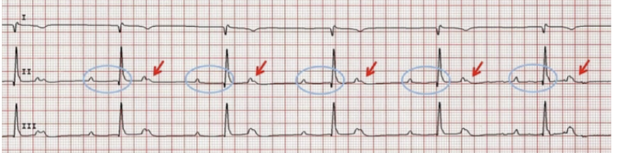
Which degree AV block is this? What is featured on this ECG?
1st degree: Prolonged PR interval
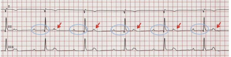
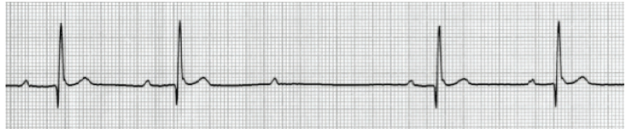
Which degree AV block is this (list the two sub types)? What is featured on this ECG?
2nd degree: Intermittent dropped QRS.
• Mobitz I (Wenckebach): Progressive PR lengthening
until a beat is dropped.
• Mobitz II: Constant PR interval with sudden dropped QRS
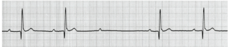
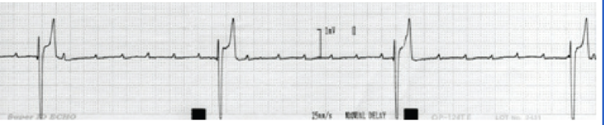
Which degree AV block is this? What is featured on this ECG?
3rd degree: No relationship between P waves and QRS;
ventricular escape rhythm.

What are the causes of AV blocks and how are they treated?
Causes:
• Physiologic: High vagal tone.
• Pathologic: Myocarditis, endocarditis, cardiomyopathy,
degenerative conduction disease.
• Drug-induced: Digitalis toxicity, β-blockers, calcium channel
blockers.
•Treatment:
• None if asymptomatic or mild
• Atropine trial may help vagally mediated cases.
• Symptomatic advanced 2nd or 3rd degree: pacemaker

This ECG is an example of ________________
(give the definition and features)
supraventricular premature contractions (tachycardia)
• Definition: Early atrial depolarizations that occur before the expected sinus beat, producing a premature “extra”
contraction.
• ECG features: premature P wave, Shortened T–P interval before premature beat.

What are the causes of supraventricular premature contractions and how are they treated?
Causes:
• Structural: Atrial disease, dilatation, or fibrosis.
• Metabolic: Electrolyte imbalances.
• Other: Sympathetic stimulation, hypoxia, systemic disease.
Treatment:
• Often none if isolated and asymptomatic.
• Treat underlying cause if frequent or associated with clinical signs.
• In some cases, antiarrhythmics may be considered.

This ECG is an example of ________________
(give the definition and features)
Atrial fibrillation (A-Fib)
• Definition: Disorganized atrial electrical activity causing ineffective atrial contraction and irregular ventricular
response.
• ECG features: No distinct P waves (replaced by fibrillatory waves), irregularly irregular R-R intervals, ventricular
rate often rapid (tachycardic).
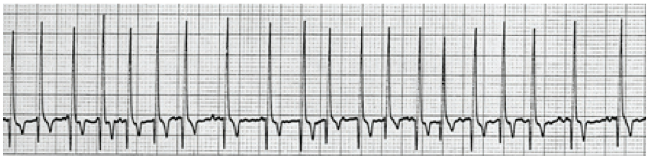
What are the causes of Atrial fibrillation (A-Fib) and how are they treated?
Causes:
• Structural: Atrial enlargement (mitral valve disease, cardiomyopathy).
• Fibrosis/scarring: Post-inflammatory or degenerative.
• Other: Hyperthyroidism, systemic disease, idiopathic in large-breed dogs.
Treatment:
• Rate control (β-blockers, Ca²⁺ channel blockers, digoxin).
• Rhythm control less common in veterinary patients (antiarrhythmics, cardioversion).
• Manage underlying cause when possible.
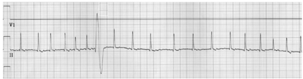
This ECG is an example of ________________
(give the definition and features)
Ventricular Premature Contraction (VPC)
• Definition: Premature ventricular depolarization originating from the ventricles, producing an abnormal,
widened QRS complex without a preceding P wave.
• ECG features: Wide, bizarre QRS complex; discordant T wave; not preceded by a P wave; may occur singly, in
couplets, or runs (ventricular tachycardia).

What are the causes of Ventricular Premature Contraction (VPC) and how are they treated?
Causes:
• Structural: Cardiomyopathy, myocarditis, ischemic or scarred myocardium.
• Metabolic/Electrolyte: Hypokalemia, hypomagnesemia, acidosis.
• Drug-induced/Toxic: Digoxin, anesthetics, stimulants.
• Other: Hypoxia, systemic disease, high sympathetic tone, stress
Treatment:
• Often none if isolated/asymptomatic.
• Address underlying cause (structural disease, electrolyte imbalance).
• Antiarrhythmic therapy if frequent, complex, or associated with clinical signs.

This ECG is an example of ________________
(give the definition and features)
Ventricular Tachycardia (V-Tach)
Definition:
A rapid ventricular rhythm (>100 bpm) caused by abnormal electrical impulses originating from the ventricles, producing consecutive wide QRS complexes.
ECG features:
Very fast ventricular rate. Wide, abnormal (“bizarre”) QRS complexes. No associated or consistent P waves. May be sustained (>30 sec) or non-sustained (<30 sec).

What are the causes of Ventricular Tachycardia (V-Tach) and how are they treated?
Causes:
• Structural: Cardiomyopathy, myocarditis, ischemic/ scarred myocardium.
• Metabolic/Electrolyte: Severe hypokalemia, hypomagnesemia, acidosis.
• Drug-induced/Toxic: Digoxin, anesthetics, stimulants.
• Other: Hypoxia, severe systemic disease, sympathetic overdrive.
Treatment:
• Emergency stabilization if unstable (antiarrhythmic drugs (lidocaine), electrical cardioversion).
• Address underlying cause (structural, metabolic, toxic).
• Long-term: antiarrhythmic therapy, implantable defibrillator in refractory cases

This ECG is an example of ________________
(give the definition and features)
Ventricular Fibrillation (V-Fib)
Definition:
Chaotic, disorganized ventricular electrical activity with no effective contraction.
ECG features:
No identifiable P waves, QRS complexes, or T waves. Random, irregular, chaotic electrical oscillations.

What are the causes of Ventricular Fibrillation (V-Fib) and how are they treated?
Causes:
• Structural heart disease: cardiomyopathy, myocardial infarction/scarring.
• Severe electrolyte disturbances: hyperkalemia.
• Drug toxicity: digitalis, anesthetics.
• Severe systemic disease: hypoxia, acidosis.
Treatment (emergency):
• Immediate CPR and electrical defibrillation.
• Correct underlying reversible causes (e.g., electrolytes, drug toxicity).
• Antiarrhythmic therapy (e.g., amiodarone, lidocaine) as adjunct
Class 0 Antiarrhythmics :
HCN channel blockers
Rate Control (slow down)
ex.Ivabradine
Class 1a Antiarrhythmics :
Moderate Nav channel blockers
Multiple Tachyarrhythmias
ex. Quinidine
Class 1b Antiarrhythmics :
Mild Nav channel blockers
Ventricular Tachyarrhythmias
ex. Lidocaine, Mexiletine
Class 2 Antiarrhythmics :
Autonomics (β-blocker)
Rate Control (slow down)
ex. Propranolol, Atenolol, Carvedilol, Esmolol…
Class 2c Antiarrhythmics :
Autonomics (M2-antagonists)
Rate Control (speed up)
ex. Atropine
Class 3 Antiarrhythmics :
Kv channel blockers
Slow repolarization (prevent reentry)
ex. Sotalol (class II/III), Amiodarone
Class 4 Antiarrhythmics :
Slow (L-type) Cav channel blockers
Ventricular Rate Control (Supraventricular Tachycardias)
ex. Diltiazem Verapamil
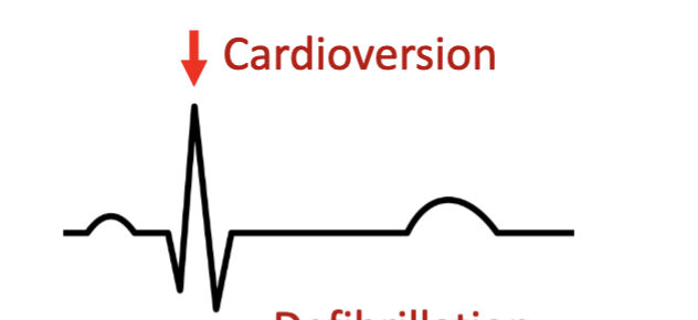
Cardioversion (synchronized shock) is timed with________
• Timed with the R wave of QRS to avoid inducing V-fib
• Used for organized tachyarrhythmias
(atrial fibrillation/flutter, supraventricular tachycardia, stable
VT)
• The shock coordinated with the R
wave may depolarize the entire heart,
allowing it to reenter the cardiac
electrical cycle together and reenter
normal rhythm